
Skin Conditions of the
Cavalier King Charles Spaniel
-
 List
of Disorders
List
of Disorders - Symptoms
- Diagnosis
- Treatment
- Veterinary Specialists
- Related Links
- Veterinary Resources
Cavalier King Charles spaniels have been reported to be pre-disposed to certain conditions affecting their skin. We list here those which have been identified and described in published veterinary literature.
We do not include all skin conditions common to many breeds, but only those specifically reported as especially affecting CKCSs.
List of Disorders
Skin conditions in the cavalier include:
- Canine atopic dermatitis (atophy)
- Cheyletiella dermatitis (Cheyletiellosis)
- Copper associated hepatopahty (CAH)
- Curly Coat / Rough Coat syndrome
- Malassezia dermatitis
- Necrolytic dermatitis
- Pemphigus foliaceus
- Piebaldism
- Sebaceous adenitis
- Skin cancers
Canine atopic dermatitis (atophy)
 Canine atopic dermatitis
(CAD), or atophy, is due to an inherited tendency to develop
IgE antibodies in response to exposure to allergens that
are inhaled or absorbed through the skin.
Canine atopic dermatitis
(CAD), or atophy, is due to an inherited tendency to develop
IgE antibodies in response to exposure to allergens that
are inhaled or absorbed through the skin.
In a December 2016 article, a team of University of California-Davis reasearchers examined the medical records from 1995 to 2010 of 90,090 dogs of all AKC-recognized breeds treated at their veterinary hospital, to determine the relation between neuter status and autoimmune diseases. They report finding that neutered dogs had a significantly greater risk of atopic dermatitis.
RETURN TO TOP
Cheyletiella dermatitis (Cheyletiellosis)
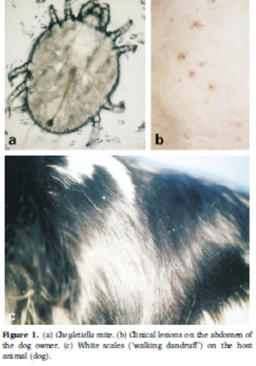
Cheyletiellosis (Cheyletiella dermatitis; walking dandruff) is a highly contagious skin disorder (dermatosis) caused by an infestation of the surface-dwelling Cheyletiella spp, a parasitic mite. They live on the dog's (and human's) skin and feed on surface debris and fluids. The females shed eggs into the host animal's hair. The primary symptoms are itching, scaling mainly on the dog's back, and redness of the skin.
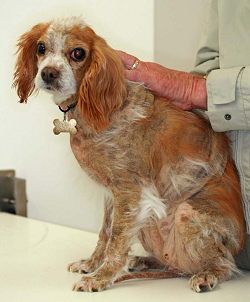
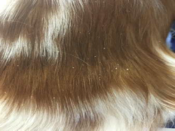
 In a
November 2000 article, Vienna, Austria veterinary dermatologists
diagnosed Cheyletiella dermatitis on the back of a cavalier King Charles
spaniel (right) with a two-month long history of excessive itching. They
observed white scales on the dog's back, although the skin looked
normal. Microscopic examination showed live mites and their eggs. The
dog's owner also was infested, with her legs and abdomen covered with
itchy red bumps. The cavalier was treated first with dips of Bayer's
phoxim (Baythion, Baymite, Sebacil, Valexone) a highly toxic
organophosphate insecticide available to veterinarians in Europe. Since
the dog continued itch somewhat, she then was treated with moxidectin
(Advantage Multi, ProHeart). The signs and itching eventually
disappeared.
In a
November 2000 article, Vienna, Austria veterinary dermatologists
diagnosed Cheyletiella dermatitis on the back of a cavalier King Charles
spaniel (right) with a two-month long history of excessive itching. They
observed white scales on the dog's back, although the skin looked
normal. Microscopic examination showed live mites and their eggs. The
dog's owner also was infested, with her legs and abdomen covered with
itchy red bumps. The cavalier was treated first with dips of Bayer's
phoxim (Baythion, Baymite, Sebacil, Valexone) a highly toxic
organophosphate insecticide available to veterinarians in Europe. Since
the dog continued itch somewhat, she then was treated with moxidectin
(Advantage Multi, ProHeart). The signs and itching eventually
disappeared.
See also this December 2002 article, which concludes that selamectin successfully treated cheyletiellosis in a family of nine cavaliers.
RETURN TO TOP
Copper associated hepatopahty (CAH)
The cavalier breed has been found to be predisposed to copper associated hepatopahty (CAH). CAH describes excessive quantities of the metal copper (Cu) in the dog's liver. Copper normally is processed by the liver into bile by certain proteins. When the dog's liver is unable to convert copper, it accumulates in the liver and causes inflammation, scarring of the liver, and death of liver cells. Symptoms of CAH rarely are evident before permanent damage to the liver's cells occur.
CAH is discussed at greater length on our Liver Disorders webpage.
RETURN TO TOP
Curly Coat / Rough Coat syndrome
Curly coat syndrome is a severe congenital condition of the skin, coat, claws, and eyes in some cavalier King Charles spaniel puppies. It is also known as rough coat syndrome and its scientific name is ichthyosis keratoconjunctivitis sicca and also as congenital keratoconjunctivitis sicca and ichthyosiform dermatosis (CKCSID).
Curly coat syndrome is discussed at greater length on our Curly Coat webpage.
RETURN TO TOP
Malassezia dermatitis
Malassezia dermatitis consists of yeast infections, for which the CKCS has been found to be at increased risk. See these veterinary reports for details about this disorder in cavaliers.
RETURN TO TOP
Necrolytic dermatitis
Superficial necrolytic dermatitis (SND) typically appears as thickened, crusting, ulcerative lesions on the dogs' paw pads, but also may appear at other pressure points. such as elbows, and at mucus-to-skin boundaries, such as the nose and lips, anal, and/or genital regions. It is a form of hyperkeratosis.
It most often is associated with a liver disorder, hepatocutaneous syndrome (HCS), in which severe lesions appear as nodules on the surface of the liver. HCS is a progressive disorder which usually is fatal. Therefore, treatment focuses upon the liver aspects of this disorder. See our Liver Disorders webpage for more information about this combination of disorders. See also this January 2023 article.
RETURN TO TOP
Pemphigus foliaceus
 Pemphigus foliaceus (PF) is an autoimmune skin disease, in which the
body’s immune system attacks the connections between the layers of skin
cells. The affected dog's immune system incorrectly recognizes a
component of skin, desmocollin-1, a component of desmosomes, as being a
foreign invader and then produces antibodies which bind to the the
desmosomes and destroy them. The destruction causes the outer layer of
skin to split and fill with fluid, leading to pustules and blisters,
which rupture, typically resulting in hair loss, scabs, and ulcers. It
is the most common autoimmune skin disease in dogs, most often observed
in middle-aged and older dogs.
Pemphigus foliaceus (PF) is an autoimmune skin disease, in which the
body’s immune system attacks the connections between the layers of skin
cells. The affected dog's immune system incorrectly recognizes a
component of skin, desmocollin-1, a component of desmosomes, as being a
foreign invader and then produces antibodies which bind to the the
desmosomes and destroy them. The destruction causes the outer layer of
skin to split and fill with fluid, leading to pustules and blisters,
which rupture, typically resulting in hair loss, scabs, and ulcers. It
is the most common autoimmune skin disease in dogs, most often observed
in middle-aged and older dogs.
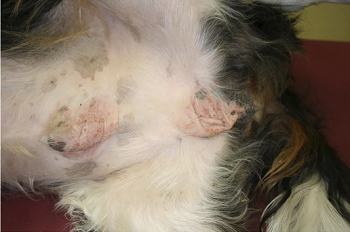
In a September 2019 article, a team of Colorado State veterinary researchers report the case of a 9 year old castrated male cavalier with a 6 month history of pruritus and crusting, affecting the paw pads, claw folds, prepuce, scrotum, ear leathers, and muzzle. Pemphigus foliaceus was confirmed by a biopsy. The cavalier had been treated unsuccessfully with prednisolone and mycophenolate, also resulting in muscle wasting and other unwanted side effects. Polysulfated glycosaminoglycan (PSGAG) then was injected under the skin for four days, along with oral doses of prednisolone and cyclosporine. After four months, the PSGAG treatments resulted in healing of the lesions after 40 days. PSGAG injections were reduced to one a week, and reduced doses of prednisolone and cyclosporine continued. Within two weeks, the PF recurred, and treatments were increased. After two injections of PSGAG, the lesions healed. Six months later, crusting appeared again on the muzzle, scrotum, and paws. Treatment frequencies were increased, and within two months the crusting healed. Frequency of treatment with prednisolone then was decreased but the dog continued to be treated with cyclosporine at the same rate. No further signs of PF were present after a total of 21 months of treatments. [See photo at right of the patient before treatment with PSGAG.]
RETURN TO TOP
Piebaldism
Piebaldism is a benign genetic disease caused by a mutation which
results in the patches of fur which lack pigment.
 It
is one of a series of defects called "neurocristopathies", some of which
result as cancer of the nervous system, deafness, digestive problems, or
holes in the heart, which are caused by cells not moving to the right
place as an embryo develops. Piebaldism is caused by a mutation in a
gene called Kit.
It
is one of a series of defects called "neurocristopathies", some of which
result as cancer of the nervous system, deafness, digestive problems, or
holes in the heart, which are caused by cells not moving to the right
place as an embryo develops. Piebaldism is caused by a mutation in a
gene called Kit.
The mutation causes melanocytes cells in the early embryo to fail to migrate correctly. Melanocytes are responsible for pigmentation of hair and of skin. These cells start at the back of the embryo and they try to migrate around through the skin and cover the whole of the embryo's skin. When arkly colored pigment cells do not proliferate enough -- not making enough daughter cells to colonize or cover the whole region of the skin that needs to be covered by the time the pigmentation pattern is set down -- regions of skin or hair result in lacking pigment, usually at the front of an animal.
In particular, regarding the association of piebaldism and deafness, cavaliers are potentially subject to pigment-associated congenital sensorineural deafness, which should be evident in the puppy age span. See our deafness webpage for more information.
In a March 2017 article, the investigators include a photo of a cavalier with axial depigmentation, a form of piebaldism. See photo at right.
RETURN TO TOP
Sebaceous adenitis
Sebaceous adenitis is an inflammatory reaction which damages the sebaceous glands of the skin. The sebaceous glands produce an oily secretion called sebum, which serves in part to hydrate the skin, particularly in haired areas. Sebaceous adenitis causes the dog's coat of hair to become dry and brittle. It has been identified in the cavalier and a few other breeds, particularly spaniels.
RETURN TO TOP
Skin cancers
Cancers of the skin include carcinoma, mastocytoma, lymphoma, mast cell tumors, and melanoma. All of these cancers are discussed at greater length on our Cancer webpage.
RETURN TO TOP
Symptoms
Several of these skin disorders have common clinical signs, particularly the crusting or scaling of the skin. Scaling consists of flaking loose skin. Crusting is an accumulation of several different things, such as dried serum, pus, exudate, blood, bacteria, and yeasts. Pus on the skin is called pyoderma.
Itchiness is called pruritis, and while not all of the disoders listed here cause itchiness, several of them do. Pruritus is measured using any of a variety of scales, all involving collecting observations from the dogs' owners.
More severe symptoms include openings through the skin surface through which fluids discharge. These may be draining tracks or ulcers and lesions.
RETURN TO TOP
Diagnosis
Because so many different skin disorders have the common clinical sign of the crusting or scaling of the skin, crusting is not a diagnosis or even specific for certain conditions. Since the external appearance of the skin is only symptom and not the disease itself, thorough general and dermatological examinations of the dog need to be performed to diagnose the underlying disease.
Hair, saliva, and/or serum testing were not reliable at diagnosing specific food reactions in dogs. Elimination diets have been found to be effective in identifying food reactions. However, food allergies are only one of several possible underlying causes of skin disorders.
RETURN TO TOP
Treatment
The courses of treatment will depend upon the specific diagnoses.
Glucocorticoids, including triamcinolone acetonide (Vetalog, Kenalog, Cortalone, TriamTabs, Aristocort), which is an anti-inflammatory and immune suppressant.
Oclacitinib (Apoquel), a novel Janus kinase (JAK) inhibitor, has been approved for the control and treatment of atopic dermatitis (AD) in dogs 12 months of age or older. In a June 2015 article, oclacitinkb has been shown to be "safe and efficacious for long-term use and improved quality of life in the 247 dogs in the study, including cavaliers. This study was sponsored by Zoetis, the maker of Apoquel. In a June 2023 article, USA specialists concluded that, "Oclacitinib has proven to be a great tool in our armamentarium and has opened countless opportunities for an alternative option to the use of glucocorticoids."
Lokivetmab (Cytopoint) is a mouse-produced antibody that is licensed in the UK for treating CAD, also manufactured by Zoetis. It provides a more targeted (to IL-31, the cytokine which prompts the itch sensation in the brain) than oclacitinib, is more fast acting, and has a better safety record. It has been found to be as effective as cyclosporin for dealing with pruritis and inflammation associated with CAD. It is injected under the skin with follow up injections no sooner than once a month.
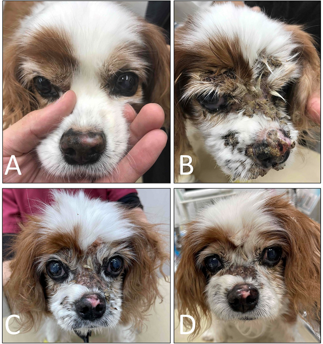
In a January 2022 article, Japanese clinicians reported the successful treatment of a 13-year-old cavalier diagnosed with cutaneous epitheliotropic lymphoma (CEL) resulting in severe facial pruritus, alopecia, erythema, erosion, and ulceration with crust. Once treatments with other medications, including prednisolone, were unsuccessful, lokivetmab was injected periodically. The lokivetmab decreased pruritus. (See photos below.) The authors concluded that anti-IL-31 therapy appears to be a potential treatment option in controlling pruritus associated with CEL in dogs.
 (A)
(A) On Day 14, nasal depigmentation had emerged. (B) On Day 105,
progression of facial alopecia with crusting was found. (C) On Day
132, facial alopecia with crusting was still present. (D) On Day
160, hair regrew on the face where alopecia was present.
(A)
(A) On Day 14, nasal depigmentation had emerged. (B) On Day 105,
progression of facial alopecia with crusting was found. (C) On Day
132, facial alopecia with crusting was still present. (D) On Day
160, hair regrew on the face where alopecia was present.
Palmitoylethanolamide (PEA) is a
N-acylethanolamine molecule in a family of long-chain fatty acid
 amides
called ALIAmides. PEA has been found in rat and mice studies to limit
hyperactvity in immune cells and thereby control inflammatory responses
and resulting tissue damage. PEA is produced by the animal's body as
needed in response to certain types of injuries. PEA is a product of
normal fatty acid synthesis from palmitic acid. It is found in many
common foods, particularly palm oil, soy beans, egg yolks, and peanuts.
The commercial version is most commonly manufactured from palm oil*.
amides
called ALIAmides. PEA has been found in rat and mice studies to limit
hyperactvity in immune cells and thereby control inflammatory responses
and resulting tissue damage. PEA is produced by the animal's body as
needed in response to certain types of injuries. PEA is a product of
normal fatty acid synthesis from palmitic acid. It is found in many
common foods, particularly palm oil, soy beans, egg yolks, and peanuts.
The commercial version is most commonly manufactured from palm oil*.
Not all PEA is alike. There are three types of PEA.
• Basic PEA, called "naive PEA", is insoluble in water and therefore the oral intake of it (rather than being injected directly into the abdomen) has very poor bioavailability, meaning that it does not get absorbed well in the dog's gut.
• Micronized PEA (m-PEA) is a patented technique that reduces the diameter of PEA particles, making them absorbable in the intestine, which has been found to be more effective than ordinary naive PEA in activating PEA levels in blood plasma in dogs. See this August 2014 article.
• Ultra-micronized PEA (um-PEA), also patented, reduces the PEA particle size further, to enable it to cross the blood-brain barrier, likewise has been found to be much more effective than naive PEA. See this August 2014 article.
 If a PEA product is not advertised as being micronized or
ultra-micronized, then
Dr. Clare Rusbridge advises that
"You
probably are wasting your money."
A variety of brands of
micronized and ultra-micronized PEA are offered on-line.
If a PEA product is not advertised as being micronized or
ultra-micronized, then
Dr. Clare Rusbridge advises that
"You
probably are wasting your money."
A variety of brands of
micronized and ultra-micronized PEA are offered on-line.
Recent research has produced evidence that ALIAmides can relieve dogs with hypersensitive skin disorders. In an August 2015 article, Italian researchers conducted an 8-week study of the effectiveness of oral ultra-micronized palmitoylethanolamide (um-PEA) in 160 dogs with moderate atopic dermatitis. Each dog received a daily dose of um-PEA at the rate of 10 mg/kg for 56 days. They report finding that um-PEA appeared to be effective and safe in reducing pruritus and skin lesions, and in improving the quality of life in dogs with moderate atopic dermatitis and moderate pruritus. This study was sponsored by patent holders of the use of PEA for inflammation and pain.
As for dosages, the studies using micronized PEA, the range was from 10 to 15 mg/kg/day, and the range for ultra-micronized was 24 mg/kg (for osteoarthritis in humans).
* Palm oil: The palm oil cultivation industry has been destroying rainforests in Sumatra and Borneo in Indonesia and Malaysia, the only habitats of orangutans. If you are going to obtain PEA, we suggest that you do so only from vendors whose PEA has been manufactured with palm oil from sustainable sources and not the deforestation of rainforests. This link connects to a "PalmOil Scan Mobile App" which will enable you to determine if the PEA vendors you select obtain their palm oil from sustainable sources.
 Cannabinol (CBD
oil) is produced from hemp and marijuana (Cannabis
Sativa) plants. CBD oil mimics the endocannabinoid molecules which the
dog’s (and our) body produces in several different organs. They play
roles in reducing pain, regulating inflammation, and affecting the
immune system, by initially binding to receptors in the brain.
Recommended dosages very widely, from 0.25 mg per kg per day to as much
as 9 mg/kg/day (for epilepsy). However, the maximum recommended dose for
adult humans is only 0.15 mg/kg/daily, the equivalent of 5 drops of 5%
CBD oil.
Cannabinol (CBD
oil) is produced from hemp and marijuana (Cannabis
Sativa) plants. CBD oil mimics the endocannabinoid molecules which the
dog’s (and our) body produces in several different organs. They play
roles in reducing pain, regulating inflammation, and affecting the
immune system, by initially binding to receptors in the brain.
Recommended dosages very widely, from 0.25 mg per kg per day to as much
as 9 mg/kg/day (for epilepsy). However, the maximum recommended dose for
adult humans is only 0.15 mg/kg/daily, the equivalent of 5 drops of 5%
CBD oil.
Varieties of CBD: Cannabidiol-based veterinary products are derived mainly from hemp (Cannabis sativa) and must contain less than 0.3% tetrahydrocannabinol (THC). This form of CBD can be processed into “full spectrum” or “broad spectrum” and also may be in the form of a “distillate”, in which all THC has been removed, or in the form of CBD “isolate”, which is a purifed powder.
• Full Spectrum: Full spectrum CBD contains other extracts found in the cannabis plant, including terpenes, and up to 0.3% THC.
• Broad Spectrum: Broad spectrum CBD also contains some other cannabis compounds but no more than trace amounts of THC.
• CBD Isolate: CBD isolate is pure CBD and contains no other cannabis plant compounds.
• Naked CBD: Naked CBD describes CBD oil by itself, as opposed to being capsultated or microcapsulated or combined with any other substance, such as deoxycholic acid (DCA).
• Liposomal CBD: This is an orally administered encapsultated CBD which is packaged within liposomes, small fatty cellular sacs which improve bioavailability of the CBD by enabling it to be withstand digesstion in the stomach and degradation in the liver. Lipsomal CBD was tested on dogs in this September 2020 article.
• Cannabidiolic acid (CBDA) is an acid precursor of CBD. It forms CBD when heated. It has been shown in some studies to be more potent that CBD for treating rats. It has been found to be more readily absorbed into the human bloodstream than CBD. Aa theory is that adding CBDA to doses of CBD may make the CBD more absorbable. In this September 2020 article, the investigators found that CBDA is absorbed at least twice as well as CBD in dogs within a 24 hour period, with some differences depending upon the medium used to deliver the oral treatment.
 Thus far, the results of published studies in which cannabinoids
(CBD) have been used to treat this skin disorder have been mixed.
Thus far, the results of published studies in which cannabinoids
(CBD) have been used to treat this skin disorder have been mixed.
CBD and cannabidiolic acid (CBDA) were involved in a 2021 pilot study invoving 17 dogs diagnosed with atopic dermatitis. None were cavaliers. All continued to be treated also with conventional medications. The dogs were given an oral dose of CBD-rich and CBDA-rich hemp in sesame oil for four weeks. Subjective measurements of inflammation using the Canine Atopic Dermatitis Scoring Index and owners subjective scaling asseessments of pruritus were performed.
The dogs were administered CBD/CBDA (approximately 2 mg/kg) twice a day with a meal for the 28-day study period. The investigators report that a bare majority of the 17 dogs in the treatment group showed measurable decreases in pruritus. They listed these symptoms as "adverse events": lethargy, increased flatulence, inconsistent appetite, somnolence, sleepiness, decreased aggression, increased calmness, and increased energy/mobility.
During the 4 weeks, since the dogs also were treated with conventional medications, the investigators concluded by suggesting tht CBD be an "adjunct therapy". They stated:
"Our results suggest that CBD as an adjunct therapy is useful in decreasing pruritus in some dogs with cAD [canine atopic dermatitis]. CBD at 2 mg/kg twice daily was well-tolerated with minimal AEs [adverse events].
An April 2022 article reports the results of testing CBD on 8 dogs suffering from canine atopic dermatitis (CAD) over a period of 8 weeks. The investigators noted clinical improvement in both CADESI-4 and pruritus scores in the dogs treated with THC-free CBD oil twice daily for 8 weeks. The dogs also continued to be treated with other medications (including oclacitinib, anixucukkub, prednisolone, and ketoconazole).
To the contrary, in an October 2023 article, 7 dogs diagnosed with canine atopic dermatitis (CAD) were treated only with full spectrum high cannabidiol (CBD) cannabis oil, 2.5 mg/kg for 6 weeks. They report finding: "This study reveals that the full-spectrum cannabis oil rich in CBD at a dosage of 2.5 mg/kg does not show therapeutic advantage when compared to olive oil. "
See our Cannabis webpage for additional details about CBD, including delivery methods, bioavailability, dosages, and adverse reactions.
Vitamin D supplementation reportedly may aid in relief of CAD.
RETURN TO TOP
Veterinary Specialists
The American College of Veterinary Dermatology (ACVD) is empowered to examine qualified candidates and confer Diplomate (board certification) status in veterinary dermatology. Board certification requires completion of a 2-3 year approved residency training program, an original research project, publication in a scientific journal, and successful completion of the certification examination. Currently there are about three hundred ACVD board certified veterinary dermatologists worldwide who work in private specialty practices, academic positions, and industry.
RETURN TO TOP
Related Links
RETURN TO TOP
Veterinary Resources
Canine atopic dermatitis:
Breed and site predispositions of dogs with atopic dermatitis: a comparison of five locations in three continents. K. Jaeger, M. Linek, H.T. Power, S.V. Bettenay, S. Zabel, R.A.W. Rosychuk, Ralf S. Mueller. Vet. Dermatology. February 2010;21(1):119-123. Quote: "The objectives of this multicentre study were to analyse and compare breed predispositions and lesion distributions of 552 dogs diagnosed with atopic dermatitis from five different dermatologic referral centres located in Australia, Germany (2) and the United States (2). Breeds were compared with the canine population in the respective locations. Breed predispositions varied from geographical site, although golden retrievers and German shepherd dogs were predisposed in three of five practices. [Cavalier King Charles spaniels were predisposed in Australia.] Lesions were present most commonly on the paws (62%), ventrum (51%), ears (48%) and face (39%). Various breeds had specific site predilections. Based on this study, breed predispositions can vary greatly both between continents and also between different locations on the same continent. In addition, some breeds showed predispositions for certain body sites which also varied in some instances with the geographical location.
Micronized/ultramicronized palmitoylethanolamide displays superior oral efficacy compared to nonmicronized palmitoylethanolamide in a rat model of inflammatory pain. Daniela Impellizzeri, Giuseppe Bruschetta, Marika Cordaro, Rosalia Crupi, Rosalba Siracusa, Emanuela Esposito, Salvatore Cuzzocrea. Neuroinflammation. August 2014; doi: 10.1186/s12974-014-0136-0. Quote: Background: The fatty acid amide palmitoylethanolamide (PEA) has been studied extensively for its anti-inflammatory and neuroprotective actions. The lipidic nature and large particle size of PEA in the native state may limit its solubility and bioavailability when given orally, however. Micronized formulations of a drug enhance its rate of dissolution and reduce variability of absorption when orally administered. The present study was thus designed to evaluate the oral anti-inflammatory efficacy of micronized/ultramicronized versus nonmicronized PEA formulations. Methods: Micronized/ultramicronized PEA was produced by the air-jet milling technique, and the various PEA preparations were subjected to physicochemical characterization to determine particle size distribution and purity. Each PEA formulation was then assessed for its anti-inflammatory effects when given orally in the carrageenan-induced rat paw model of inflammation, a well-established paradigm of edema formation and thermal hyperalgesia. Results: Intraplantar injection of carrageenan into the right hind paw led to a marked accumulation of infiltrating inflammatory cells and increased myeloperoxidase activity. Both parameters were significantly decreased by orally given micronized PEA (PEA-m; 10 mg/kg) or ultramicronized PEA (PEA-um; 10 mg/kg), but not nonmicronized PeaPure (10 mg/kg). Further, carrageenan-induced paw edema and thermal hyperalgesia were markedly and significantly reduced by oral treatment with micronized PEA-m and ultramicronized PEA-um at each time point compared to nonmicronized PeaPure. However, when given by the intraperitoneal route, all PEA formulations proved effective. Conclusions: These findings illustrate the superior anti-inflammatory action exerted by orally administered, micronized PEA-m and ultramicronized PEA-um, versus that of nonmicronized PeaPure, in the rat paw carrageenan model of inflammatory pain.
Review: Clinical and histological manifestations of canine atopic dermatitis. Petra Bizikova, Domenico Santoro, Rosanna Marsella, Tim Nuttall, Melissa N. C. Eisenschenk, Cherie M. Pucheu-Haston. Vet. Dermatology. February 2015. Quote: "Background: Many studies focusing on clinical and histological signs of canine atopic dermatitis (AD) have been published since its early descriptions decades ago. Findings of these studies contributed to our current knowledge about the disease pathogenesis and allowed establishment of diagnostic criteria used by clinicians and researchers. Objectives: This review serves as an update on the clinical and histological features of canine AD published by the American College of Veterinary Dermatology Task Force on Canine Atopic Dermatitis in 2001 and summarizes the recent discoveries in these fields. Results: The overall findings of studies focusing on clinical features mirrored those published by the Task Force in 2001. The novelty was the larger number of animals included in these studies, which allowed establishment of a new set of diagnostic criteria that exceeded the sensitivity and specificity of the previous criteria. The same study uncovered some clinical differences between dogs with food-induced and nonfood-induced AD; however, the authors concluded that these two entities cannot be distinguished based on clinical signs only. Another study demonstrated some major breed-specific phenotypes. Several publications addressed the histological features of canine AD skin lesions in experimental models of AD, but none of those addressed naturally occurring lesions. Nevertheless, the histopathological description of the skin reactions was generally similar to that published by the Task Force in 2001. Conclusions: Considerable work has been done in recent years to provide a better definition of the clinical appearance and histopathology of canine AD. New sets of diagnostic criteria have been developed, and additional breed-associated differences in phenotypes have been demonstrated.
Increased numbers of peripheral blood CD34+ cells in dogs with canine atopic dermatitis. Vincent Bruet, Blandine Lieubeau, Julie Herve, Anne Roussel, Laëtitia Imparato, Jean-Claude Desfontis, Patrick Bourdeau. Vet. Dermatology. June 2015;26(3):160-e33. Quote: "Background: The bone marrow may be involved in human atopic diseases, as shown by the release of CD34+ cells into the peripheral blood. Hypothesis/Objectives: The aim was to determine the numbers of CD34+ cells in atopic dogs. Animals: The following three groups of dogs were studied: 27 dogs with nonfood-induced atopic dermatitis (NFICAD)[including a cavalier King Charles spaniel]; 16 dogs with nonallergic inflammatory diseases; and 13 healthy control dogs [including a cavalier King Charles spaniel]. Methods: Dogs with NFICAD were selected after fulfilment of Favrot's criteria and exclusion of other pruritic dermatoses, including flea infestation and adverse reaction to foods. The Canine Atopic Dermatitis Extent and Severity Index (CADESI)-03 and a Visual Analog Scale (VAS) score for pruritus were used to quantify clinical signs. A phycoerythrin-conjugated anticanine CD34 antibody was used to stain peripheral blood CD34+ cells, and these were enumerated using a flow cytometer. The CD34+ cell counts were compared between groups and tested (in the NFICAD group) for correlation with the severity of clinical signs. Results: The numbers of peripheral CD34+ cells in dogs with NFICAD (median 1.7) were statistically higher than in dogs with other nonallergic inflammatory diseases (median 1.0; P = 0.01) and healthy control dogs (median 0.9; P = 0.009). In dogs with NFICAD, there was no correlation between CD34+ cell numbers and CADESI-03 scores or owner-assessed pruritus (VAS score). Conclusions and clinical importance: The results of this study suggest the possible involvement of CD34+ cells in dogs with NFICAD. The role of CD34+ cells in the aetiopathogenesis of canine atopic dermatitis remains to be determined."
Long-term compassionate use of oclacitinib in dogs with atopic and allergic skin disease: safety, efficacy and quality of life. Sallie B. Cosgrove, Dawn M. Cleaver, Vickie L. King, Amy R. Gilmer, Anne E. Daniels, Jody A. Wren, Michael R. Stegemann. Vet. Dermatology. June 2015;26(3):171-e35. Quote: Background: Oclacitinib is safe and effective for treating dogs with pruritus associated with allergic and atopic dermatitis, based on randomized clinical trials of up to 4 months duration. Hypothesis/Objectives: This study assessed long-term safety, efficacy and quality of life of oclacitinib-treated dogs enrolled in a compassionate use programme. Animals: Two hundred and forty-seven client-owned dogs with allergic skin disease that had previously benefited from oclacitinib therapy [including cavalier King Charles spaniels]. Methods: Dogs were enrolled in an open-label study at 26 veterinary clinics. Dogs received 0.4–0.6 mg/kg oclacitinib twice a day for 14 days, then once a day for up to 630 days. Assessments were performed at ~90 day intervals. Owners completed a quality-of-life survey and assessed pruritus using a Visual Analog Scale (VAS) at each clinic visit. Veterinarians assessed dermatitis using a similar VAS. Abnormal health events, concomitant medication and clinical pathology results were summarized. Results: Visual Analog Scale scores showed improvement from baseline at all time points. The percentage of dogs showing ≥50% reduction from baseline on day 90 was 63.9% for pruritus and 66.4% for dermatitis. Owners saw a positive impact on quality of life in >91% of all dogs. Urinary tract infection/cystitis, vomiting, otitis, pyoderma and diarrhoea were the most frequently reported (>5% of dogs) abnormal clinical signs. Haematology and serum chemistry means remained within the normal reference ranges. Concomitant medications were well tolerated. Conclusions and clinical importance: Results indicated that oclacitinib was safe and efficacious for long-term use and improved the quality of life for dogs in this study.
Efficacy of ultra-micronized palmitoylethanolamide in canine atopic
dermatitis: an open-label multi-centre study. Chiara Noli,
M. Federica della Valle, Alda Miolo, Cristina Medori, Carlo
Schievano. Vet. Dermatol. August 2015; doi: 10.1111/vde.12250.
Quote: Background: Palmitoylethanolamide is a naturally occurring
bioactive lipid, produced on-demand by damage-exposed cells.
Palmitoylethanolamide is documented to counteract inflammation, itch
and pain. Objective: The aim of this 8-week study was to evaluate
the efficacy of oral ultra-micronized palmitoylethanolamide (PEA-um)
in dogs with moderate atopic dermatitis. Animals: Clinicians from 39
veterinary clinics enrolled 160 dogs with nonseasonal atopic
dermatitis and moderate pruritus. Methods: This was a multi-centre
open-label study. On days 0 (D0) and 56 (D56), owners evaluated
pruritus with a Visual Analog Scale (VAS) and completed a validated
Quality of Life (QoL) questionnaire. Veterinarians assessed the
severity of skin lesions using the Canine Atopic Dermatitis Lesion
Index (CADLI). Results: Mean pruritus VAS score decreased from 5.7 ±
0.08 cm (range 3.8–7.9 cm) to 3.63 ± 0.19 cm (range 0.1–9.2 cm) (P <
0.0001). At D56, 58% of dogs showed a greater than 2 cm reduction
from baseline and 30% showed an absent-to-very mild pruritus (VAS ≤
2 cm). Mean total CADLI at D56 decreased significantly (P < 0.0001);
in 62% of dogs this score reached a value in the remission range
(≤5). Mean total QoL score was significantly decreased (P < 0.0001)
with 45% of dogs reaching QoL values described for healthy animals.
Tolerability was good-to-excellent with only four dogs reporting
treatment associated reversible adverse events. Conclusions and
clinical importance:
PEA-um appears to be effective and safe in
reducing pruritus and skin lesions, and in improving QoL in dogs
with moderate atopic dermatitis and moderate pruritus.
Lokivetmab improved pruritus in a dog with cutaneous epitheliotropic lymphoma. Kiyohiko Inai, Keita Kitagawa, Mami Murakami, Toshiroh Iwasaki. J. Vet. Med. Sci. January 2022; doi: 10.1292/jvms.21-0346. Quote: A 13-year-old spayed female Cavalier King Charles Spaniel presented with chronic swelling and pruritus on the palmar aspect of the left forepaw and on the tail. Cutaneous epitheliotropic lymphoma (CEL) was diagnosed by histopathology and immunocytochemistry. Prednisolone was initially used alone as an alternative treatment for CEL. Despite long-term corticosteroid therapy, the patient’s physiological (pruritus) and dermatological signs (alopecia, erythema, erosion, and ulceration with crust) progressed and showed no evidence of improvement. ... In the hope of mitigating this ongoing pruritus, lokivetmab (1.25 mg/kg, SC, once) was initiated in combination with prednisolone (1.5 mg/kg, PO, q24 hr). On Day 112, facial alopecia with crusting was still present. The ulcerative lesions remained unchanged with no remarkable improvement. Despite the patient’s minimal clinical response, the [pruritus visual analogue scale] (PVAS) score was found to be 7. A decision was made to further taper the dose of prednisolone to 1 mg/kg, PO, q24 hr. The PVAS score fell from 5 to 3 in two weeks. On Day 132, one month after starting lokivetmab, the pruritus subsided. To prevent the possibility of relapse, lokivetmab (1.25 mg/kg, SC, once) was re-administered. On Day 153, despite the persistence of the facial alopecia with crusting, the PVAS score had dropped to 2, suggesting the pruritus was now well-controlled. On Day 160, the pruritus was controlled with a PVAS score of 1. Since the pruritus was negligible, active monitoring was initiated without further treatment of lokivetmab. Hair started to regrow on the face where alopecia was previously reported. On Day 167, the pruritus remained stable with a PVAS score of 1. Since the last visit, the patient sustained an excellent quality of life and eventually passed away due to progressive lymphoma on Day 183.
(A)
(A) On Day 14, nasal depigmentation had emerged. (B) On Day 105,
progression of facial alopecia with crusting was found. (C) On Day
132, facial alopecia with crusting was still present. (D) On Day
160, hair regrew on the face where alopecia was present.
In this dog, there was no history of CAD or cutaneous adverse food reactions before the age of 8, and the patient’s pruritus worsened as skin lesions progressed over time despite the use of prednisolone. Therefore, in this case the pruritus was considered to be related to CEL. IL-31 is produced mainly by activated T cells, and it is a critical pruritogenic cytokine associated with atopic dermatitis in humans and dogs. It has been reported that the serum concentrations of IL-31 were not statistically different between non-pruritic dogs with CEL and healthy controls. However, the contribution of IL-31 to pruritus in dogs with cutaneous lymphoma has not been fully understood in veterinary medicine. ... Lokivetmab, a monoclonal antibody used to treat CAD, is reported to neutralize IL-31 and mitigate canine pruritus. Although lokivetmab is not indicated for the treatment of CEL, it was used in the hope of reducing pruritus. In this case of CEL, lokivetmab decreased pruritus. Thus, anti-IL-31 therapy appears to be a potential treatment option in controlling pruritus associated with CEL in dogs. ... Further investigation on the critical role of IL-31 in the pruritus pathway of dogs with CEL is required.
Effects of cannabidiol without delta-9-tetrahydrocannabinol on canine atopic dermatitis: a retrospective assessment of 8 cases. Chie Mogi, Masanori Yoshida, Koji Kawano, Takaaki Fukuyama, Toshiro Arai. Can. Vet. J. April 2022;63(4):423-426. Quote: Objective: We aimed to examine the effects of cannabidiol (CBD)-containing hemp oil without delta-9-tetrahydrocannabinol (THC) as a supplemental treatment for canine atopic dermatitis (CAD), as well as its adverse effects, and effects on concurrent drug use in dogs. Animal: In this retrospective case series, 8 dogs with CAD [none being cavaliers] were diagnosed by veterinary dermatologists certified by the Japanese Society of Veterinary Dermatology. Procedure: The medical records of dogs supplemented with CBD-containing hemp oil were evaluated with respect to signalment, physical examination, plasma C-reactive protein concentrations, pharmacologic management, the CAD Extent and Severity Index (4th iteration), and the Pruritus Visual Analog Scale. ... We administered a 10% CBD-containing broad-spectrum hemp oil (Mary’s Tails Hemp Extract Tincture; Mary’s Nutritionals, Denver, Colorado, USA) to the 8 dogs with CAD for at least 8 wk. Each dog received oral administrations q12h for the entire study period at a dose of 0.14 to 1.43 mg/kg/d. These CBD products were certified to not contain THC. ... According to the CBD manufacturer’s information, all dogs were started at a dose of approximately 0.07 to 0.25 mg/kg of body weight twice daily. During the testing period, the dose was increased depending on the skin condition of each dog and the observed response at 0.125 mg/kg. The dose was increased if no apparent change was observed with the previous dose. Concomitant medications were allowed during the study period, and their doses could be maintained or reduced by the certified dermatologist on Day 0. ... No adverse events were reported following ingestion of the CBD oil. Improvements were noticed in each dog, as described in the following paragraphs. Due to the absence of a control group, we could not conclude whether the improvements were caused fully or in part by the CBD. The CADESI values decreased in 5 dogs, remained unchanged in 1 dog, and increased in 1 dog. The PVAS values decreased in 7 dogs but increased in 1 dog. ... In the present study, improvements in pruritic behavior were observed in all dogs, which may have been related to the use of CBD. However, the lack of a control group is one of the main limitations of this study, which restricted the conclusions regarding the efficacy of CBD. ... Results: Overall, CBD, used as a supplement in combination with other drugs, was well-tolerated over a wide dose range and decreased the occurrence of pruritus in dogs with CAD when ingested twice a day. ... Although the number of cases was limited, we tested a new treatment method with the aim of examining the safety of combining a CBD supplement with therapeutic drugs, with a lack of adverse events following administration of CBD. Furthermore, there was a decrease in the frequency of concomitant medication use and an improvement in quality of life and symptoms, suggesting that a new approach using supplementary CBD could improve the quality of life for dogs as well as their owners. ... Conclusion: This study provides the first report of supplementation with CBD without THC that was effective in controlling pruritic behavior in dogs with CAD. Clinical relevance: Further controlled studies are required to investigate the dose range, efficacy, and safety.
The effect of a mixed cannabidiol and cannabidiolic acid based oil on client-owned dogs with atopic dermatitis. Melissa Loewinger, Joseph J. Wakshlag, Daniel Bowden, Jeanine Peters-Kennedy, Andrew Rosenberg. Vet. Derm. May 2022; doi:10.1111/vde.13077. Quote: Background: Cannabidiol (CBD) and cannabidiolic acid (CBDA) are reported to have antinociceptive, immunomodulatory and anti-inflammatory actions. Objectives: To determine if CBD/CBDA is an effective therapy for canine atopic dermatitis (cAD). Animals: Thirty-two privately owned dogs with cAD. Materials and methods: Prospective, randomised, double-blinded, placebo-controlled study. Concurrent therapies were allowed if remained unchanged. Dogs were randomly assigned to receive either 2 mg/kg of an equal mix of CBD/CBDA (n = 17) or placebo for 4 weeks. On Day (D)0, D14 and D28, Canine Atopic Dermatitis Extent and Severity Index, 4th iteration (CADESI-04) and pruritus Visual Analog Scale (pVAS) scores were determined by investigators and owners, respectively. Complete blood count, serum biochemistry profiles and cytokine bioassays were performed on serum collected on D0 and D28. Results: There was no significant difference in CADESI-04 from D0 to D14 (p = 0.42) or D28 (p = 0.51) in either group. pVAS scores were significantly lower for the treatment group at D14 (p = 0.04) and D28 (p = 0.01) and a significant change in pVAS from baseline was seen at D14 (p = 0.04) and not D28 (p = 0.054) between groups. There was no significant difference in serum levels of interleukin (IL)-6, IL-8, monocyte chemoattractant protein -1, IL-31 or IL-34 between groups at D0 or D28. Elevated alkaline phosphatase was observed in four of 17 treatment group dogs. Conclusions and clinical relevance: CBD/CBDA as an adjunct therapy decreased pruritus, and not skin lesions associated with cAD in dogs.
Oclacitinib 10 years later: lessons learned and directions for the future. Rosanna Marsella, Katherine Doerr, Andrea Gonzales, Wayne Rosenkrantz, Jennifer Schissler, Amelia White. JAVMA. June 2023; doi:10.2460/javma.22.12.0570. Quote: Oclacitinib was approved in the United States 10 years ago for the management of atopic dermatitis (AD) and allergic skin disease in dogs. Many studies and case reports have been published in the past 10 years on the efficacy and safety of this medication, both at labeled doses to treat allergic dogs and off label to treat other diseases and given to other species. Concerns and confusion have occurred for both clinicians and owners regarding the long-term safety of this drug. The purpose of this review is to present the current knowledge on the efficacy, speed of action, effects on the immune system, and clinical safety of oclacitinib, based on evidence and published literature. We also aim to summarize the lessons learned in the past 10 years and to propose directions for the future. ... We have learned about the safety, efficacy, and speed of action of oclacitinib compared to glucocorticoids. Instinctively we had assumed that injectable glucocorticoids would be faster than an oral medication, but the current evidence proves the opposite. ... While initially oclacitinib was considered a quick drug to stop itch, we now realize how blocking the signaling of so many cytokines involved in allergic inflammation affects a cascade of events that affect inflammation and not just pruritus. Yet, no study has specifically addressed this topic in a controlled fashion. Another question is whether oclacitinib could be of help for stenotic ear canals in allergic patients, a presentation for which the majority of dermatologists rely on glucocorticoids to decrease the swelling and inflammation. ... As the effect of oclacitinib on the various JAKs is concentration dependent and an issue of threshold, frequency of administration besides dose is important. Most case reports on the use of oclacitinib for autoimmune cases required twice-daily administration for extended periods of time to keep the diseases into remission highlighting the ability to immunosuppress by giving the medication more frequently and selecting the high end of the approved dose. As clinicians, we need to continue to be observant to provide the best care for our patients to maximize benefits and minimize unwanted adverse effects. Oclacitinib has proven to be a great tool in our armamentarium and has opened countless opportunities for an alternative option to the use of glucocorticoids.
Dermatological evaluation in dogs with atopic dermatitis treated with full-spectrum high cannabidiol oil: a pre study part 1. Carollina Mariga1, Ana Lűcia Souza Silva Mateus, Ângela Isabel dos Santos Dullius, Ana Paula da Silva, Mariana Martins Flores, Andrě Vasconcelos Soares, Erik Amazonas, Saulo Tadeu Lemos Pinto Filho. Front. Vet. Sci. October 2023; doi: 10.3389/fvets.2023.1285384. Quote: Introduction: Dermatological consultations represent a great part of the small animal medical clinic routine. Canine atopic dermatitis (CAD) is a common skin disease that affects a significant amount of dogs, making it a relevant consideration in clinical practice. The role of the endocannabinoid system on skin homeostasis has been described and its deregulation contributes to dermatopathies. Its function in specialized skin cells reveals an expressive therapeutic potential. Due to the difficulties and the growing scientific evidence of the therapeutic benefits of cannabis on animals, this work aimed to evaluate the anti-inflammatory effects of cannabis-derived oil in the treatment of CAD. Methods: Fourteen canines [none were cavaliers] diagnosed with CAD were divided into two groups: T: full spectrum high cannabidiol (CBD) cannabis oil, 2,5 mg/kg; and C: control group (treated with olive oil alone). The effectiveness was evaluated based on the degree of pruritus, dermatological evaluation (CADESI-4) and histopathological evaluation of the skin including mast cell count. Results: Despite the theoretical basis, there were no significant results obtained between the compared treatments. Discussion: Thus, it can be concluded that although full spectrum high cannabinoids therapy presents a promising approach to immunological diseases, further research is required in order to establish the actual effective cannabinoid ratio within the myriad possible combinations and for multi-target therapy of CAD.
RETURN TO TOP
Cheyletiella dermatitis (Cheyletiellosis):
Cheyletiella dermatitis in humans, dogs and cats.
 R. Wagner
N. Stallmeister. Brit. J. Dermatology. November
2000;143(5):1110-1112. Quote: In animals, Cheyletiella dermatitis
(walking dandruff) is usually a highly contagious but mild,
non-suppurative mite-induced dermatitis produced by Cheyletiella
spp. These parasites live on the skin of cats, dogs, rabbits and
humans. The non-burrowing mites live in the keratin layer of the
epidermis and feed on surface debris and tissue fluids. Their ova
are smaller than louse nits and are attached to hairs by fine
fibrillar strands. The egg-larval-nymphal-adult stages are completed
within 21 days on one host. Adult females may live free of their
host for up to 10 days. Eggs that are shed with the animals' hair
into the environment are important sources of reinfestation. Humans
may serve as an accidental or transitory host. The prevalence may be
underestimated because of false-negative samples. ... Case 3. A
7-year-old female Cavalier King Charles spaniel was brought to the
[Vienna, Austria] Dermatology Service with a 2-month history of
itch. One month after onset of the dog's pruritus [itching], the
owner's abdomen (Fig. 1b [right]) and legs were covered with pruritic red
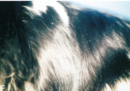
papules. Examination of the dog revealed a few white scales over the
dorsum [back] (Fig. 1c). The skin looked normal. Acetate samples
from scales revealed many living Cheyletiella mites and eggs. For
the dog, dips with phoxim three times weekly were prescribed. After
two baths the owner reported decreased pruritus in the dog; in
addition, the owner's lesions and pruritus were decreased. After
three baths the dog was still a little itchy. Additional therapy was
given with moxidectin, two treatments 8 days apart. Lesions and
pruritus disappeared in both the dog and the owner. There are
numerous reports of skin disease due to Cheyletiella spp. in humans.
The mites are highly contagious, and in households with infested
animals 20% of pet owners are also infected.
R. Wagner
N. Stallmeister. Brit. J. Dermatology. November
2000;143(5):1110-1112. Quote: In animals, Cheyletiella dermatitis
(walking dandruff) is usually a highly contagious but mild,
non-suppurative mite-induced dermatitis produced by Cheyletiella
spp. These parasites live on the skin of cats, dogs, rabbits and
humans. The non-burrowing mites live in the keratin layer of the
epidermis and feed on surface debris and tissue fluids. Their ova
are smaller than louse nits and are attached to hairs by fine
fibrillar strands. The egg-larval-nymphal-adult stages are completed
within 21 days on one host. Adult females may live free of their
host for up to 10 days. Eggs that are shed with the animals' hair
into the environment are important sources of reinfestation. Humans
may serve as an accidental or transitory host. The prevalence may be
underestimated because of false-negative samples. ... Case 3. A
7-year-old female Cavalier King Charles spaniel was brought to the
[Vienna, Austria] Dermatology Service with a 2-month history of
itch. One month after onset of the dog's pruritus [itching], the
owner's abdomen (Fig. 1b [right]) and legs were covered with pruritic red
papules. Examination of the dog revealed a few white scales over the
dorsum [back] (Fig. 1c). The skin looked normal. Acetate samples
from scales revealed many living Cheyletiella mites and eggs. For
the dog, dips with phoxim three times weekly were prescribed. After
two baths the owner reported decreased pruritus in the dog; in
addition, the owner's lesions and pruritus were decreased. After
three baths the dog was still a little itchy. Additional therapy was
given with moxidectin, two treatments 8 days apart. Lesions and
pruritus disappeared in both the dog and the owner. There are
numerous reports of skin disease due to Cheyletiella spp. in humans.
The mites are highly contagious, and in households with infested
animals 20% of pet owners are also infected.
Efficacy of selamectin in the treatment of canine cheyletiellosis. R. S. Mueller, S. V. Bettenay. Vet. Rec. December 2002; doi:10.1136/vr.151.25.773. Quote: This short communication describes the efficacy of selamectin in the treatment of cheyletiellosis in two multidog households. Three breeding colonies in two households were treated. One household (household 1) consisted of nine cavalier King Charles spaniels. The referring veterinarian had identified Cheyletiella mites in superficial skin scrapings from one dog. This dog had been pruritic for several weeks, as had its owner. ... All of the dogs were treated by one of the authors (R. M.) with selamectin every other week at the recommended dose of 6 to 12 mg/kg for a total of four treatments. After spreading the hair between each dog's shoulder blades, the selamectin was applied as a spot-on. In household 1, which had fewer dogs, methoprene was used in the environment immediately after the first treatment of the dogs. ... Superficial skin scrapings, tape preparations and hair and scale samples obtained with a flea comb from each of the dogs, were evaluated microscopically immediately before the first and after the last treatments. ... In household 1, four dogs showed mild to moderate scaling of the trunk, and one dog and the owner were pruritic. Before therapy with selamectin, C yasguri mites were identified in a number of dogs with the various diagnostic tests used. Skin scrapings, tape preparations and hair plucks of all dogs were negative after four treatments with selamectin. The pruritus subsided in all dogs and the affected owner. ... On the basis of the findings of this study, selamectin is a safe and effective means of treating C yasguri in multidog households.
RETURN TO TOP
Malassezia dermatitis:
Malassezia dermatitis in the dog: a retrospective histopathological and immunopathological study of 86 cases (1990-95). Elizabeth A. Mauldin, Danny W. Scott, William H. Miller, Jr., Christina A. Smith. Vet. Derm. September 1997;8(3):191-202. Quote: In our study, West Highland White Terriers, English Setters, Shih Tzus, Basset Hounds and American Cocker Spaniels were at increased risk for developing Malassezia dermatitis. ... Other breeds reported to be at increased risk [for developing Malassezia dermatitis] include ... Cavalier King Charles spaniels... .
Malassezia Dermatitis. Jennifer L. Matousek, Karen L. Campbell. Compendium. March 2002;24(3):224-232. Quote: Other breeds of dogs predisposed to Malassezia dermatitis and otitis include the ... cavalier King Charles spaniel ... . Breed prevalence may suggest an inherited predilection for Malassezia dermatitis or may be a reflection of a hereditary predisposition for underlying disorders (e.g., hypersensitivity).
The method of application and short term results of tympanostomy tubes for the treatment of primary secretory otitis media in three Cavalier King Charles Spaniel dogs. Corfield GS, Burrows AK, Imani P, Bryden SL. Aust Vet J. March 2008;86(3):88-94. Quote: Case 1: A 4-year-old desexed female Cavalier King Charles Spaniel was presented to the Murdoch University Veterinary Hospital with a history of chronic, non-seasonal, bilateral otitis externa of approximately 2 years duration in conjunction with a recent onset of pedal and facial pruritus. ... Cytological examination of otic exudates retrieved from both ears ... revealed Malassezia organisms ... predominantly adhering to corneocytes. Cytological examination of tape strip samples collected from the interdigital and interpad region of all four feet, examined as above, revealed cocci and Malassezia organisms 1 to 3/OIF. A provisional diagnosis of bilateral Malassezia otitis externa and bacterial and Malassezia pododermatitis secondary to underlying hypersensitivity dermatitis (atopic dermatitis or adverse food reaction) was made. ... Case 3: A 6-year-old desexed female Cavalier King Charles Spaniel presented for assessment of presumed bilateral deafness, apparent for the previous 6 months. ... Cytological examination of samples of otic discharge taken from the left and right horizontal ear canals revealed Malassezia 4 to 6/OIF, and 6 to 8/OIF respectively. ... Oral ketoconazole 5 mg/kg every 12 hours was prescribed for 4 weeks to treat the Malassezia infection. ... In addition the first dog received topical betamethasone otic drops, a topical medicated shampoo and a systemic antifungal agent for the treatment of Malassezia otitis externa and bacterial and Malassezia pododermatitis. Dog 3 received a systemic antifungal agent for treatment of bilateral Malassezia otitis externa.
RETURN TO TOP
Necrolytic dermatitis:
Treatment of Superficial Necrolytic Dermatitis with Copper Chelationina Dog with Copper-Associated Hepatitis. Cindy Talbot, Shawn Kearns, Pamela J. Mouser. J. Am. Hosp. Assn. January 2023; doi: 10.5326/JAAHA-MS-7217. Quote: A 7 year old castrated male Cavalier King Charles spaniel presented for evaluation of liver enzyme elevations. Abdominal ultrasound revealed a small liver with mixed echogenicity, small hypoechoic nodules, and an irregular surface. Histologic examination and copper quantification of the liver obtained by laparoscopy diagnosed copper-associated hepatitis. One month later the dog developed hyperkeratosis of all four foot pads and ulcerations of feet, legs, and rectum. Punch biopsies confirmed superficial necrolytic dermatitis. After a total of 2 mo of chelation with no changes to medications, skin lesions began to improve, continuing over the following 6 wk to almost complete resolution. At this point the skin lesions returned and had minimal response to four amino acids infusions. The dog was switched from penicillamine to trientine. Zinc acetate was initiated 6 wk after the switch to trientine, and skin improvement was noted soon thereafter. At the time of death, skin lesions were improving and the dog was clinically comfortable. Copper-associated hepatitis should be considered as a possible etiology for superficial necrolytic dermatitis. Treatment of superficial necrolytic dermatitis is often unrewarding, and copper chelation, when copper-associated hepatitis has been confirmed, represents another therapeutic option.
RETURN TO TOP
Pemphigus foliaceus:
Polysulfated Glycosaminoglycan as a Novel, Adjunctive Therapy for
Pemphigus Foliaceus in Three Dogs. Andrew Simpson, Rod
Rosychuck, Jennifer Schissler, Clarissa Souza. J. Am. Anim. Hosp.
Assn. September 2019; doi: 10.5326/JAAHA-MS-6750.
Quote:
Three dogs who were presented with cutaneous lesions and had
histopathologic findings consistent with pemphigus foliaceus were
treated with injectable polysulfated glycosaminoglycan as an
adjunctive to systemic immune-modulatory therapy. ... Case 1: A 9 yr
old, 12.6 kg castrated male Cavalier King Charles spaniel
was presented with a 6 mo history of pruritus and crusting.
Multifocal crusts and moderate hyperkeratosis affected the paw pad
margins. There was severe crusting on all claw folds. A
well-demarcated erythematous plaque with mild peripheral crusting
involved the prepuce and skin within a 2 cm radius around the
prepuce. There was moderate crusting of the scrotum. Severe
symmetric crusting and erythema affected the perianal region,
periocular region, medial aspects of both pinnae, and the lateral
muzzle. Histopathology of the skin obtained via punch biopsies
confirmed a diagnosis of PF [pemphigus foliaceus]. [See
photograph.] ... These patients were not adequately controlled
with oral glucocorticoids in conjunction with cyclosporine,
azathioprine, and/or mycophenolate. Polysulfated glycosaminoglycan
contributed to induction of remission and reduced glucocorticoid
doses in all dogs.
RETURN TO TOP
Piebaldism:
Analogs
of human genetic skin disease in domesticated animals.
Justin Finch, Stephanie Abrams, Amy Finch. Int'l J. Women's Derm.
March 2017. Quote: Genetic skin diseases encompass a vast, complex,
and ever expanding field. Recognition of the features of these
diseases is important to ascertain a correct diagnosis, initiate
treatment, consider genetic counseling, and refer patients to
specialists when the disease may impact other areas. Because
genodermatoses may presentwith a vast array of features, it can be
bewildering to memorize them. This manuscript will explain and
depict some genetic skin diseases that occur in both humans and
domestic animals and offer a connection and memorization aid for
physicians. In addition,we will explore howanimal diseases serve as
a model to uncover the mechanisms of human disease. The genetic skin
diseases we will review are pigmentary mosaicism, piebaldism,
albinism, Griscelli syndrome, ectodermal dysplasias, Waardenburg
syndrome, and mucinosis in both humans and domesticated animals. ...
Cavalier King Charles Spaniel dog demonstrates
axial depigmentation. (Right)
CONNECT WITH US