
Corneal Ulcers and the
Cavalier King Charles Spaniel
-
 What It Is
 - Symptoms
- Diagnosis
- Treatment
- What You Can Do
- Research News
- Related Links
- Veterinary Resources
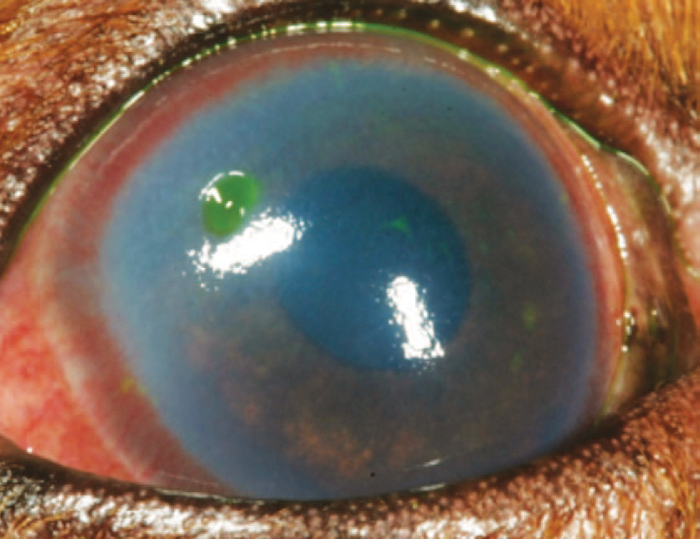
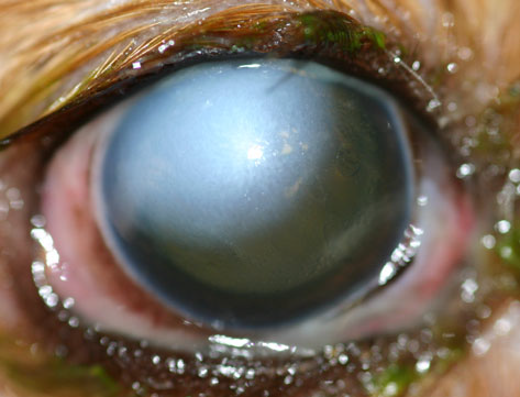
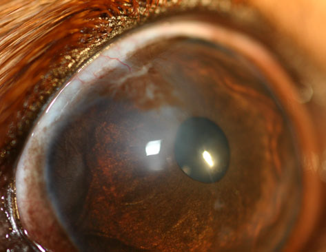
Some cavalier King Charles spaniels may develop deep abrasions -- ulcers -- in the corneas of their eyes, as a result of their short muzzles and head shapes. Such brachycephalic dogs, including the CKCS, may also be predisposed to ulcers of their corneas, due their apparent "bulging" eye balls (called "macropalpebral fissures") and cases of inability to blink fully ("lagophthalamos"), which make them vulnerable to scratches and infections. Untreated dry eye conditions also can lead to ulceration. This condition is referred to as "corneal ulcerative keratitis". The photo at right is of the eye of a cavalier with an ulcer -- the greenish area -- just above the white mark in the upper left of the eyeball, which resulted from an untreated dry eye condition (Photo by Dr. Christine Heinrich).
Untreated cases of corneal ulceration can cause more severe conditions, including "corneal melting" which presents a high risk of permanent blindness. See our Corneal Melting webpage.
RETURN TO TOP
What It Is
The cornea is the transparent membrane outer surface of the eyeball, and is a smooth, moist, and
usually transparent structure when healthy. It consists of three layers:
the outer layer is called the epithelium; beneath it is the stroma; and the
deepest layer is Descemet's membrane.
Corneal ulceration is the loss of a
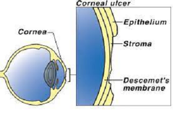 portion of the outer or more layers of the cornea.
Corneal ulceration is one of the most common eye diseases in domestic
dogs, and is a major cause of blindness due to either scarring or
corneal perforation.
Damage to the cornea can cause substantial pain.
portion of the outer or more layers of the cornea.
Corneal ulceration is one of the most common eye diseases in domestic
dogs, and is a major cause of blindness due to either scarring or
corneal perforation.
Damage to the cornea can cause substantial pain.
Corneal ulcers vary in severity, and can be classified into grades based on their depth. (See diagram at left.) Superficial (simple) corneal ulcers affect the epithelium. Deeper ones affect the stroma. A complicated ulcer, also called a descemetocele ulcer, is so deep that it extends to the low level of what is called Descemet’s membrane. More superficial lesions tend to be more painful as the nerve endings within the cornea are close to the surface.
Ulcers also may be noninfected or infected with bacteria. Corneal melting is more apt to occur in infected ulcers.
Corneal ulcers may be caused by a direct injury, tear abnormalities such as dry eye, external irritants, eyelid or eyelash abnormalities such as distichiasis or entropion, immune-mediated or allergic inflammation, foreign bodies, or the inability to blink.
RETURN TO TOP
Symptoms
Corneal ulceration causes extreme pain, redness, light sensitivity, watering eyes, and twitching eyelids. The ulcer may also be visible to the naked eye when examined closely. If the ulceration is severe enough to also include corneal melting, see details about that disorder on our Corneal Melting webpage.
RETURN TO TOP
Diagnosis
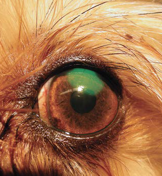 Corneal ulcers are detected by applying a stain, such as
fluorescein. Fluorescein is a
water-soluble stain applied to the cornea to detect corneal ulcers.
Either sterile fluorescein strips or sterile vials of fluorescein dye
are used to stain the cornea. An ulcer in the epithelial surface appears
bright green (see photo at right), especially under an ultraviolet or cobalt blue light. If the ulcer very deep, a
sample may be taken for a culture and cell study before applying the
stain or medication.
Corneal ulcers are detected by applying a stain, such as
fluorescein. Fluorescein is a
water-soluble stain applied to the cornea to detect corneal ulcers.
Either sterile fluorescein strips or sterile vials of fluorescein dye
are used to stain the cornea. An ulcer in the epithelial surface appears
bright green (see photo at right), especially under an ultraviolet or cobalt blue light. If the ulcer very deep, a
sample may be taken for a culture and cell study before applying the
stain or medication.
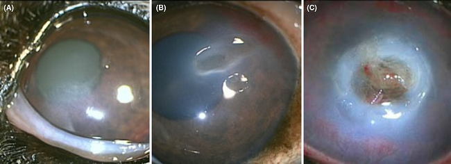
Ulcerative keratitis is classified according to the depth of the ulcer (lesion) in the cornea. They are:
A or Grade 1: superficial
B or Grade 2: stromal
C or Grade 3: descemetoceles/ perforations.
(See Figure 1 below from this July 2020 article.)
In addition to fluorescein staining and measurement of ulcer size, the dog also should receive a thorough ophthalmic examination, including evaluation of the menace response, dazzle reflex, pupillary light reflex, slit-lamp examination, Schirmer Tear Test I, and rebound tonometry.
RETURN TO TOP
Treatment
Medications
Medication includes antibiotic drops or ointment to
prevent bacterial infections*, and atropine drops or
ointment to relieve the pain and dilate the pupil to prevent spasm and
adhesion of the iris to the lens. The pain medication must be applied daily,
and the antibiotic even more
 frequently.
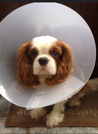
The injured eye must be
protected during the healing process, so usually the dog will be
required to wear a cone -- Elizabethan collar -- attached around its
neck.
frequently.
The injured eye must be
protected during the healing process, so usually the dog will be
required to wear a cone -- Elizabethan collar -- attached around its
neck.
* In a September 2015 article, researchers report that a cavalier with a corneal ulcer but no history of trauma had developed a fungal organism which was treated with antifungal treatment, including 1% voriconazole solution or 1% itraconazole ointment.
Infected ulcers will require frequent application of antibiotics, such as cefazolin and tobramycin or fluoroquinolones such as ciprofloxacin.
Usually after a week to ten days, the eye is re-examined to determine if it has healed, again using the fluoresein stain. If healing is not progressing normally, or if a complication develops, your veterinarian will recommend additional medical treatment or surgery.
Some corneal ulcers, called indolent ulcers, such as spontaneous chronic corneal epithelial defects (SCCEDs), may not respond to medications. SCCEDs are non-infected and superficial ulcers, surrounded by a ring.
RETURN TO TOP
Surgeries
A minor surgical procedure is grid keratotomy, which involves pricking or scratching the eye down to the stroma to stimulate the epithelium to heal by making it easier to attach back to the stroma. General anesthesia is not required for this procedure, as a combination of sedation and local anesthesia is applied.
Diamond burr debridement (DBD) is a surgerical procedure with reportedly faster healing times than grid keratomy.
Corneal thermal cautery (CTC) is the use of a heat source delivered through an electrolysis needle to treat persisent ulcers such as SCCEDs.
Other surgical procedures are important options, especially for deep ulcers. In each, tissue from other parts of the patient's eyes or elsewhere is inserted into the void and then sutured to hold the repair in place. Conjunctival grafts involve taking tissue from the patient' conjunctiva, a pale membrane which covers the white of the eye and includes blood vessels. A piece of conjunctiva is moved and rotated so that it covers the ulcer and is then sutured into place. Corneal conjunctival transposition (CCT) includes both the conjunctiva and cornea. Healthy corneal tissue, with attached conjunctiva, adjacent to the ulcer is moved into the wound, with the conjunctiva providing a functioning blood supply. Other types of grafts include tissue from pig bladder, pig intestines, donor dog or pig cornea, and amnion (an innermost fetal membrane).
In a November 2016 article, French and UK ophthalmologists reported the 100% successful use of pig bladder grafts for corneal reconstruction in 27 dogs (and three cats), including two cavaliers suffering from deep melting corneal ulcers. After 90 days following surgery, all eyes were sighted. They concluded that "Use of a porcine urinary bladder acellular matrix appears to be effective in the surgical management of deep corneal ulcers and feline corneal sequestra."
RETURN TO TOP
Home Care
Healing times for dogs with ulcers, particularly deep ones into the
stroma or Descemet’s membrane, may take weeks. Therefore, dogs which
have been treated for corneal ulcers should have their eyes protected to
assure they heal properly. Elizabethan collars or similar devices should
be worn, and the dogs should be prevented
 from
rubbing their faces on the floor or pawing at their eyes.
from
rubbing their faces on the floor or pawing at their eyes.
They also need to avoid pressure on their necks. Neck pressure relates directly to pressure on the eyes. So, no leashes attached to neck collars should be used. Instead, the leash may be attached to a sturdy harness. One of the best harnesses for cavaliers is the BRILLIANT K9 "Lucy Small" harness. It is easy to put on and easy to take off. Watch the videos: "Opening the harness" and "Walking the dog with the harness".
RETURN TO TOP
What You Can Do
 In
a
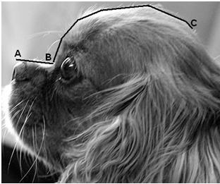
May 2015 report, UK researchers found that cavaliers were among the
breeds most commonly affected by corneal ulcers. They calculated a
measurement called the craniofacial ratio (CFR), which was the muzzle
length (A - B in the photo at right) divided by the cranial
length (C - D). They determined that
brachycephalic dogs (including the pug,
Shih Tzu, bulldog, as well as the cavalier), had a craniofacial ratio of <0.5 and
were twenty times more likely to be affected than non-brachycephalic
dogs (The cavalier in the photo has a craniofacial ratio of 0.27.)
In addition, they found that: (a) a 10% increase in relative eyelid
aperture width more than tripled the ulcer risk, and (b) exposed
eye-white was associated with a nearly three times increased risk.
They concluded that:
In
a
May 2015 report, UK researchers found that cavaliers were among the
breeds most commonly affected by corneal ulcers. They calculated a
measurement called the craniofacial ratio (CFR), which was the muzzle
length (A - B in the photo at right) divided by the cranial
length (C - D). They determined that
brachycephalic dogs (including the pug,
Shih Tzu, bulldog, as well as the cavalier), had a craniofacial ratio of <0.5 and
were twenty times more likely to be affected than non-brachycephalic
dogs (The cavalier in the photo has a craniofacial ratio of 0.27.)
In addition, they found that: (a) a 10% increase in relative eyelid
aperture width more than tripled the ulcer risk, and (b) exposed
eye-white was associated with a nearly three times increased risk.
They concluded that:
"The results demonstrate that artificially selecting for these facial characteristics greatly heightens the risk of corneal ulcers, and such selection should thus be discouraged to improve canine welfare."
RETURN TO TOP
Research News
October 2020:
Cavaliers' eyes with corneal ulcers were among the most included
in review of corneo-limbo-conjunctival surgeries.  In
an
October 2020 article, UK ophthalmologists (Prado Cebrian [right],
Natalia Escanilla, Robert C. Lowe, Charlotte Dawson, Rick F. Sanchez)
reviewed the surgical results of 418 eyes of dogs with corneal ulcers,
using the corneo-limbo-conjunctival transportation procedure. The most
commonly affected breeds were, first, pugs (116 dogs -- 27.75%), and
then Shih Tzus (64 -- 15.31%), cavalier King Charles spaniels (34 --
8.13%), and French bulldogs (34 -- 8.13%). The investigators report a
97.13% success rate, but with a less desirable outcome when treating
perforations and pugs. (For an explanation of this surgical process,
see our Surgeries section above.)
In
an
October 2020 article, UK ophthalmologists (Prado Cebrian [right],
Natalia Escanilla, Robert C. Lowe, Charlotte Dawson, Rick F. Sanchez)
reviewed the surgical results of 418 eyes of dogs with corneal ulcers,
using the corneo-limbo-conjunctival transportation procedure. The most
commonly affected breeds were, first, pugs (116 dogs -- 27.75%), and
then Shih Tzus (64 -- 15.31%), cavalier King Charles spaniels (34 --
8.13%), and French bulldogs (34 -- 8.13%). The investigators report a
97.13% success rate, but with a less desirable outcome when treating
perforations and pugs. (For an explanation of this surgical process,
see our Surgeries section above.)
July 2020:
Cavaliers were the 10th most prevalent breed for corneal ulcers
in a 10-year Japanese study.
 In
a
July 2020 article, a team of Japanese veterinary opthalmologists
(Hiroko Iwashita, Shinsuke Wakaiki, Yoshiyuki Kazama, Akihiko Saito
[right]) reviewed ten years (2008 - 2017) of records of 1,081 dogs
(1,109 eyes) diagnosed with ulceative keratitis (UK) at one
opthalmologist clinic -- Triange Animal Eye Clinic -- in Tokyo, Japan.
The severity of the UK was classified according to the depth of the
ulcer (lesion) in the cornea, as:
In
a
July 2020 article, a team of Japanese veterinary opthalmologists
(Hiroko Iwashita, Shinsuke Wakaiki, Yoshiyuki Kazama, Akihiko Saito
[right]) reviewed ten years (2008 - 2017) of records of 1,081 dogs
(1,109 eyes) diagnosed with ulceative keratitis (UK) at one
opthalmologist clinic -- Triange Animal Eye Clinic -- in Tokyo, Japan.
The severity of the UK was classified according to the depth of the
ulcer (lesion) in the cornea, as:
A or Grade 1: superficial
B or Grade 2: stromal
C or Grade 3: descemetoceles/ perforations.
The breeds of dogs were divided into brachycephalic (BC) and non-BC. Cavalier King Charles spaniels were included in the BC category and were the tenth breed in terms of number of UK-affected eyes (451 eyes). The investigators found that BC dogs were significantly more frequently affected by Grades 2 and 3 and less frequently by Grade 1 than non-BC dogs. They concluded that, "Brachycephalic dogs are more likely to have deeper corneal involvement in UK [ulcerative keratitis]."
 June 2017:
Cavalier breed ranked 4th in number of corneal ulcer cases at
the UK primary care clinics in 2013. In a
June 2017 article by Royal Veterinary College researchers (Dan G.
O’Neill [right], Monica M. Lee, Dave C. Brodbelt, David B. Church), they
reviewed the records of 104,233 dogs treated at 110 UK primary care
clinics during 2013, under the VetCompass Programme. They found 834
cases of dogs treated for corneal ulcers. Of those, 58 dogs were
cavalier King Charles spaniels, the fourth highest number per breed. The
top three were pugs, boxers, and Shih Tzus. In general, the
investigators found that brachycephalic breeds had 11.18 times the odds
of crossbreeds, and spaniel types had 3.13 times those odds.
June 2017:
Cavalier breed ranked 4th in number of corneal ulcer cases at
the UK primary care clinics in 2013. In a
June 2017 article by Royal Veterinary College researchers (Dan G.
O’Neill [right], Monica M. Lee, Dave C. Brodbelt, David B. Church), they
reviewed the records of 104,233 dogs treated at 110 UK primary care
clinics during 2013, under the VetCompass Programme. They found 834
cases of dogs treated for corneal ulcers. Of those, 58 dogs were
cavalier King Charles spaniels, the fourth highest number per breed. The
top three were pugs, boxers, and Shih Tzus. In general, the
investigators found that brachycephalic breeds had 11.18 times the odds
of crossbreeds, and spaniel types had 3.13 times those odds.
 May
2017:
Dr. David Williams finds pain from corneal ulceration causes
increased tear production and may mask dry eye. In a
May 2017 report, UK ophthalmologist Dr. David Williams (right)
examined corneal ulceration in 100 dogs, including six cavaliers. He
found that ulcers result in an increase in tear production and a
decrease in intraocular pressure (IOP) in dogs' eyes. He also noted that
the study shows the importance of measuring tear production in both eyes
of dogs with corneal ulceration since the higher tear production in the
ulcerated eye may produce an apparently normal Schirmer tear test (STT)
reading and mask an underlying case of dry eye
(keratoconjunctivitis sicca).
May
2017:
Dr. David Williams finds pain from corneal ulceration causes
increased tear production and may mask dry eye. In a
May 2017 report, UK ophthalmologist Dr. David Williams (right)
examined corneal ulceration in 100 dogs, including six cavaliers. He
found that ulcers result in an increase in tear production and a
decrease in intraocular pressure (IOP) in dogs' eyes. He also noted that
the study shows the importance of measuring tear production in both eyes
of dogs with corneal ulceration since the higher tear production in the
ulcerated eye may produce an apparently normal Schirmer tear test (STT)
reading and mask an underlying case of dry eye
(keratoconjunctivitis sicca).
November
2016:
Pig bladder grafts successfully return sight to two cavaliers
with corneal ulcers.
 In a
November 2016 article, French and UK ophthalmologists (Olivier
Balland [right], Anne-Sophie Poinsard, Frank Famose, Frédéric
Goulle, Pierre-François Isard, Iona Mathieson, Thomas Dulaurent) report
the 100% successful use of pig bladder grafts for corneal reconstruction
in 27 dogs (and three cats), including two cavalier King Charles
spaniels suffering from deep melting corneal ulcers. After 90 days
following surgery, all eyes were sighted. They concluded that "Use of a
porcine urinary bladder acellular matrix appears to be effective in the
surgical management of deep corneal ulcers and feline corneal
sequestra."
In a
November 2016 article, French and UK ophthalmologists (Olivier
Balland [right], Anne-Sophie Poinsard, Frank Famose, Frédéric
Goulle, Pierre-François Isard, Iona Mathieson, Thomas Dulaurent) report
the 100% successful use of pig bladder grafts for corneal reconstruction
in 27 dogs (and three cats), including two cavalier King Charles
spaniels suffering from deep melting corneal ulcers. After 90 days
following surgery, all eyes were sighted. They concluded that "Use of a
porcine urinary bladder acellular matrix appears to be effective in the
surgical management of deep corneal ulcers and feline corneal
sequestra."
August 2015:
UK researchers opine that corneal ulcers in cavaliers may be due to the
breed standard favoring large eyes.
 In a
May 2015 study by
a team of researchers (Rowena M. A. Packer [right], Anke Hendricks, Charlotte C.
Burn) from the UK's Royal Veterinary College, they measured eleven
conformational features demonstrated to be breed-defining (muzzle
length, cranial length, head width, eye width, neck length, neck girth,
chest girth, chest width, body length, height at the withers and height
at the base of tail) of 700 dogs, 31 dogs of which were affected with
corneal ulcers, including three cavalier King Charles spaniels.
They specifically criticized the CKCS breed standard for considering
"large" eyes as a desirable feature, and also noted that the cavalier's
predisposition to dry eye can lead to corneal ulcers. They stated:
In a
May 2015 study by
a team of researchers (Rowena M. A. Packer [right], Anke Hendricks, Charlotte C.
Burn) from the UK's Royal Veterinary College, they measured eleven
conformational features demonstrated to be breed-defining (muzzle
length, cranial length, head width, eye width, neck length, neck girth,
chest girth, chest width, body length, height at the withers and height
at the base of tail) of 700 dogs, 31 dogs of which were affected with
corneal ulcers, including three cavalier King Charles spaniels.
They specifically criticized the CKCS breed standard for considering
"large" eyes as a desirable feature, and also noted that the cavalier's
predisposition to dry eye can lead to corneal ulcers. They stated:
"Several brachycephalic breeds have been identified as being predisposed to dry eye, including the Bulldog, Lhasa Apso, Shih Tzu, Pug, Pekingese, Boston Terrier and Cavalier King Charles Spaniel. Even moderately lowered tear production associated with dry eye may produce clinical signs in brachycephalic dogs, as a larger portion of the globe is exposed. In a UK based study, a higher proportion of brachycephalic dogs that were affected by dry eye were also affected by ulcers, than were non-brachycephalic dogs with dry eye, e.g. 36% of Shih Tzus and 30% of Cavalier King Charles Spaniels versus 17% of dogs in the overall study population."
They devised morphometric predictors for corneal ulcers, including
the "craniofacial ratio" (CFR), "the
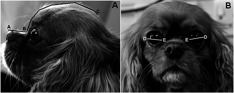 muzzle
length divided by the cranial length, which quantifies the degree of
brachycephaly", to differentiate skull morphologies. [Photos at
right: "This Cavalier King Charles Spaniel has a craniofacial ratio of
0.27 (muzzle length 28mm / cranial length 102mm), and a relative
palpebral fissure width value of 33.3% ((palpebral fissure width 34mm /
cranial length 102mm) *100".]
muzzle
length divided by the cranial length, which quantifies the degree of
brachycephaly", to differentiate skull morphologies. [Photos at
right: "This Cavalier King Charles Spaniel has a craniofacial ratio of
0.27 (muzzle length 28mm / cranial length 102mm), and a relative
palpebral fissure width value of 33.3% ((palpebral fissure width 34mm /
cranial length 102mm) *100".]
They found that brachycephalic dogs (craniofacial ratio <0.5) were twenty times more likely to have corneal ulcers than non-brachycephalic dogs. In addition, they found that: (a) a 10% increase in relative eyelid aperture width more than tripled the ulcer risk, and (b) exposed eye-white was associated with a nearly three times increased risk. They concluded that:
"The results demonstrate that artificially selecting for these facial characteristics greatly heightens the risk of corneal ulcers, and such selection should thus be discouraged to improve canine welfare."
 April
2007:
Researchers find cavaliers are more likely to acquire ulcerative
dry eye. Drs. Rick F.
Sanchez (England) (right), G. Innocent (Scotland), J.
Mould (England), and F. M. Billson (Australia)
reported in an April 2007
report that the cavalier King Charles spaniels in their study
"showed a more acute disease pattern with a biphasic age distribution at 0 to
less than two years of age, and four to less than six and six to less than eight
years of age, respectively, with more males affected than females and a
significantly higher incidence of ulcerative keratitis in some cases resulting
in corneal perforation."
April
2007:
Researchers find cavaliers are more likely to acquire ulcerative
dry eye. Drs. Rick F.
Sanchez (England) (right), G. Innocent (Scotland), J.
Mould (England), and F. M. Billson (Australia)
reported in an April 2007
report that the cavalier King Charles spaniels in their study
"showed a more acute disease pattern with a biphasic age distribution at 0 to
less than two years of age, and four to less than six and six to less than eight
years of age, respectively, with more males affected than females and a
significantly higher incidence of ulcerative keratitis in some cases resulting
in corneal perforation."
RETURN TO TOP
Related Links
Veterinary Resources
Surgical repair of deep melting ulcers with porcine small intestinal submucosa (SIS) graft in dogs and cats. Maria Vanore, Sabine Chahory, Guillaume Payen, Bernard Clerc. Vet. Ophtal. March 2007;10(2):93-99. Quote: Abstract Purpose: To evaluate the efficacy of using a porcine small intestinal submucosa (SIS) graft for the surgical repair of deep melting ulcers in dogs and cats. Methods: Two cats and five dogs [incluidng one cavalier King Charles spaniel, diagnosed with hypopion and melting ulcer] presented with deep and large melting ulcers of the cornea. In each case, the necrotic and collagenolytic tissue of the cornea was removed by keratectomy. A SIS graft, 1 mm greater than the corneal defect, was rehydrated in sterile saline and sutured to the edges of the ulcer with a simple interrupted pattern of 9/0 polyglactin 910. A nictitating membrane flap was utilized in two cats and four dogs for 2 weeks. All cases were treated postoperatively with topical and systemic antibiotics, a systemic anti‐inflammatory drug and topical atropine. All animals were re‐evaluated 15 days, 4 weeks, 35–45 days, 2–3 months and 6 months postsurgery. Results: At 15 days postsurgery, a superficial intense corneal neovascularization surrounded the SIS graft. No ocular discomfort was present and fluorescein staining was negative in all cases. At 4 weeks the SIS graft was thick and opaque in all cases, although in one cat the SIS graft had partially detached. Between 35 and 45 days, SIS graft integration was evident in all eyes, and corneal neovascularization had decreased progressively. All eyes healed without complications and retained corneal transparency. This occurred even in the presence of corneal perforation in two cases: one prior to and one during surgery. Conclusion: Results of our study suggest the SIS graft may be an effective alternative surgical treatment to the traditional conjunctival grafts commonly used to repair melting ulcers in dogs and cats. The advantages of using a SIS graft include good corneal transparency, preservation of corneal integrity and maintenance of vision.
Canine keratoconjunctivitis sicca: disease trends in a review of 229 cases. R. F Sanchez, G Innocent, J Mould, F. M Billson. J.Sm.Anim.Pract.; April 2007;48(4): 211-217. Quote: There were 44 breeds in the study, with four breeds, English cocker spaniels [45 dogs], cavalier King Charles spaniels [44 dogs], West Highland white terriers [37 dogs] and shih-tzus, making up 58 per cent of the cases. ... In contrast, cavalier King Charles spaniels and shih-tzus showed a more acute disease pattern with a biphasic age distribution at 0 to less than two years of age, and four to less than six and six to less than eight years of age, respectively, with more males affected than females and a significantly higher incidence of ulcerative keratitis in some cases resulting in corneal perforation. ... In the USA, predisposed breeds include cavalier King Charles spaniels (CKCS), English bulldogs, Lhasa apsos, shih-tzus, West Highland white terriers (WHWT), pugs, bloodhounds, American cocker spaniels, Pekingeses, Boston terriers, miniature schnauzers and Samoyeds (Kaswan and Salisbury 1990).
Evaluation of accelerated collagen cross-linking for the treatment of melting keratitis in eight dogs. Frank Famose. Vet. Ophth. September 2014;17(5):358-367. Quote: Objective: Melting keratitis is serious condition presenting a high risk of permanent blindness and is caused by infectious or noninfectious factors. In humans, the clinical efficacy of collagen cross-linking (CXL) has been described in the treatment of refractory infectious keratitis by arresting keratomalacia. The aim of this study was to evaluate the efficacy of accelerated CXL for the treatment of melting keratitis in dogs. Animal studied and Procedure: Eight dogs ... two were Cavalier King Charles Spaniels with macropalpebral fissures ... were treated for unilateral melting keratitis by accelerated CXL. Corneas were irradiated by UVA (370 nm) at 30 mW/cm² irradiance for 3 min after soaking by 0.1% riboflavin in 20% dextran for 30 min. Follow-up was conducted 3, 7, 14, and 30 days after treatment. Results: Pain improvement was observed for all cases within 3 days after treatment. Epithelial healing was observed within 15 days for all cases. Disappearance of cellular infiltration was observed for all cases at day 7. The corneal vascularization disappeared for 4 of 8 dogs and was reduced for 4 of 8 dogs within 1 month. At 1 month, all cases presented a variable degree of corneal scarring, but all eyes had visual function. ... In the present study, at 30 days after the treatment, corneal melanosis was observed in 3 of 5 brachycephalic dogs, but not in the two Cavalier King Charles Spaniels that presented with macropalpebral fissures nor in the Great Dane with euryblepharon. ... No recurrent infection was observed. Conclusions: The main observation of this study is that all the cases have presented with the same clinical result regardless of the presence and the sensitivity of the infectious agents and regardless of the duration of the condition prior to CXL treatment. Accelerated CXL appears to be a valuable option for the treatment of melting keratitis.
Advances in treating ocular issues. Christine Heinrich. Vet. Times. November 24, 2014:8-12. Quote: "Canine dry eye is a painful and blinding disease, which, unfortunately, is not usually successfully managed with only the use of tear replacements – even if the client has the time and inclination to comply with complex treatment schedules. In the past, it must have been disheartening – even with frequent applications of tear replacements – for colleagues to watch patients with severe dry eye continuing to suffer from excessive amounts of tenacious ocular discharge, progressive corneal pigmentation, vision loss and, at times, devastating corneal ulceration."
Ocular alkaline injury in four dogs – presentation, treatment, and
follow-up – a case series. Claudia Busse, Claudia Hartley,
Cristiane Kafarnik, Mauro Pivetta. Vet.Ophth. March
2015;18(2):127-134. Quote: Purpose: To describe presentation,
treatment, and follow-up after unilateral alkaline injuries to the
eye in four dogs. Material and method: The case notes of four
patients that suffered from alkaline injuries to the eye were
included in this series. Results: Acute clinical signs included
blepharospasm and edema of the eyelids, chemosis and conjunctival
hyperemia, conjunctival ischemia, destruction of the corneal
epithelium, a whitish haze of the corneal stroma, mild corneal
edema, and uveitis. Two patients showed depigmentation of the
eyelids. Presumed endothelial cell damage resulted in severe corneal
edema in two dogs. Long-term complications included phthisis bulbi,
scarring of the eyelids and damage to the meibomian glands,
symblepharon formation, conjunctivalization of the cornea, corneal
vascularization, pigmentation, and fibrosis. Persisting corneal
edema was seen in the dogs with presumed endothelial
 cell
damage. One dog developed a mild bullous keratopathy with
superficial corneal ulcerations 4½ years after the injury and had a
reduced anterior chamber depth on ultrasound. Conclusion: The damage
to the ocular structures described here mainly affects the ocular
surface. One patient presumably suffered an injury to the ciliary
body epithelium resulting in a phthisical globe. Chronic corneal
edema, conjunctivalization, and scarring can result in permanent
visual impairment. Healing of the ocular surface can take weeks and
is associated with a dramatic vascular response. However, a severely
vascularized cornea has the potential to clear and allow a good
visual outcome long term. Ongoing discomfort was only seen in one
case with persistent corneal edema and a secondary bullous
keratopathy. ... Case 1: A 4-week-old cavalier King Charles
spaniel was presented to the Animal Health Trust (AHT)
after her right eye was exposed to caustic soda (sodium hydroxide).
The eye was flushed by the owner with tap water. The referring
veterinarian (RV) treated the dog with the following medications:
systemic meloxicam, buprenorphine, amoxicillin and clavulanic acid,
topical tetracaine eye drops, and artificial tears. An ophthalmic
examination at the AHT 12 h after the injury revealed mild
blepharospasm and mucoid discharge in the right eye, with moderate
chemosis and conjunctival hyperemia. The dorsolateral aspect of the
right cornea showed a superficial ulceration and a whitish haze in
the stroma and mild corneal edema (Fig. 1 -- above left).
cell
damage. One dog developed a mild bullous keratopathy with
superficial corneal ulcerations 4½ years after the injury and had a
reduced anterior chamber depth on ultrasound. Conclusion: The damage
to the ocular structures described here mainly affects the ocular
surface. One patient presumably suffered an injury to the ciliary
body epithelium resulting in a phthisical globe. Chronic corneal
edema, conjunctivalization, and scarring can result in permanent
visual impairment. Healing of the ocular surface can take weeks and
is associated with a dramatic vascular response. However, a severely
vascularized cornea has the potential to clear and allow a good
visual outcome long term. Ongoing discomfort was only seen in one
case with persistent corneal edema and a secondary bullous
keratopathy. ... Case 1: A 4-week-old cavalier King Charles
spaniel was presented to the Animal Health Trust (AHT)
after her right eye was exposed to caustic soda (sodium hydroxide).
The eye was flushed by the owner with tap water. The referring
veterinarian (RV) treated the dog with the following medications:
systemic meloxicam, buprenorphine, amoxicillin and clavulanic acid,
topical tetracaine eye drops, and artificial tears. An ophthalmic
examination at the AHT 12 h after the injury revealed mild
blepharospasm and mucoid discharge in the right eye, with moderate
chemosis and conjunctival hyperemia. The dorsolateral aspect of the
right cornea showed a superficial ulceration and a whitish haze in
the stroma and mild corneal edema (Fig. 1 -- above left).
 There
was no aqueous flare, but the right pupil was slightly miotic
relative to the left. Menace response, dazzle reflex, and the
consensual pupillary light reflex from right to left were present.
The pH of the lower conjunctival sac was assessed using a
Combur-Test urine test strip (Roche, Mannheim, Germany) and was pH7
and therefore within normal limits. The remainder of the ophthalmic
examination was within normal limits. Intraocular pressure
measurements were not documented. The dog was admitted to the
hospital for intensive medical treatment. The dog was discharged
after 2 days as the corneal ulcer was starting to epithelialize. At
reexamination 7 days after the initial presentation, corneal
vascularization was present from the dorsal limbus and 90% of the
corneal ulcer was re-epithelialized. Three weeks after the injury,
the cornea was fluorescein negative with vascularization extending
over the entire affected area. A reexamination 4 months after the
trauma revealed only mild perilimbal pigmentation in the dorsal
quadrant of the right cornea (Fig. 2 -- at right).
There
was no aqueous flare, but the right pupil was slightly miotic
relative to the left. Menace response, dazzle reflex, and the
consensual pupillary light reflex from right to left were present.
The pH of the lower conjunctival sac was assessed using a
Combur-Test urine test strip (Roche, Mannheim, Germany) and was pH7
and therefore within normal limits. The remainder of the ophthalmic
examination was within normal limits. Intraocular pressure
measurements were not documented. The dog was admitted to the
hospital for intensive medical treatment. The dog was discharged
after 2 days as the corneal ulcer was starting to epithelialize. At
reexamination 7 days after the initial presentation, corneal
vascularization was present from the dorsal limbus and 90% of the
corneal ulcer was re-epithelialized. Three weeks after the injury,
the cornea was fluorescein negative with vascularization extending
over the entire affected area. A reexamination 4 months after the
trauma revealed only mild perilimbal pigmentation in the dorsal
quadrant of the right cornea (Fig. 2 -- at right).
Impact of Facial Conformation on Canine Health: Corneal
Ulceration. Rowena M. A. Packer, Anke Hendricks,
Charlotte C. Burn. PlosOne. May 2015. Quote: "Concern has arisen
in recent years that selection for extreme facial morphology in
the domestic dog may be leading to an increased frequency of eye
disorders. Corneal ulcers are a common and painful eye problem
in domestic dogs that can lead to scarring and/or perforation of
the cornea, potentially causing blindness. Exaggerated
juvenile-like craniofacial conformations and wide eyes have been
suspected as risk factors for corneal ulceration. ... Several
brachycephalic breeds have been identified as being predisposed
to dry eye, including the Bulldog, Lhasa Apso, Shih Tzu, Pug,
Pekingese, Boston Terrier and Cavalier King Charles
Spaniel. Even moderately lowered tear production
associated with dry eye may produce clinical signs in
brachycephalic dogs, as a larger portion of the globe is
exposed. In a UK based study, a higher proportion of
brachycephalic dogs that were affected by ulcers, than were
non-brachycephalic dogs with dry eye, e.g. 36% of Shih Tzus and
30% of Cavalier King Charles Spaniels versus
17% of dogs in the overall study population. ... This study
aimed to quantify the relationship between corneal ulceration
risk and conformational factors including relative eyelid
aperture width, brachycephalic (short-muzzled) skull shape, the
presence of a nasal fold (wrinkle), and exposed eye-white. A 14
month cross-sectional study of dogs entering a large UK based
small animal referral hospital for both corneal ulcers and
unrelated disorders was carried out. Dogs were classed as
affected if they were diagnosed with a corneal ulcer using
fluorescein dye while at the hospital (whether referred for this
disorder or not), or if a previous diagnosis of corneal ulcer(s)
was documented in the dogs’ histories. Of 700 dogs recruited, measured and clinically examined, 31 were affected by corneal
ulcers. Most cases were male (71%), small breed dogs (mean± SE
weight: 11.4±1.1 kg), the most common being the Pug (n = 12
affected), the Shih Tzu (n = 4), the Bulldog and the
Cavalier King Charles Spaniel (n = 3). ... Morphometric
data were collected for each dog using previously defined
measuring protocols, measuring 11 conformational features
that were demonstrated to be breed-defining: muzzle length,
cranial length, head width, eye width, neck length, neck girth,
chest girth, chest width, body length, height at the withers and
height at the base of tail (all in cm). ... A further
morphometric predictor of interest for corneal ulcers was
craniofacial ratio, (CFR): the muzzle length divided by the
cranial length, which quantifies the degree of brachycephaly,
was used to differentiate skull morphologies. [Photos at right:
"This Cavalier King Charles
Spaniel
has a craniofacial ratio of 0.27 (muzzle length 28mm / cranial
length 102mm), and a relative palpebral fissure width value of
33.3% ((palpebral fissure width 34mm / cranial length 102mm)
*100"] ...
[B]rachycephalic dogs (craniofacial ratio <0.5) were twenty
times more likely to be affected than non-brachycephalic dogs. A
10% increase in relative eyelid aperture width more than tripled
the ulcer risk. Exposed eye-white was associated with a nearly
three times increased risk. The results demonstrate that
artificially selecting for these facial characteristics greatly
heightens the risk of corneal ulcers, and such selection should
thus be discouraged to improve canine welfare."
Key Points in Complicated Canine Corneal Ulcers. Diane Hendrix, Georgina Newbold. Vet. Brief. September 2015. Quote: Complicated corneal ulcers are epithelial defects that remain unresolved after several days, become infected, or show progressive deepening into the stroma. Complete ocular examination—including a Schirmer’s tear test and a light examination of the eyelids and conjunctiva—may or may not reveal the underlying cause. Recognizing a complicated ulcer is critical for appropriate management of these potentially vision-threatening keratopathies. Clinical Findings: An ulcer is considered complicated based in part on its depth and duration. Even without a detailed history, the cornea offers clues during the course of the disease. Corneal vessels generally appear at the limbus after 7-10 days, then grow approximately 1 mm per day, so corneal vascularization is a sign of chronicity. Etiologies: An ulcer generally becomes complicated because of infection, indolence (ie, failure of the epithelium to adhere to the underlying stroma), or lack of treatment for the primary cause.
Outcome of phacoemulsification following corneal and lens laceration in cats and dogs (2000–2010). Barbara K. Braus, Alexander Tichy, Heidi J. Featherstone, Peter W. Renwick, Michael Rhodes, Christine L. Heinrich. Vet. Ophthalmology. December 2015. Quote: "Objective: To investigate the success rate of phacoemulsification following corneal and lens laceration in dogs and cats. Procedure: Retrospective review of cats and dogs presenting with corneal and lens laceration and treated with phacoemulsification. Results: The records of 33 patients (33 eyes: six feline, 27 canine) presenting to a private referral center were reviewed. ... Dogs: English Springer Spaniels (7/27; 25.9%), Crossbreeds (4/27; 14.8%), and Cavalier King Charles spaniels (3/27; 11.1%) were most commonly affected by corneal and lens laceration. ... Affected dogs were younger (median 18 months) than affected cats (median 30 months). The lacerations were caused by cat scratch trauma (9/33), thorn injury (6/33), and glass shards (1/33); the cause was unknown in 17/33 cases. All cats and 85.2% of all dogs were visual at the last examination. The median follow-up was 4 and 8 months for cats and dogs, respectively. In all canine cases that developed vision loss, this occurred within the first 14 weeks postoperatively. The ultimate cause for vision loss in dogs was secondary glaucoma (4/4) and retinal detachment (1/4). Conclusion: Cats have an excellent outcome and dogs a very good outcome following surgery for corneal and lens laceration. The cause of the trauma, the size of the lesion, the time interval between the ocular trauma and surgery, and the type of surgery were not found to have an influence on the outcome of patients in this study. We postulate that vision loss might develop more often in cases with complications associated with the initial corneal laceration wound."
Keratomycosis in five dogs. Jessica C. Nevile, Simon D. Hurn, Andrew G. Turner. Vet. Ophthalmology. September 2016;19(5):432-438. Quote: "Five cases of canine keratomycosis were diagnosed and treated at a private Veterinary Ophthalmology Practice in Melbourne, Australia. Clinical presentations varied between dogs. Predisposing factors were identified in 4 of 5 cases. Diagnostic modalities utilized were corneal cytology and fungal culture. ... Case 1. A 10-year 7-month-old spayed female Cavalier King Charles Spaniel was referred for a corneal ulcer in the right eye (OD). The dog was systemically healthy, there was no known history of trauma, and the dog had not been treated with topical or systemic corticosteroids prior to presentation. ... Corneal cytology confirmed the presence of fungal organisms in all five cases. Aspergillus, Scedosporium, and Candida were cultured from three cases, respectively. Specific antifungal treatment included 1% voriconazole solution or 1% itraconazole ointment. Keratectomy and conjunctival grafting surgery was performed in two patients. Resolution of infection and preservation of vision were achieved in 4 of 5 patients."
Use of a porcine urinary bladder acellular matrix for corneal reconstruction in dogs and cats. Olivier Balland, Anne-Sophie Poinsard, Frank Famose, Frédéric Goulle, Pierre-François Isard, Iona Mathieson, Thomas Dulaurent. Vet. Ophthal. November 2016;19(6):454-463. Quote: Objective: To describe the use of a porcine urinary bladder acellular matrix for surgical reconstruction of the cornea in cases of canine and feline deep corneal ulcers, and feline corneal sequestra. Materials and methods: Twenty‐seven dogs [including two cavalier King Charles spaniels, both with deep, melting corneal ulcers] and three cats with deep corneal ulcers and seven cats with corneal sequestra were included in the study with overall 38 eyes. For each patient, the necrotic material (ie corneal sequestrum or collagenolytic tissue) was removed by circular lamellar keratectomy. The collagen graft was then cut and prepared to match the stromal defect and then sutured into the lamellar keratectomy bed using interrupted and continuous patterns of absorbable polyglactin 9–0 sutures. Postoperative medical treatment consisted of topical and systemic administration of antibiotics, combined with topical administration of atropine sulfate. The animals were examined 18, 45, and 90 days after the surgery. Results: Postoperative examination revealed complete integration of the biomaterial in 93.5% of ulceration cases in both species and in 100% of feline corneal sequestrum cases. In two cases of ulceration (1 dog and 1 cat), progression of the collagenolytic process at the graft periphery required an additional conjunctival graft 7 days after the first surgery. At 90 days post‐op, 100% of the eyes were sighted. Conclusion: Use of a porcine urinary bladder acellular matrix appears to be effective in the surgical management of deep corneal ulcers and feline corneal sequestra.
Tear production and intraocular pressure in canine eyes with corneal ulceration. David L. Williams, Philippa Burg. Open Vet. J. May 2017;7(2):117-125. Quote: This study aimed to evaluate changes in lacrimation and intraocular pressure (IOP) in dogs with unilateral corneal ulceration using the Schirmer tear test (STT) and rebound (TonoVet®) tonometry. IOP and STT values were recorded in both ulcerated and non-ulcerated (control) eyes of 100 dogs [including 6 cavalier King Charles spaniels] diagnosed with unilateral corneal ulceration. Dogs presented with other ocular conditions as their primary complaint were excluded from this study. The mean ± standard deviation for STT values in the ulcerated and control eyes were 20.2±4.6 mm/min and 16.7±3.5 mm/min respectively. The mean ± standard deviation for IOP in the ulcerated and control eyes were 11.9±3.1 mmHg and 16.7±2.6 mmHg respectively. STT values were significantly higher (p<0.000001) in the ulcerated eye compared to the control eye while IOP was significantly lower (p<0.0001). There is an increase in lacrimation and a decrease in IOP in canine eyes with corneal ulceration. ... The current study supports previous findings that ocular pain, in this case resulting from corneal ulceration, will cause an increase in tear production due to stimulation of the lacrimal functional unit. ... The higher tear production in ulcerated eyes shows the importance of measuring STT in both eyes in cases of corneal ulceration, since this increased lacrimation may mask an underlying keratoconjunctivitis sicca only evident in the contralateral eye. The lower IOP in ulcerated eyes is likely to relate to mild uveitic change in the ulcerated eye with a concomitant increase in uveoscleral aqueous drainage. ... The present study documents that corneal ulceration in the dog is associated with an increase in tear production and a decrease in IOP. ... This study shows the importance of measuring tear production in both eyes of dogs with corneal ulceration since the higher tear production in the ulcerated eye may produce an apparently normal STT reading and mask an underlying case of KCS.
Corneal ulcerative disease in dogs under primary veterinary care in England: epidemiology and clinical management. Dan G. O’Neill, Monica M. Lee, Dave C. Brodbelt, David B. Church, Rick F. Sanchez. Canine Genetics & Epidem. June 2017. Quote: Background: Corneal ulcerative disease (CUD) has the potential to adversely affect animal welfare by interfering with vision and causing pain. The study aimed to investigate for the first time the prevalence, breed-based risk factors and clinical management of CUD in the general population of dogs under primary veterinary care in England. Results: Of 104,233 dogs attending 110 clinics participating within the VetCompass Programme from January 1st to December 31st 2013, there were 834 confirmed CUD cases (prevalence: 0.80%). Breeds with the highest prevalence included Pug (5.42% of the breed affected), Boxer (4.98%), Shih Tzu (3.45%), Cavalier King Charles Spaniel (2.49% [based upon 58 affected dogs out of a total of 2332]) and Bulldog (2.41%). Purebred dogs had 2.23 times the odds of CUD compared with crossbreds. Brachycephalic types had 11.18 and spaniel types had 3.13 times the odds for CUD compared with crossbreds. Pain was recorded in 385 (46.2%) cases and analgesia was used in 455 (54.6%) of dogs. Overall, 62 (7.4%) cases were referred for advanced management and CUD contributed to the euthanasia decision for 10 dogs. Conclusions: Breeds such as the Pug and Boxer, and conformational types such as brachycephalic and spaniels, demonstrated predisposition to CUD in the general canine population. These results suggest that breeding focus on periocular conformation in predisposed breeds should be considered in order to reduce corneal disease.
Breed prevalence of canine ulcerative keratitis according to depth of corneal involvement. Hiroko Iwashita, Shinsuke Wakaiki, Yoshiyuki Kazama, Akihiko Saito. Vet. Ophthal. July 2020; doi: 10.1111/vop.12808. Quote: Objective: To investigate the breed prevalence of canine ulcerative keratitis (UK) according to the depth of corneal involvement. Procedures: Dogs diagnosed with ulcerative keratitis from 2008 to 2017 at the Triangle Animal Eye Clinic were included in this study. Only breeds with more than 20 eyes affected were selected [including 451 eyes of cavalier King Charles spaniels]. UK lesions were classified as superficial (Grade 1), stromal (Grade 2) or descemetoceles and perforations (Grade 3) and compared between brachycephalic (BC) and non-BC dog breeds. [CKCSs were classified as BC.] Results: Of 8877 dogs evaluated at Triangle Animal Eye Clinic from 2008 to 2017, 1109 eyes of 1018 dogs (male, 326 eyes; neutered male, 253 eyes; female, 211 eyes; spayed female, 316 eyes; and unknown sex, 3 eyes) aged between 0.1 and 19.2 years (mean ± standard deviation [SD], 8.33 ± 4.24 years) were diagnosed with UK. The number of eyes that was classified as Grade 1 was 359 eyes (187 non-BC and 172 BC), Grade 2 was 373 eyes (60 non-BC and 313 BC) and Grade 3 was 377 eyes (47 non-BC and 330 BC). Significant differences were observed between BC and non-BC dogs for all grades of UK. BC dogs were significantly more frequently affected by Grades 2 and 3 and less frequently by Grade 1 UK (P < .01). [The CKCS ranked 10th out of the top 20 breeds that attended the Triange Animal Eye Clinic from 2008 to 2017.] French bulldogs are more likely to be affected with Grade 1. Conclusions: Brachy-cephalic dogs are more likely to have deeper corneal involvement in UK. This study provides novel data on the prevalence of superficial UK, which was low in BC dogs and high in non-BC breeds.
Corneo-limbo-conjunctival transposition to treat deep and perforating corneal ulcers in dogs: A review of 418 eyes and corneal clarity scoring in 111 eyes. Prado Cebrian, Natalia Escanilla, Robert C. Lowe, Charlotte Dawson, Rick F. Sanchez. Vet. Ophthal. October 2020; doi: 10.1111/vop.12833. Quote: Purpose: To report surgical and corneal clarity scores (CCSs) of corneo‐limbo‐conjunctival transpositions (CLCTs) in a large number of canine cases. Methods: Retrospective review of records that underwent CLCT to repair deep ulcers or perforations between 2002 and 2018. Signalment, concurrent eye disease, additional procedures, pathogenesis, medication, graft orientation, follow‐up, and CCSs were recorded. Results: 418 eyes of 399 dogs were included. Brachycephalics were most commonly affected, comprising 325/418 (77.75%) of the eyes. The most commonly affected breeds were Pugs, Shih Tzus, Cavalier King Charles Spaniels, and French Bulldogs, with 116/418 (27.75%), 64/418 (15.31%), 34/418 (8.13%), and 34/418 (8.13%) ulcerated eyes, respectively. Mean age at surgery was 5.5 years (range 59 days-17.7 years), and median follow‐up time was 100 days (range 3 days-7.64 years). The most common etiopathogenesis was spontaneous ulceration in 205/418 eyes (49.04%) of which 191 (93.17%) occurred in brachycephalics. Primary keratoconjunctivitis sicca affected 122/418 eyes (29.19%) and injury 39/418 eyes (9.33%). Mean ulcer width was 3.5 mm (0.5-10 mm). Success rate was 97.13% (406/418 eyes). Failure end points recorded included no menace response, secondary glaucoma, and endophthalmitis. Pre-existing perforation was found in 101/418 (24.16%) of the eyes and significantly increased failure rate. The median CCS was G3 (G0-G4), which was lower for Pugs (G2). Graft orientation affected CCS, but did not reach statistical significance. Conclusion: The high success rate and CCS for CLCT in dogs make it a good technique to treat deep ulcers but a less desirable outcome is anticipated when treating perforations and Pugs.
CONNECT WITH US