
Distichiasis Can Damage Corneas in Cavaliers
-
 What It Is
 - Symptoms
- Diagnosis
- Treatment
- Breeders' Responsibilities
- What You Can Do
- Research News
- Related Links
- Veterinary Resources
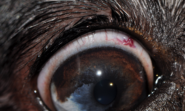
Distichiasis* is the growth of hairs (distichia or cilia) from the glands of the margin of the upper or lower eyelid, which normally is hairless. It is considered to be inherited, and the cavalier King Charles spaniel is predisposed as a breed to this disorder. See this 2022 book.
* The term "distichiasis" is from the Greek words di and stichos, meaning "two rows".
What It Is
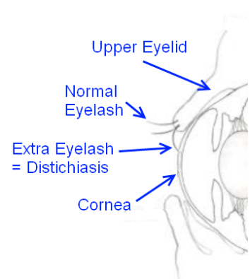 A follicle develops deep within the glands rather than on
the skin surface of the eyelid. As the follicle grows, it follows the duct of
the gland and grows out of the gland opening along the eyelid as a set of
eyelashes. (See diagram at right.)
A follicle develops deep within the glands rather than on
the skin surface of the eyelid. As the follicle grows, it follows the duct of
the gland and grows out of the gland opening along the eyelid as a set of
eyelashes. (See diagram at right.)
Distichiasis is presumed to be inherited in the cavalier King Charles spaniel, according to the American College of Veterinary Ophthalmologists (ACVO).* It is most common in English bulldogs. Dr. Keith Barnett has described distichiasis as the most common hereditary eye disease in canines.
* See also, Ophthalmic Disease in Veterinary Medicine.
All CKCSs should be examined at least annually by a board certified veterinary ophthalmologist. They are listed on the website of the American College of Veterinary Ophthalmologists (ACVO).
RETURN TO TOP
Symptoms
Soft, fine distichia and those which do ot come in contact with the cornea may cause no discomfort. Dr. Keith Barnett has observed that distichiasis may produce only a slight increase in tear production and not present a clinical problem. However, when stiff or multiple distichia curve inward and come in contact with the conjunctiva or cornea, they will cause chronic irritation, resulting in various degrees of abnormal contractions or twitches of the eyelid (blepharospasm), excessive tearing (epiphora), inflammation (conjunctivitis; keratitis), ulceration, blepharitis, and infection.
The irriation can cause the dog to keep the affected eye closed, or to involuntarily blink constantly. The eye may appear red and sore from the inflammation. The dog may paw at or rub its eyes, and otherwise whine and appear agitated.
The symptoms are very similar to a more serious optical disorder, ectopic cilia, in which one or more eyelashes protrude through the inside of the eye, causing similar injury to the affected eyes.
RETURN TO TOP
 Diagnosis
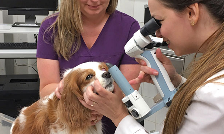
During a thorough eye examination, the emphasis will be on focal illumination and magnification using a slit-lamp bio microscope (see photo at right), to identify and locate the distichia.
Because the distichia may have damaged the conjunctiva and/or cornea, the ophthalmologist must also diagnose any resulting disorders, such as conjunctivitis, ulceration, blepharitis, and/or infection.
RETURN TO TOP
Treatment
Some cases of distichiasis are soft and non-irritating hairs which require no treatment. The goal is to treat the clinical signs and injuries to the eyes, remove the distichia, and preserve the functions of the eyelids.
Treatment usually starts with the application of ophthalmic lubricants, to protect the cornea and coat the eyelashes with an oily film. Surgical correction may remove the eyelashes -- which may be performed manually -- and kill the hair follicles, if they are causing corneal changes. Regrowth of hairs is a common problem and may require repeated surgeries. The appearance of new follicles at new locations may also occur after surgery.
Also, cryoepilation (cryotherapy or cryosurgery) has been used by veterinary ophthalmologists to remove the distichiatic lashes without damaging the normal lashes. Cryoepilation is the application of a liquid nitrogen probe which freezes the hair follicles, which then are removed. It has been reported that with cryoepilation, up to 90% of the treated distichiatic lashes do not regrow, and repeat surgical treatment is seldom required. However, this form of ophthalmologic surgery may be very expensive -- from $1,000.00 to over $2,000.00.
In this December 2023 article, a specific cryotherapy technique -- "a triple freeze-thaw cycle via a transconjunctivally applied, liquid nitrogen-cooled, 2-mm diameter closed cryoprobe" -- was employed in 125 dogs with distichiasis, totaling 234 eyes, including cavaliers. The authors concluded:
"This cryotherapy technique for treating canine distichiasis is relatively simple and safe to perform, has a high success rate and a low recorded complication rate, but would benefit from further assessment of any potential detrimental effects on qualitative tear film components."
Meibomian glands are oil glands along the edge of the eyelids where the eyelashes are found. These glands produce the oily layer on the outside of the tear film that prevents tears from drying too quickly. In a December 2022 article, Australian veterinary ophthalmologists report finding that cryoepilation for distichaiasis appears to be a risk factor for permanently damaging the meibomian glands, resulting in tear film breakup times significantly shorter than for untreated dogs.
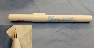 Transconjunctival
thermal electrocautery (TCEC) is a process which destroys hair follicles
using a thermal cautery pen and magnification. (See photo of pen at
right.) The heated pen is inserted into the
conjunctiva to the distichia and then immediately removed. The
conjunctival tissue is treated repeatedly until it is charred in the
area of the distichia, and they are removed individually using forceps.
See this
January 2019 article for more details.
Transconjunctival
thermal electrocautery (TCEC) is a process which destroys hair follicles
using a thermal cautery pen and magnification. (See photo of pen at
right.) The heated pen is inserted into the
conjunctiva to the distichia and then immediately removed. The
conjunctival tissue is treated repeatedly until it is charred in the
area of the distichia, and they are removed individually using forceps.
See this
January 2019 article for more details.
Blend electrolysis is an electrolysis technique which
combines electrical and thermal energy. using a fine epilating
electrode/needle inserted into the gland opening beside the hair shaft.
The electrode supplies a verey low current of electricity for 15 to 30
seconds. the needle is rotated until the cilia can be removed with
forceps.
See this
October 2020 article.
RETURN TO TOP
Breeders' Responsibilities
The Genetics Committee of the ACVO classifies distichiasis as a "breeder option" for CKCSs. Therefore, the Canine Eye Registration Foundation (CERF) does not deny certification to cavalier King Charles spaniels which are affected with the disorder. However, the Canine Inherited Disorders Database recommends that cavalier King Charles spaniels affected with distichiasis should not be bred. At the very least, dogs both affected with the disorder should not be bred to each other. Any littermates of breeding stock having distichiasis should be taken into consideration. All cavalier breeding stock should be examined by board certified veterinary ophthalmologists to determine if the dogs are affected with distichiasis. See The Blue Book.
The Cavalier King Charles Spaniel Club, USA recommends that, prior to breeding any Cavalier, the dog have a normal rating or be within CERF "breeder options" from a screening by a board certified veterinary ophthalmologist.
The Canine Health Information Center (CHIC) is a centralized canine health database sponsored by the AKC/Canine Health Foundation (AKC/CHF) and OFA. The CHIC, working with participating parent clubs, provides a resource for breeders and owners of purebred dogs to research and maintain information on the health issues prevalent in specific breeds.
AKC's national breed clubs establish the breed specific testing protocols. Dogs complying with the breed specific testing requirements are issued CHIC numbers. The ACKCSC requires that, to qualify for CHIC certification, cavaliers must have a CERF eye examination, recommending that an initial CERF exam be performed at 8 to 12 weeks, with a follow up exam once the dog reaches 12 months, and annual exams thereafter until age 5 years, and every other year until age 9 years. However, all that is required to qualify for a CHIC certificate is that the breeding stock be examined by a veterinary ophthalmologist. It does not require that the results of the examination show no eye disorders.
RETURN TO TOP
 What You Can Do:
All CKCSs should be examined at least annually by a board certified veterinary ophthalmologist. They are listed on the website of the ACVO.
Ocu-GLO Rx is a nutraceutical containing several natural antioxidants in a combination blend formulated specifically for canine eye health. Many veterinary ophthalmologists recommend this product to maintain healthy eyes. Even if your dog has not been diagnosed with a vision disorder, antioxidants contained in Ocu-GLO Rx are considered helpful in keeping dogs' eyes healthy.
RETURN TO TOP
Research News
April 2023:
Cavaliers rank third in French study of breeds diagnosed with
distichiasis.
 In
an
April 2023 article, French ophthalmologists (Coline Jondeau
[right], M. Gounon, A. Bourguet, S. Chahory) examined the extent of
distichiasis in canines in France between 2010 and 2019. They obtained
the records of 291 dogs in one specialty clinic, all dagnosed with
distichiasis. In addition to breed status, they analyzed the dogs' sex,
skull conformation, coat type, age at the time of diagnosis, reason for
presentation, clinical examination findings, and affected eyelid(s).
They found that the breeds with the highest prevalence of this disorder
were English bulldogs (35.2%), American cocker spaniels (19.4%), and
cavalier King Charles spaniels (17.8%), followed by boxers (17.0%) and
English cocker spaniels (12.2%). Most of the dogs had the disorder
in both eyes. Distichiasis was non-irritating in 85% of the affected
dogs. Dogs with clinical signs had various types of corneal ulcers --
superficial and deep stromal.
In
an
April 2023 article, French ophthalmologists (Coline Jondeau
[right], M. Gounon, A. Bourguet, S. Chahory) examined the extent of
distichiasis in canines in France between 2010 and 2019. They obtained
the records of 291 dogs in one specialty clinic, all dagnosed with
distichiasis. In addition to breed status, they analyzed the dogs' sex,
skull conformation, coat type, age at the time of diagnosis, reason for
presentation, clinical examination findings, and affected eyelid(s).
They found that the breeds with the highest prevalence of this disorder
were English bulldogs (35.2%), American cocker spaniels (19.4%), and
cavalier King Charles spaniels (17.8%), followed by boxers (17.0%) and
English cocker spaniels (12.2%). Most of the dogs had the disorder
in both eyes. Distichiasis was non-irritating in 85% of the affected
dogs. Dogs with clinical signs had various types of corneal ulcers --
superficial and deep stromal.
RETURN TO TOP
Related Links
Veterinary Resources
Inherited eye disease in the dog and cat. K. C. Barnett. J. Sm. Anim. Pract. July 1988; doi: 10.1111/j.1748-5827.1988.tb03514.x Quote: The most common hereditary eye disease in the dog must be distichiasis, a clinical problem in many breeds and one to which there is no really satisfactory answer. In some breeds very many individuals are affected, for example the American cocker spaniel, the Shetland sheepdog, the miniature longhaired dachshund and the Pekingese. Distichiasis may produce only a slight increase in tear production and hence not present a clinical problem. However, a related condition, ectopic cilia, almost invariably produces severe clinical signs including blephar.ospasm, epiphora and corneal ulceration. It is rare to find a case of ectopic cilia without concurrent distichiasis and the two together.
Control of Canine Genetic Diseases. Padgett, G.A., Howell Book House 1998, pp. 198-199, 239.
Ocular Disorders Presumed to be Inherited in Purebred Dogs. Genetics Committee, A.C.V.O. 1999.
Guide to Congenital and Heritable Disorders in Dogs. Dodds WJ, Hall S, Inks K, A.V.A.R., Jan 2004, Section II(88).
Breed Predispositions to Disease in Dogs & Cats. Alex Gough, Alison Thomas. 2004; Blackwell Publ. 44-45.
Diseases and Surgery of the Canine Eyelid. In: Veterinary Ophthalmology, 4th ed. Stades FC, Gelatt KN. Blackwell Publishing; 2007; 563-617. Summary: Distichiasis occurs frequently in the dog and is presumed to be inherited in several breeds, although the mode of inheritance is unknown. Certain breeds are overrepresented, including the American and English Cocker Spaniel, Cavalier King Charles Spaniel, Boxer, English Bulldog, Pekingese, Shih Tzu, and Dachshunds.
Ophthalmic Disease in Veterinary Medicine. Charles L. Martin. Manson Publ. 2009; page 475, table 15.1. Quote: "Presumed Inherited Ocular Diseases: Table 15.1: Breed predisposition to eye disease in dogs: Cavalier King Charles Spaniel: ... Distichiasis".
Breed Predispositions to Disease in Dogs & Cats (2d Ed.). Alex Gough, Alison Thomas. 2010; Blackwell Publ. 53.
Ocular Disorders Presumed to be inherited in purebred dogs. Genetics Committee of the American College of Veterinary Ophthalmologists. Blue Book 6th Ed. 2013. pp. 241-247. Quote: "Cavalier King Charles Spaniel: Disorder: D. Distichiasis. Inheritance: Not defined."
Prevalence and heritability of distichiasis in the English Cocker spaniel. Tanja Petersen, Helle Friis Proschowsky, Tommy Hardon, Søren Nyhuus Rasch, Merete Fredholm. Canine Genetics & Epidemiology. August 2015;2:11. Quote: "Background: Canine distichiasis is a well-known cause of ocular irritation and excessive lacrimation (secretion of tears) in the dog. The term distichiasis originates from the Greek words di and stichos meaning two and rows, respectively, and as the name implies, the condition is characterized by an additional row of cilia, which erupts on the eyelid margin. Many purebred dogs are known to be predisposed to the condition, with many affected individuals within the populations. Even though the problem is widespread, the exact mode of inheritance and the heredity has not been studied extensively. However, some degree of genetic influence has been assumed, due to the high incidences within specific breeds. In the present study we have examined a cohort of English Cocker spaniels in Denmark to determine the prevalence and heritability of the disease. Results: Data from English Cocker spaniels with an ECVO eye examination registered between 2004-2013 were included in the study. The number of dogs examined during this period was 799, and the prevalence of distichiasis within this cohort was estimated at 49.31 % with a gender predisposition that females are more likely to get distichiasis than males. The correlation between the distichiasis status of the parents and their offspring revealed a significant association between the breeding combination of the parents and the occurrence of distichiasis in the offspring (p <0.0001). A relative risk (RR) ranging from 1.3 to 1.8 demonstrates that offspring of two affected parents are more likely to be affected than offspring descending from either one or two unaffected parents. The heritability was estimated to be moderate to high, i.e., 0.22 to 0.51. Conclusions: The prevalence of distichiasis in English Cocker spaniels from Denmark, examined in 2004-2013 was shown to be extremely high. The relative risk of developing the disease was 1.3 and 1.8 for offspring of one or two affected parents respectively. This together with the moderate to high heritability of the condition indicates that selective breeding could be used to reduce the incidence of distichiasis."
Distichiasis. Caryn Plummer. Clinicians Brief. March 2016;39-41. Quote: Distichiasis is the abnormal presence of cilia (eg, eyelashes) in the meibomian glands. Distichia hair follicles are found in the base of the meibomian gland, where the cilia exit through its orifice; the cilia may contact the cornea and cause irritation. ... Any individual patient may present with distichiasis; however, certain breeds are more prone to the condition: American and English cocker spaniels, Welsh springer spaniels, Cavalier King Charles spaniels, flat-coated retrievers, English bulldogs, shih-tzus, poodles, dachshunds, and Parson Russell terriers. ... The majority of dogs with distichia do not require any treatment; however, in cases associated with discomfort, therapeutic intervention should be pursued. If the hairs emerge from the conjunctival surface as ectopic cilia, therapeutic removal is usually necessary. Distichiasis is thought to be inherited, but the mechanism of transmission is unknown.
Evaluation of transconjunctival thermal electrocautery for treatment of canine distichiasis: 88 eyelids (2013-2016). Kelli L Zimmerman, Shelby L Reinstein. Vet. Ophthal. January 2019;22(1):50-60. Quote: Objective: To describe a successful, simple treatment for canine distichiasis. Animals studied: Client‐owned dogs presenting to Veterinary Specialty and Emergency Center. Procedure: Retrospective analysis of medical records for canine patients that underwent transconjunctival thermal electrocautery treatment (TCEC) for distichiasis alone or with concurrent eyelid surgery between 2013 and 2016. Fifty eyes of 26 dogs (n = 88 eyelids) were included in the study. Sixty‐five eyelids (74%) were treated for distichia only, while 23 eyelids (26%) underwent concurrent eyelid surgery. Successful treatment was defined as resolution of clinical signs attributable to distichiasis. Forty‐eight of 50 eyes (96%) were successfully treated with a single TCEC treatment (mean follow‐up 187 ± 222 days). Sixty‐one of 88 eyelids (69%) had no distichia at any follow‐up time. Twenty‐two eyelids (25%) had recurrence at or near a previously treated site (mean 150 ± 152 days). Of the eyes with recurrent distichia, all but 2 (91%) remained asymptomatic, requiring no further treatment. One dog with extensive TCEC treatment had significant recurrence on all eyelids requiring retreatment that resulted in focal entropion of 1 eyelid. Transient eyelid margin pigment loss and mild‐to‐moderate eyelid swelling were noted in all treated eyelids postoperatively. Suspected treatment site infection occurred 2 days postoperatively in 2/50 eyes (4%) of 1 patient. Two of 10 eyes (20%) with extensive eyelid treatment developed qualitative tear film deficiency OU (554 days postoperatively) and responded to topical tear stimulant therapy. Conclusion: TCEC is a successful, simple treatment for canine distichiasis.
A description of blend electrolysis for treatment of canine distichiasis; 78 cases (2012 - 2017). Joy Ioannides, Richard Mark Everson, Màrian Matas Riera, Charlotte Dawson. Vet. Rec. October 2020; doi: 10.1136/vr.105816. Quote: Objective: To describe a population of dogs treated with blend electrolysis for distichiasis at The Royal Veterinary College and report the complications seen. Methods: In part 1, records were reviewed from 2012 to 2017 and a population of 78 dogs [including one cavalier King Charles spaniel] with distichiasis treated using blend electrolysis (Sterex SX-B blend epilator) analysed. In part 2, 18 dogs treated with blend electrolysis were re-examined prospectively by a diplomate of the European College of Veterinary Ophthalmologists (ECVO). Results: In part 1, brachycephalic breeds accounted for 62 per cent. English bulldog was the most common breed (42 per cent). In this population, 88 per cent of dogs were successfully treated with one treatment of electrolysis (successful treatment defined as resolution of clinical signs). Forty-five dogs had recurrent distichia on follow-up, mostly fine distichia without clinical discomfort. Twelve per cent required repeat electrolysis. Complications were infrequent: five dogs had scarring or hypopigmentation of the eyelid margin. In part 2, 18 dogs were re-examined. Ten had distichia recurrence, six had eyelid scarring and five had depigmentation associated with electrolysis. Two dogs had occasional clinical signs thought to be related to distichiasis. All owners perceived their dogs' ocular comfort to be improved following blend electrolysis. Conclusions: Brachycephalic breeds, most notably English bulldogs, are over-represented in this population. Blend electrolysis appears an effective treatment for resolution of clinical signs.
Essentials of Veterinary Ophthalmology (4th ed.). Gelatt, Kirk N.; Plummer, Caryn E. Wiley-Blackwell. 2022. Quote: Distichiasis is considered to be inherited, and predisposed breeds include ... Cavalier King Charles Spaniel (24%).
An investigation into the development of qualitative tear film disorders in dogs following cryoepilation for distichiasis. Benjamin D. Reynolds, Cameron Whittaker, Kelly Caruso, Matthew J. Annear, Negar Hamzianpour, William Irving, Paul M. G. McCarthy, Jeffrey S. Smith. Vet. Ophthal. December 2022; doi: 10.1111/vop.13047. Quote: Objective: The aim of this prospective study was to compare tear film quality between dogs who have previously undergone cryoepilation for distichiasis to a reference population. Animals Studied: Nine dogs (17 eyes) were recruited after surgery and were compared to a reference population of 21 dogs (42 eyes). Procedures: Canine patients who had previously undergone cryoepilation for distichiasis for a minimum of 1 month prior to examination were recruited. A complete ophthalmic examination was performed by an ABVO resident (BDR), with additional tear tests, including tear film interferometry, infra-red meibography, and a tear film break-up time (TFBUT) performed. The tear test results were compared to a reference population obtained from client-owned dogs with no history of ophthalmic complaints, a normal ophthalmic examination performed by an ABVO resident (BDR) and a Schirmer Tear Test-1>15mm/min. Statistical analysis was performed of the results obtained. Results: The treated group was significantly more affected with meibomian gland dropout (MG-dropout) in 11/17 (64.7%) cases, compared to the reference population of 2/21 (9.5%) . The treated group had an odds ratio of 23.8 to develop MG-dropout compared to the reference population. Tear film breakup time (TFBUT) was significantly shorter in the treatment group (5.8±2.6 s) compared to the reference population (10.1±1.1 s). In the treatment group, 12/17 (70.5%) of treated eyes had a TFBUT<5 s compared to 2/21 (9.5%) of the reference population. Conclusion: Cryoepilation for distichaiasis appears to be a risk factor for developing MG-dropout and qualitative tear film disorders post-operatively in canines.
Epidemiology and clinical significance of canine distichiasis: A retrospective study of 291 cases. C. Jondeau, M. Gounon, A. Bourguet, S. Chahory. Vet. Ophthal. April 2023; doi: 10.1111/vop.13091. Quote: Objective: To describe the epidemiological factors and clinical significance of canine distichiasis. Animals studied: Two hundred and ninety-one client-owned dogs. Methods: Retrospective study of medical records for canine patients diagnosed with distichiasis between 2010 and 2019 in an ophthalmology specialty practice. The breed, sex, skull conformation, coat type, age at the time of diagnosis, reason for presentation, clinical examination findings, and affected eyelid(s) were reviewed. Results: The prevalence of distichiasis was 5.5% (95% confidence interval (CI): 4.9-6.1) in the population of dogs presented to an ophthalmology specialty practice. The breeds with the highest prevalence of distichiasis were English bulldogs (35.2%, 95% CI: 26.7-43.7), American cocker spaniels (19.4%, 95% CI: 8.3-30.5), Cavalier King Charles spaniels (17.8%, 95% CI: 11.2-24.4), Boxers (17.0%, 95% CI: 9.1-24.9), and English cocker spaniels (12.2%, 95% CI: 6.5-17.9). The prevalence was significantly higher in brachycephalic dogs (11.9%, 95% CI: 9.8-14.0) than in non-brachycephalic dogs (4.6%, 95% CI: 4.0-5.3) and in short-haired dogs (8.2%, 95% CI: 6.8-9.6) than in dogs with other coat types (5.3%, 95% CI: 4.5-6.1). Most dogs were affected bilaterally (63.6%, 95% CI: 58.0-69.1). Among dogs with clinical signs, 39.0% (95% CI: 26.5-51.4) exhibited corneal ulceration, including superficial ulcers (28.8%, 95% CI: 17.3-40.4) and deep stromal ulcers (10.2%, 95% CI: 2.5-17.8). Distichiasis was non-irritating in 85.0% (95% CI: 80.6-89.4) of affected dogs. Conclusion: This study reports the largest cohort of canine distichiasis to date. In a large proportion of dogs, distichiasis was a non-irritating condition. However, brachycephalic breeds, especially English bulldogs, were the most frequently and severely affected.
The Blue Book. Ocular Disorders Presumed to be inherited in purebred dogs. Genetics Committee of the American College of Veterinary Ophthalmologists. Blue Book 15th Ed. 2023. pp. 297-302.
Canine distichiasis cryoepilation using a liquid nitrogen-cooled, closed probe transconjunctival triple-freeze technique: Outcomes in 125 dogs (234 eyes). Alexander Verloop, Tony Read. Vet. Ophthal. December 2023; doi: 10.1111/vop.13174. Quote: Purpose: To describe a simple cryosurgical technique for treating canine distichiasis, its success rate and any associated complications. Methods: Clinical records from canine patients undergoing treatment for distichiasis using a specific cryotherapy technique were retrospectively reviewed. The technique employed a triple freeze-thaw cycle via a transconjunctivally applied, liquid nitrogen-cooled, 2-mm diameter closed cryoprobe. Results: The cryotherapy technique was employed in 125 dogs with distichiasis over a period of 13 years, with 109 (87%) bilaterally and 16 (13%) unilaterally affected, totaling 234 eyes. The mean age at diagnosis was 1.9 years (4 months to 7.5 years), with Staffordshire bull terriers the most common breed (31%). Treatment was deemed successful if clinical signs related to distichiasis resolved, including blepharospasm, conjunctival hyperemia and excess lacrimation. A single triple-freeze cryotherapy treatment eliminated distichiae in 195/234 (83%) eyes at a mean follow-up of 4 months (2 weeks to 10 years). Minor recurrence of distichiae in previously treated regions was observed in 39/234 (17%) eyes. Repeat cryotherapy was required in 25/234 (11%) eyes, with the remaining eyes asymptomatic. The two most common postoperative complications were mild to moderate eyelid swelling in all eyes immediately following surgery and eyelid margin depigmentation in 59/234 eyes (25%) between two and 4 weeks postoperatively, both of which were temporary. Conclusion: This cryotherapy technique for treating canine distichiasis is relatively simple and safe to perform, has a high success rate and a low recorded complication rate, but would benefit from further assessment of any potential detrimental effects on qualitative tear film components.
CONNECT WITH US