
Lungworm (heartworm) in the
Cavalier King Charles Spaniel
-
 What It Is
 - Symptoms
- Diagnosis
- Treatment
- Other Lung Parasites
- Other Heartworms
- What You Can Do
- Research News
- Related Links
- Veterinary Resources
The lungworm is a parasitic nematode that can live in small pulmonary (lung) arteries and the hearts of dogs -- the cardio-respiratory system. It also is known as a heartworm. Significantly higher occurrences of these parasites have been found and frequently reported in cavalier King Charles spaniels* than in most other breeds, and especially in the cavaliers' eyes as well as their hearts and lung blood vessels.
* See this September 2004 article and this June 2009 article and this December 2010 article and this September 2011 article and this June 2014 article and this September 2014 article and this July 2015 article and this September 2015 article and this October 2016 article ("Also, some purebred dogs, and in particular Cavalier King Charles Spaniels and Staffordshire Bull Terriers ... are reported to be at a higher risk than crossbreeds.") and this March 2020 article.
RETURN TO TOP
What It Is
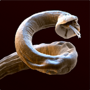
The most common lungworm affecting canines' respiratory systems is the Angiostrongylus vasorum. It also is called the French heartworm and the small heartworm and has a medical title: Canine pulmonary angiostrongylosis (CPA) . It is a thread-like worm as big as an inch long which can live and thrive in the heart, pulmonary (lung) blood vessels, liver, pancreas, brain, spinal fluid, and even the eyes of its host canines. Cavaliers have accounted for a high percentage of reported cases of A. vasorum found in dogs' eyes.
A more commonly known canine heartworm is Dirofilaria immitis, which has not been reported as frequently in CKCSs. D. immitis is discussed briefly, below.
A. vasorum are particularly common in large areas of the United Kingdom and Ireland
and
 have been found in
Belgium, Denmark, France, Germany, Greece, Hungary, Italy, Portugal, Spain, and
elsewhere in Europe. This lungworm also has been found in tropical,
subtropical, and temperate regions of North and South America and
Africa. It is believed to be far more prevalent in foxes than in dogs.
Other wildlife mammals have been identified as hosts, including weasels
and meerkats.
have been found in
Belgium, Denmark, France, Germany, Greece, Hungary, Italy, Portugal, Spain, and
elsewhere in Europe. This lungworm also has been found in tropical,
subtropical, and temperate regions of North and South America and
Africa. It is believed to be far more prevalent in foxes than in dogs.
Other wildlife mammals have been identified as hosts, including weasels
and meerkats.
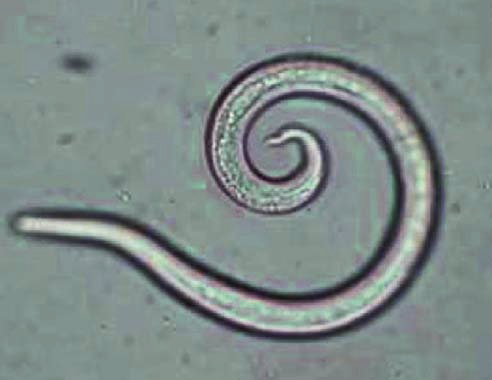
Initially, these worms are carried by slugs and snails and frogs, called transport or intermediate hosts. They then can spread through puddles of water and wet grass to their ultimate hosts, the dogs, or dogs could eat the transport hosts or lick their slime. Once ingested by canines, they penetrate the intestine walls and reach the heart. lungs, and other organs. They also are excreted in feces, where they can survive for several days. Slugs and snails then feast on the feces containing A. vasorum larvae. (Photo at right is of a microscopic view of a first stage larva [L1], courtesy of Bayer Animal Health).
Left untreated, the infectious diseases the A. vasorum carries can be fatal to affected dogs. Further, their presence in the lung's blood vessels can cause pulmonary hypertension, which in turn can lead to severe damage to the two right chambers of the heart and the tricuspid valve.
RETURN TO TOP
Symptoms
The symptoms of lungworm infection can be numerous and widespread. Typical signs displayed by dogs which are acutely infected include:
• Hacking coughing
• Anorexia
• Gagging
• Shortness of breath (dyspnea)
• Depression
• Hemorrhage
• Anemia
• Eating disorders
• Weight loss
•Syncope
• Harsh lung sounds
• Exercise intolerance
• Bleeding disorders
• Pulmonary arterial hypertension (PAH)
Other categories of signs include neurological ones and right-side heart enlargement, and even heart failure.
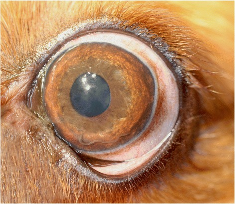
Oddly, in four reported cases, a cavalier was found to have a live and moving A. vasorum in a chamber of the dog's eye. The symptoms in each case were related to discomfort within the affected eye. (See the photo at right of a cavalier's eye with the worm visible in the lower left corner of the cornea.)
RETURN TO TOP
Diagnosis
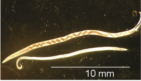
The sooner affected dogs are diagnosed, the more likely they will have a successful treatment. Mild cases are likely to be overlooked until the progress to life-threating levels. Because symptoms may be common to other disorders, mis-diagnosis and therefore failure to properly treat promptly can be a serious clinical problem. Careful auscultation (stethoscopic examination) of the lungs is an important first diagnositic step in even asymptomatic dogs. (Photo below is a microscopic view of adult female lungworms.)
 Chest x-rays
(thoracic radiographs) can show broncial thickening and an interstitial pattern
with alveolar infiltrates.
Chest x-rays
(thoracic radiographs) can show broncial thickening and an interstitial pattern
with alveolar infiltrates.
One of the most certain methods of diagnosing the presence of A. vasorum larvae L1s is testing the dog's feces using the Baermann technique (if properly performed). Multiple fecal samples should be collected over three consecutive days. The Baermann technique is not perfect, and its negative results should not be relied upon as conclusive without other diagnostic steps. Fecal smear exams may also detect larvae. Because the fecal samples must be sent to an external laboratory for Baermann technique analysis, it may take two or three days to obtain the results.
Antigen ELISA blood chemistry tests may show abnormalities. The Angiodetect blood test by IDEXX (Angio Detect™) comes in a kit which analyses the dog's blood serum or plasma and can produce results within 15 minutes. It is an enzyme-linked immunosorbent assay (ELISA) which measures antigens in the samples. However, in a December 2021 article, in which eight other published reports were reviewed, the authors concluded that "There is weak evidence supporting Angio Detect as a highly specific and moderately sensitive diagnostic test when compared to Baermann coprology [feces analysis]. ... As Angio Detect™ has moderate sensitivity, if a negative result were obtained in the clinical scenario (i.e. a patient with high clinical suspicion of angiostrongylosis) additional diagnostic procedures, such as BAL [see below], would be advised."
Quantitative polymerase Chain Reaction (qPCR) assay is molecular test which can detect the presence of worm DNA in blood, feces, and saliva and thus identify the species of the larvae.
Echocardiography is recommended in a January 2021 article in which pulmonary arterial hypertension (PAH) was detected in 58+% of 17 dogs diagnosed with A. vasorum. Some of the common symptoms of angiostrongylosis, including exercise intolerance, syncope, and ascites, may be attributable to PAH.
High resolution computed tomography (CAT) can show the presence of lung lesions. In cases with neurological symptoms,, magnetic resonance imaging (MRI) or myelography may be useful diagnostic tools. If right-side heart disorders are suspected, echocardiography would be appropriaate.
Bronchoalveolar lavage (BAL cytology) may be used. It involves passing a bronchoscope through the mouth or nose to the airway in the lungs. The qPCR test is used to determine the species of larvae found in BAL samples.
RETURN TO TOP
Treatment
The sooner affected dogs are diagnosed, the more likely they will have a successful treatment. Treatment typically consists of medicating with anti-parasitic drugs (anthelmintics) and symptomatic care. Drugs include:
• moxidectin (ProHeart)
• moxidectin + sarolaner + pyrantel (Simparica Trio)
• fenbendazole* (Panacur)
• moxidectin + imidacloprid (Advovate)
• milbemycin oxime (Milbenax)
• levamisole
• ivermectin
* Fenbendazole has reported side effects, including causing renal birth defects in offspring of a pregnant CKCS, and bone marrow hypoplasia/aplasia. See this May 2007 article and this November 2019 article.
Treatment of the symptoms, of course, depends upon those symptoms. PAH can be treated with sildenafil (Viagra). Respiratory treatment might include oxygen supplementation, blood transfusions for bleeding disorders, steroids of immune-medidated thrombocytopenia and to reduce inflammation and fibrois in the lungs, especially to reduce an immune response to dead, decomposing lungworms following effective treatment.
In the cases of A. vasorum in the eye, surgical removal of the worm will be necessary.
RETURN TO TOP
Other Lung Parasites
In addition to the A. vasorum discussed above, cavaliers have been reported to have been diagnosed with other parasites infecting their lungs. An example is the Paragonimus, a parasitic flatworm, also known as a fluke or lung fluke. The sources of ingesting this worm are raw or undercooked meats, especially shellfish and deer. It causes an infection, primarily in the lungs, called paragonimiasis.
In this March 2023 article, a cavalier in Japan consumed raw deer meat four months prior to being examined for a chronic cough and acute dyspnea (difficulty breathing and shortness of breath). X-rays and computed tomography (CT scan) showed several hollow lesions in one of the lobes of the lungs, resulting in pneumothorax (leakage of air from the lungs into the chest cavity). The lesions were surgically removed in a thoracotomy. Examination of the lesions confirmed paragonimiasis. The dog then was treated with praziquantel (Biltricide). The dog survived and was found to be healthy a year after the surgery.
RETURN TO TOP
Other Heartworms
 Dirofilaria immitis is a canine parasite,
a nematode which is the
primary heartworm causing which affects the cardio-pulmonary system of
dogs worldwide (and cats
and other pets). Fortunately, cavaliers have not been found highlt
vulnerable to this heartworm.
Dirofilaria immitis is a canine parasite,
a nematode which is the
primary heartworm causing which affects the cardio-pulmonary system of
dogs worldwide (and cats
and other pets). Fortunately, cavaliers have not been found highlt
vulnerable to this heartworm.
These heartworms are carried by mosquitos which have drawn blood from infected animals and then are transmitted to the dogs by the infected mosquitos (particularly the Aedes albopictus – Asian tiger mosquito), in turn biting them. The bite transmits larval worms, which then reach the dog’s lynph vessels. These larvae will grow into adult worms in about 6 months.
The pre-larval worms (microfilariae) will be produced, which can be detected with microscopic examination of a native blood smear or with Knott’s test. Antigen testing on a blood sample is standard method to screen dogs for the presence of adult female worms. The test is an ELISA immunoassay that detects the circulating antigens released by the reproductive tract of the adult female worms.
According to the American Heartworm Society: ALL DOGS MUST BE TESTED FOR MICROFILARIAE ANNUALLY. Annual testing is an integral part of ensuring that prophylaxis is achieved and maintained. Administering a heartworm preventive without performing appropriate diagnostic tests to determine a dog’s heartworm status is NOT RECOMMENDED. Should the dog be infected with heartworms, that would be initiating a sub-optimal “slow-kill” protocol without consideration for activity restriction or doxycycline and could pose additional safety risks to the dog.
Heartworm preventatives are designed to work in concert with the
dog’s immune system to
kill susceptible larval stages. They are
intended to be adminisered orally, topically, or injected. Oral
heartworm preventatives approved by the US Food & Drug Administration
(FDA) for dogs include:
• Ivermectin (Heartgard)
• Ivermectin + pyrantel pamoate (Heartgard Plus, Iverhart Plus, Tri-Heart Plus)
• Ivermectin + pyrantel pamoate + praziquantel (Iverhart Max)
• Milbemycin oxime (Interceptor, MilbeGuard)
• Milbemycin oxime + praziquantel (Interceptor Plus)
• Milbemycin oxime + lufenuron (Sentinel)
• Milbemycin oxime + lufenuron + praziquantel (Sentinel Spectrum)
• Milbemycin oxime + spinosad (Trifexis)
• Moxidectin + Afoxolaner + Pyrantel (Nexgard Plus)
• Moxidectin + sarolaner + pyrantel pamoate (Simparica® Trio)
Infected dogs may be treated with injected melarsomine and orally administered doxycycline. For more information, see the Canine Heartworm Guidelines published by the American Heartworm Society. See also the Society's Heartworm Treatment Guidelines for the Pet Owner.
RETURN TO TOP
What You Can Do
Cavalier owners in high-risk areas can take preventative steps to avoid letting their dogs become infected. They include keeping their dogs current on worming regimes, including monthly treatments with preventative anit-parasitic drugs (moxidectin, milbeycin oxime). In an August 2020 article, an international team of ressearchers report finding that monthly oral treatment with the combination of sarolaner, moxidectin and pyrantel (Simparica Trio) was 100% effective in the prevention of natural infection with A. vasorum in dogs in highly endemic areas.
Be certain to not overdose the dogs with any pesticides and watch carefully for undesirable side effects. Check the warnings on the container labels to see what side effectes may occur.
• Dispose of the dogs' feces daily, to break the lungworm's life cycle.
• Reduce access by foxes, slugs, and snails.
• Avoid allowing the dogs to take off-lead walks in damp areas or during wet weather, particularly after dark, when slugs are most active.
Consider having dogs' feces examined periodically using the Baermann technique, particularly in high-risk areas.
RETURN TO TOP
Research News
February 2024:
Bleeding indicates a more severe case of Angiostrongylus vasorum
infection in 180 dog study.
 In
a
February 2024 article, a team of Danish researchers (A. S. Thomsen,
M. P. Petersen, J. L. Willesen, M. B. T. Bach, I. N. Kieler, A. T.
Kristensen, J. Koch, Lise Nikolic Nielsen [right]) studied the
symptoms of 180 dogs infected with Angiostrongylus vasorum, finding that
bleeding, of all sorts, was an important sign of the severity of A.
vasorum infection and an indicator of a lower survival rate than in
non-bleeding dogs. Survival rate of bleeding dogs was lower at hospital
discharge (76.9%) and 1 month after diagnosis (66.0%) than in dogs
without signs of bleeding (94.8% and 90.1% at discharge and at 1 month,
respectively).
In
a
February 2024 article, a team of Danish researchers (A. S. Thomsen,
M. P. Petersen, J. L. Willesen, M. B. T. Bach, I. N. Kieler, A. T.
Kristensen, J. Koch, Lise Nikolic Nielsen [right]) studied the
symptoms of 180 dogs infected with Angiostrongylus vasorum, finding that
bleeding, of all sorts, was an important sign of the severity of A.
vasorum infection and an indicator of a lower survival rate than in
non-bleeding dogs. Survival rate of bleeding dogs was lower at hospital
discharge (76.9%) and 1 month after diagnosis (66.0%) than in dogs
without signs of bleeding (94.8% and 90.1% at discharge and at 1 month,
respectively).
September 2023:
Cavalier contracts lungworm from eating raw deer meat.
 In
a
March 2023 article, Japanese clinicians (Aritada Yoshimura, Daigo
Azakami, Miori Kishimoto, Takahiro Ohmori, Daiki Hirao, Shohei Morita,
Shinogu Hasegawa, Tatsushi Morita, Ryuji Fukushima [right])
report a cavalier King Charles spaniel which had consumed raw deer meat
four months prior to being examined for a chronic cough and acute
dyspnea (difficulty breathing and shortness of breath). X-rays and
computed tomography (CT scan) showed several hollow lesions in one of
the lobes of the lungs, resulting in pneumothorax (leakage of air from
the lungs into the chest cavity). The lesions were surgically removed in
a thoracotomy. Examination of the lesions confirmed paragonimiasis. The
dog then was treated with praziquantel. The dog survived and was found
to be healthy a year after the surgery.
In
a
March 2023 article, Japanese clinicians (Aritada Yoshimura, Daigo
Azakami, Miori Kishimoto, Takahiro Ohmori, Daiki Hirao, Shohei Morita,
Shinogu Hasegawa, Tatsushi Morita, Ryuji Fukushima [right])
report a cavalier King Charles spaniel which had consumed raw deer meat
four months prior to being examined for a chronic cough and acute
dyspnea (difficulty breathing and shortness of breath). X-rays and
computed tomography (CT scan) showed several hollow lesions in one of
the lobes of the lungs, resulting in pneumothorax (leakage of air from
the lungs into the chest cavity). The lesions were surgically removed in
a thoracotomy. Examination of the lesions confirmed paragonimiasis. The
dog then was treated with praziquantel. The dog survived and was found
to be healthy a year after the surgery.
January 2021:
Over half of dogs infected with
Angiostrongylus vasorum also had pulmonary
arterial hypertension, in an Italian study.
 In a
January 2021 article, a team of Italian veterinary researchers
(Paola Paradies, Mariateresa Sasanelli, Antonio Capogna, Angelica
Mercadante, Giuseppe Tommaso Roberto Rubino [right], Claudio Maria Bussadori) examined the medical records of 17 dogs diagnosed with
Angiostrongylus
vasorum infection, resulting in canine angiostrongylosis. All patients had been examined by echocardiography to
determine if they had pulmonary arterial hypertension (PAH) as a result
of their infections. PAH was diagnosed in 10 (58.8%) of the 17 dogs.
They concluded:
In a
January 2021 article, a team of Italian veterinary researchers
(Paola Paradies, Mariateresa Sasanelli, Antonio Capogna, Angelica
Mercadante, Giuseppe Tommaso Roberto Rubino [right], Claudio Maria Bussadori) examined the medical records of 17 dogs diagnosed with
Angiostrongylus
vasorum infection, resulting in canine angiostrongylosis. All patients had been examined by echocardiography to
determine if they had pulmonary arterial hypertension (PAH) as a result
of their infections. PAH was diagnosed in 10 (58.8%) of the 17 dogs.
They concluded:
"PAH associated with angiostrongylosis could therefore be considered a more common condition innaturally infected animals than previously reported. ... This data suggests that PAH can contribute to the worsening of the clinical picture in infected dogs, thus investigating its presence and severity is warranted in order to optimize the therapeutic management of single cases (i.e. treatment with sildenafil). Studiesto understand the role of sildenafil to improve the clinical conditions of these dogs in short termfollow up would be beneficial."
September 2020:
A. vasorum-infected dog develops tricuspid valve disease and
irreversible pulmonary hypertension.
 In
an
August 2020 article, Dutch veterinary clinician Viktor Szatmári
(right) reports treating an English Cocker spaniel infected by
adult Angiostrongylus vasorum worms in its pulmonary artery. He found
pulmonary hypertension due to obstruction and inflammation of the
artery's branches. The hypertension in turn had caused damage to the
tricuspid valve -- including ruptured chordae tendineae -- and severe
enlargement of the right ventricle and enlargement of the right atrium,
resulting in congestive heart failure (CHF). The CHF led to ascites.
Symptoms included abdominal distension and exercise intolerance. Dr.
Szatmári treated the A. vasorum infection with fenbendazole for two
weeks along with sildenafil indefinitely for the heart issues. After six
weeks, the infection was cured and the heart issues had improved. Daily
doses of sildenafil continued to be prescribed to manage the pulmonary
hypertension and keep the dog free of ascites.
In
an
August 2020 article, Dutch veterinary clinician Viktor Szatmári
(right) reports treating an English Cocker spaniel infected by
adult Angiostrongylus vasorum worms in its pulmonary artery. He found
pulmonary hypertension due to obstruction and inflammation of the
artery's branches. The hypertension in turn had caused damage to the
tricuspid valve -- including ruptured chordae tendineae -- and severe
enlargement of the right ventricle and enlargement of the right atrium,
resulting in congestive heart failure (CHF). The CHF led to ascites.
Symptoms included abdominal distension and exercise intolerance. Dr.
Szatmári treated the A. vasorum infection with fenbendazole for two
weeks along with sildenafil indefinitely for the heart issues. After six
weeks, the infection was cured and the heart issues had improved. Daily
doses of sildenafil continued to be prescribed to manage the pulmonary
hypertension and keep the dog free of ascites.
September 2020:
Study of 100 southeast England dogs infected by
A. vasorum finds cavaliers out of the high-risk category.
 In an
August 2020 article, UK researchers (Sarah Holmes, Phillip Paine, Ian
Wright, Eric R. Morgan, Hany M. Elsheikha [right]) studied the records
of 100 dogs infected with Angio-strongylus vasorum between 2003 and 2009.
They report finding that dogs less than a year old were 4.2 times more
likely to fall into the infected group, but tht gender was not a
significant risk factor for infection. Also, they report:
In an
August 2020 article, UK researchers (Sarah Holmes, Phillip Paine, Ian
Wright, Eric R. Morgan, Hany M. Elsheikha [right]) studied the records
of 100 dogs infected with Angio-strongylus vasorum between 2003 and 2009.
They report finding that dogs less than a year old were 4.2 times more
likely to fall into the infected group, but tht gender was not a
significant risk factor for infection. Also, they report:
"Other breeds found by Blehaut et al (2014) and Chapman et al (2004) to be at higher risk of the disease, i.e. Springer Spaniels, Jack Russell Terriers and Cavalier King Charles Spaniels, were not associated with increased risk in the current study."
February 2020:
ACVIM issues a Consensus Statement on pulmonary hypertension in
dogs.
 In
a
February 2020 article, a team of veterinary specialists, including
five cardiologists (Carol Reinero [right], Lance C. Visser,
Heidi B. Kellihan, Isabelle Masseau, Elizabeth Rozanski, Cécile Clercx,
Kurt Williams, Jonathan Abbott, Michele Borgarelli, Brian A. Scansen)
have issued an ACVIM Consensus Statement of guidelines for the
diagnosis, classification, treatment, and monitoring of pulmonary
hypertension (PH) in dogs. They group the PH into six categories, of which Group
5 is "PH secondary to parasitic disease (heartworm or angiostrongylus
infection)." They state that:
In
a
February 2020 article, a team of veterinary specialists, including
five cardiologists (Carol Reinero [right], Lance C. Visser,
Heidi B. Kellihan, Isabelle Masseau, Elizabeth Rozanski, Cécile Clercx,
Kurt Williams, Jonathan Abbott, Michele Borgarelli, Brian A. Scansen)
have issued an ACVIM Consensus Statement of guidelines for the
diagnosis, classification, treatment, and monitoring of pulmonary
hypertension (PH) in dogs. They group the PH into six categories, of which Group
5 is "PH secondary to parasitic disease (heartworm or angiostrongylus
infection)." They state that:
"In a small retrospective study of dogs with angiostrongylosis and PH, treatment with sildenafil did not impact survival.162 However, because moderate to severe PH is recognized in approximately 15% of dogs with Angiostrongylus vasorum infection, and these dogs have a worse prognosis than thosewithout PH, treatment using a PDE5i may be considered."
 June
2018:
French clinicians find a lungworm/heartworm in a cavalier's eye.
In a
June 2018 article, a team of French clinicians (Mario Cervone [right]
and Laurent Bouhanna) discovered an adult Angiostrongylus vasorum
nematode living in the front chamber of the left eye of a cavalier King Charles
spaniel which was having ocular discomfort. They performed the standard
Baermann method of stool examination and found A. vasorum larvae. The
parasite was removed surgically. The dog was treated with oral
fenbendazole for three weeks. The ocular signs had disappeared after
seven days, and the stool was negative for larvae after a month.
June
2017:
Italian case study of two CKCSs infected with heartworm
Angiostrongylus vasorum brings total to 12 published reports on
cavaliers diagnosed with this parasite since 2004.
 In a
June 2017 article, a team of Italian veterinarians (Olivieri E,
Zanzani SA, Gazzonis AL, Giudice C, Brambilla P, Alberti I, Romussi S,
Lombardo R, Mortellaro CM, Banco B, Vanzulli FM, Veronesi F, Manfredi MT
) report case
studies of two cavalier King Charles spaniels infected with the nematode
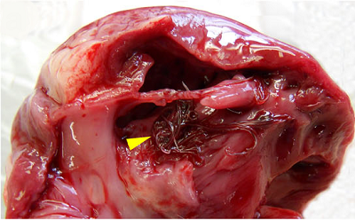
parasite, Angiostrongylus vasorum, a deadly lungworm/heartworm. One of
the cavaliers did not survive despite treatment, and the worms were
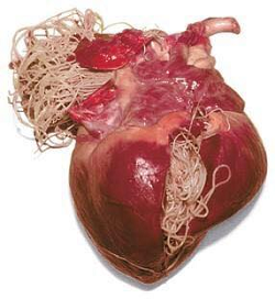
found in the dog's heart. (See arrow in photo at right.) This
article brings to a total of
12 such publications of CKCSs suffering from Angiostrongylosis, the
disease caused by this parasite, since 2004 -- far more reports than for
any other breed.
In a
June 2017 article, a team of Italian veterinarians (Olivieri E,
Zanzani SA, Gazzonis AL, Giudice C, Brambilla P, Alberti I, Romussi S,
Lombardo R, Mortellaro CM, Banco B, Vanzulli FM, Veronesi F, Manfredi MT
) report case
studies of two cavalier King Charles spaniels infected with the nematode
parasite, Angiostrongylus vasorum, a deadly lungworm/heartworm. One of
the cavaliers did not survive despite treatment, and the worms were
found in the dog's heart. (See arrow in photo at right.) This
article brings to a total of
12 such publications of CKCSs suffering from Angiostrongylosis, the
disease caused by this parasite, since 2004 -- far more reports than for
any other breed.
March 2016:
Paris, France cavalier is diagnosed with a lungworm in its eye.
In
a
March 2016 article, a team of European ophthalmologists report that
a 2-year-old male cavalier King Charles spaniel was found to have a
wiggly, thread-like organism in the front chamber of its left eye (see
photo). the organism was surgically removed, and a Baermann
technique test confirmed it to be an A. vasorum female nematode. The
cavalier displayed no signs of respiratory infection. The dog was
treated with fenbendazol for three weeks and with prednisolone. Two
other dogs diagnosed with ocular infection with A. vasorum were included
in the report. The investigators noted that: "Interestingly, ... of the
few reports now available in literature, three ... involved Cavalier
King Charles Spaniel dogs. However, additional epidemiological data are
needed for a clear assessment of risk factors (e.g. breed and age)
related to the occurrence of canine ocular angiostrongylosis."
RETURN TO TOP
RETURN TO TOP
Veterinary Resources
Angiostrongylus vasorum in the anterior chamber of a dog's eye. M. C. A. King, R. M. R. Grose, G. Startup. J. Small Anim. Prac. June 1994;35:326–8. Quote: "An unusual case of Angiostrongylus vasorum infestation occurred in a three-year-old female cavalier King Charles spaniel. The dog presented with signs consistent with right otitis interna, followed by the appearance of a free-swimming nematode in the anterior chamber of the right eye. The dog died of acute heart failure before surgical removal of the parasite was possible. Post mortem examination confirmed the presence of large numbers of worms in the pulmonary artery and right ventricle. These worms were identified histologically as A vasorum." Age, sex and breed: 3-year-old female Cavalier King Charles Spaniel. Location: UK. Clinical presentation: Otitis interna, head tilt, submaxillary lymph node enlargement. Presence of a motile worm in the anterior chamber of the eye on a one-day follow up visit. The dog died from a sudden attack of acute respiratory distress. Diagnosis: Numerous A. vasorum adults in the right ventricle and pulmonary artery and one adult in the anterior chamber detected at post-mortem examination.
Crenosoma vulpis, the fox lungworm, in a dog in Ireland. G. A. C. Reilly, J. W. McGarry, M. Martin, C. Belford. Vet. Rec. June 2000;146:764-765. Quote: Crenosoma vulpis is a metastrongylid parasite of worldwide distribution, which inhabits the trachea, bronchi and bronchioles of many Canidae. The pathogenesis of crenosomiosis in the dog has been described by Stockdale and Hulland (1970). They note that transmission of infection is effected by ingestion of land snails or slugs harbouring the infective third-stage larvae, and that larval migration to the lungs is via the portal circulation and the liver. Since the first report in a domestic dog in the UK (Cobb and Fisher 1992) there have been very few cases of crenosomiosis in dogs recorded in the literature. This short communication reports what the authors believe is the first case in a dog in Ireland. A one-year-old cavalier King Charles spaniel from Northern Ireland was examined ... for a suspected infectious tracheobronchitis of several days duration. The dog had a productive cough and dribbled saliva, which was occasionally mucopurulent in character. Its respiratory rate was elevated (35/minute) and it was slightly hyperpnoeic. On examination, both tonsils were markedly enlarged, and a soft moist cough was elicited by tracheal palpation. On auscultation, the dog had increased lung sounds over the entire lung fields. ... Under general anaesthesia, plain lateral and ventrodorsal thoracic radiographs were obtained with the lungs inflated. These revealed a normal cardiac outline and the presence of an ill-defined and irregular increase in patchy infiltrates throughout the lungs which was most striking in the diaphragmatic lobes. The predominant pattern was alveolar, with some small nodular-like densities in places. Bronchoscopic examination revealed a hyperaemic trachea, and a mucopurulent exudate affecting the lower bronchi. No parasites were visible grossly, but cytological examination of fluid collected by bronchoalveolar lavage confirmed the presence of a parasitic bronchitis. The airway cytology revealed a moderate to severe, chronically active inflammation with a significant eosinophil component, associated with large numbers of larvae. These were identified as the larvae of C vulpis (McGarry and others 1995). Faecal examination by the Baermann technique failed to identify any larvae. Therapy with fenbendazole (Panacur; Hoescht) at 50 mg/kg orally once daily, was initiated. After seven days, the dog was depressed, vomiting and coughing much more severely. The peripheral eosinophil count had risen threefold to 3·1 x 109 (normal range 0·5 to 1·5 x 109). The dose of fenbendazole was reduced to 20 mg/kg once daily, for the next seven days, and prednisolone at a dose of 1 mg/kg bodyweight once daily, was given concurrently. The dog made a rapid recovery, with resolution of all symptoms within two weeks. Repeat thoracic radiographs at this stage showed that the radiographic lung changes visible on presentation were no longer evident.
Clinical signs, diagnosis and treatment of three dogs with angiostrongylosis in Ireland. Sheila F. Brennan, Grainne McCarthy, Hester McAllister, Hugh Bassett, Boyd R. Jones. Irish Vet. J. February 2004;57(2):103-109. Quote: "Infection with Angiostrongylus vasorum was diagnosed at necropsy on a dog [20-month-old male Cavalier King Charles spaniel] that died from acute pulmonary haemorrhage, and on recovery of L1 larvae by Baermann examination of faeces from two dogs, one of which had abdominal pain and retroperitoneal haemorrhage, while the other had right-sided heart failure due to corpulmonale. The presenting signs included syncope (one dog), exercise intolerance (two dogs), cough (two dogs), abdominal pain (one dog) and depression (one dog). Onestage prothrombin time and activated partial thromboplastin time were prolonged in two dogs, buccal mucosal bleeding time was prolonged in one dog and globulin was elevated in all three dogs. Two dogs were treated with fenbendazole and recovered."
Radiographic findings in 16 dogs infected with Angiostrongylus vasorum. A. K. Boag, C. R. Lamb, P. S. Chapman, A. Boswood. Vet. Rec. April 2004;154:426-430. Quote: Thoracic radiographs of 16 dogs infected naturally with Angiostrongylus vasorum showed signs of bronchial thickening, an interstitial pattern and a multifocal and/or peripheral alveolar pattern. In dogs treated with fenbendazole, follow-up radiographs showed that the alveolar pattern had resolved and a mild, hazy interstitial pattern had developed. In contrast with dogs with heartworm (Dirofilaria immitis), no pulmonary vascular lesions were identified. ... The 16 dogs with angiostrongylosis included three Staffordshire bull terriers, two cavalier King Charles spaniels, two boxers, one mixed breed and one each of eight other breeds. ... The 49 control dogs included a wide range of breeds, including six boxers, five cavalier King Charles spaniels and three Staffordshire bull terriers.
Angiostrongylus vasorum infection in 23 dogs (1999–2002). P. S. Chapman, A. K. Boag, J. Guitian, A. Boswood. J. Small Anim. Prac. September 2004;45(9):435-440. Quote: "Angiostrongylosis was diagnosed in 23 dogs presenting Queen Mother Hospital for Animals between June 1999 and 2002. The animals' clinical records were reviewed retrospectively and certain risk factors were compared with a control population of 3407 dogs. Twenty-two of the 23 dogs were from south-England and dogs from Surrey (n=8) were significantly overrepresented. There were also significantly more Cavalier Charles spaniels (n=5) and Staffordshire bull terriers (n=5) among the affected dogs than in the control group. The median affected dogs was 10 months (range five to 90 months). common presenting signs were cough (65 per cent), dyspnoea (43 per cent), haemorrhagic diathesis (35 per cent) and (26 per cent). Four dogs were thrombocytopenic and eight significant prolongations in prothrombin time and/or activated partial thromboplastin time. Thoracic radiographs were abnormal 18 of 19 dogs. A variety of changes were observed, the most being a patchy alveolar-interstitial pattern affecting the dorsocaudal lung fields. Angiostrongylus vasorum larvae were found in 10 bronchoalveolar lavage specimens and 19 of 19 faecal Three dogs died shortly after admission to the hospital. The remainder were successfully treated with fenbendazole at of 50 mg/kg for five to 21 days. A vasorum should now be considered endemic to south-east England."
Angiostrongylus vasorum at a preadult phase in the anterior chamber of a young dog's eye. G. Payen. Vet. Ophthal. October 2004;7:434 (Abst. #47). Quote: The present clinical observation shows intraocular infestation by a worm, of an 8-month-old Cavalier King Charles dog, which presented with a strong and fitful cough. Signs of uveitis, associated with the presence of the parasite, were moderate. Hematologic exams, thorax radiographies, and cytologic exam of a broncho-alveolar washing, suggested a pulmonary parasitic infestation. Etiologic diagnosis was established by surgical removal of a worm from the anterior chamber of the right eye, indicating the presence of an immature female adult Angiostrongylus vasorum . The search for parasites in stools by the Baermann method, as well as in the liquid issued from a broncho-alveolar washing, was, however, unsuccessful. A fenbendazole-based antiparasite treatment was implemented after worm removal, and a 2-week follow-up of the dog showed a satisfactory outcome, without observable ophthalmologic after effects. Cases of aberrant migration of Angiostrongylus vasorum at preadult stages in eyes are rare, and the presence of this parasite in the anterior chamber of the eye has only been described four times, including two cases before 1930. The present clinical report offers an original description of ophthalmologic signs associated with the intraocular presence of Angiostrongylus vasorum.
Canine Angiostrongylosis. Sheila Brennan. UK Vet. April 2008;13(3):1-4. Quote: The diagnosis of canine angiostrongylosis appears to have been increasing over recent years. Whether this is due to an increased awareness or a true increase in incidence is largely unknown. Importantly, infected dogs are now being recognised outside of traditional endemic foci. This article aims to provide relevant and practical information on canine angiostrongylosis, particularly concerning the diagnosis and treatment of this potentially fatal parasitic infection. ... The majority of reported cases of canine angiostrongylosis have been in dogs less than 24 months of age. In one report Cavalier King Charles Spaniels and Staffordshire Bull Terriers were over-represented (Chapman et al, 2004). Conclusion: The reasons for the seemingly increased numbers of clinical cases on canine angiostrongylosis are unclear. A combination of increased awareness coupled with the spread of the urban fox, warmer wetter winters, ineffective anthelmintic prophylaxis and changes in domestic dog husbandry may account for the increase. Many questions remain unanswered. However through increased clinical observation, knowledge and screening, answers may be found and unnecessary deaths avoided.
Canine pulmonary angiostrongylosis: An update. J. Koch, J.L. Willesen. Vet. J. March 2009;179(3):348-359. Quote: Canine pulmonary angiostrongylosis is an emerging snail-borne disease causing verminous pneumonia and coagulopathy in dogs. The parasite is found in Europe, North and South America and Africa, covering tropical, subtropical and temperate regions. Its distribution has been characterised by isolated endemic foci, with only sporadic occurrences outside these areas. In the last two decades, the literature has been dominated by several case reports and small case series describing sporadic disease in old or new endemic areas. Case reports and experimental studies with high doses of infective third stage larvae may not reflect what happens under field conditions. There is insufficient understanding of the spread of infection and the dynamic consequences of this parasite in the canine population. This review discusses the biology, epidemiology, clinical aspects and management of canine pulmonary angiostrongylosis.
Angiostrongylus vasorum: a parasite on the move? Ian Wright. UK Vet. June 2009;14(5):1-3. Quote: Angiostrongylus vasorum, also known as the ‘small’ or ‘French’ heartworm, is a metastrongylid roundworm of canids. Until recently it had largely been forgotten or ignored by veterinary surgeons in most of the UK as it was not perceived to be as pathogenic as its larger cousin Dirofilaria and was limited in the UK to a few small pockets in the south. The last few years however have revealed Angiostrongylus either to have been vastly under reported or, as recent research would suggest, to be spreading across and up the country. ... Miniature breeds are under-represented for Angiostrongylus infection, possibly due to rare ingestion of the intermediate host. The exception to this rule is the Cavalier King Charles Spaniel which is over-represented. This presents a further challenge in early recognition of the disease, as so many Cavaliers are already predisposed to congestive heart failure.
Angiostrongylus vasorum infection in dogs: continuining spread and developments in diagnosis and treatment. Morgan E., Shaw S. December 2010. J. Small Animal Practice. 2010;51:616-621. Quote: "Disease caused by Angiostrongylus vasorum is increasingly diagnosed in dogs, as the geographic range of the parasite increases along with awareness among clinicians. ... There are limited data on risk factors for infection, but these appear to include age (younger dogs more likely to be infected), recent worming history and breed (Staffordshire bull terriers and cavalier King Charles spaniels marginally over-represented). ... Diagnosis, treatment and prevention are not always straightforward, although recent developments offer hope for improved options in future. Understanding of the epidemiology and pathogenesis remains poor. This paper provides an overview of the current state of knowledge of this parasitic disease, focussing on the most recent developments and advances."
Canine Angiostrongylus vasorum. I. Moeremans, D. Binst, E. Claerebout, I. Van de Maele, S. Daminet. Vlaams Diergeneeskundig Tijdschrift. September 2011;80(5):319-326. Quote: "The French heartworm Angiostrongylus vasorum is a parasitic nematode that lives in the pulmonary vessels and the heart of canids. Transmission occurs through ingestion of infected intermediate hosts, such as snails and slugs. There are increasing reports of autochthonous infections in our neighbouring countries. ... In the Danish survey, no breed predisposition was reported, although others have found a higher occurrence in Cavalier King Charles spaniels, Staffordshire Bull terriers and beagles. The last author attributed this fact to the use of this breed as a hunting dog. Dogs used for hunting seem to be more at risk because of their exposure to infection from the fox-snail life cycle during training. ... Clinical signs usually relate to the respiratory system, coagulopathy and the neurologic system. Anorexia, gastrointestinal dysfunction and weight loss are also frequently observed. Diagnosis is not straightforward, but abnormalities detected by thoracic radiography, echocardiography, magnetic resonance imaging (MRI) or computed tomography (CT) scan can be helpful. Eosinophilia, regenerative anemia and thrombocytopenia with or without abnormalities in the coagulation profile can occur. Definitive diagnosis is made by demonstrating the parasite in the cerebrospinal fluid, in faeces (Baermann technique) and/or in bronchoalveolar lavage fluid. Treatment consists of anthelmintic drugs and supportive care if necessary."
Canine Angiostrongylosis in Naturally Infected Dogs: Clinical Approach and Monitoring of Infection after Treatment. Paola Paradies, Manuela Schnyder, Antonio Capogna, Riccardo Paolo Lia, Mariateresa Sasanelli. Sci. World. J. November 2013; doi: 10.1155/2013/702056. Quote: Canine angiostrongylosis is an increasingly reported disease in Europe which can be fatal if left untreated. The wide range of clinical presentation along with the absence of pathognomonic alterations can make the diagnosis challenging; thus any additional information that may provide clues to an early diagnosis may be of value, in order to ensure adequate anthelmintic treatment. Aim of the study was to assess a clinicopathological scoring system associated with natural Angiostrongylus vasorum infection diagnosed in canine patients during clinical practice, to clinically and paraclinically monitor infected dogs after treatment, and to monitor the presence of L1 larvae in faecal samples by Baermann's test. Of the total 210 enrolled animals A. vasorum infection was diagnosed in 7 dogs. These dogs were clinically and paraclinically investigated and monitored after specific treatment. Further 3 symptomatic dogs were retrospectively included in the monitoring. Results suggest that the computed scoring system can help to increase the clinical suspicion of infection particularly in asymptomatic dogs before the onset of potentially lethal lesions. Data of faecal monitoring suggested that treatment may control parasite burden but be unable to eradicate infection. Thus, a continued faecal monitoring after treatment is advisable for identification of still infected or reinfected dogs.
Spatial, demographic and clinical patterns of Angiostrongylus vasorum infection in the dog population of southern England. T. R. W. Blehaut, J. L. Hardstaff, P. S. Chapman, D. U. Pfeiffer, A. K. Boag, F. J. Guitian. Vet. Rec. June 2014. Quote: "A retrospective study was carried out to provide updated knowledge of the spatial pattern of Angiostrongylus vasorum infection in Southern England and to investigate associations between selected host characteristics (age, breed, sex), risk of infection and clinical presentation (cardiorespiratory signs v haemorrhagic diathesis). One hundred and fortyone cases diagnosed between April 1999 and July 2012 were compared with a control population of dogs referred to the same hospital. A significant association was found between haemorrhagic diathesis and breed but not for other host characteristics and clinical presentations. Younger dogs and certain breeds of dog (Jack Russell terriers, Cocker Spaniels, Springer Spaniels, Cavalier King Charles spaniels and Staffordshire Bull Terriers) had significantly higher odds of angiostrongylosis than other breeds in the study. A significant cluster of cases was found in Southern England. Animals presenting with cardiorespiratory signs or haemorrhagic diathesis in Southern England, especially if they are young or of a breed associated with angiostrongylosis, should be given special consideration with regards to possible A. vasorum infestation. Our results should be interpreted bearing in mind that they are based on the retrospective exploration of dogs seen at a referral centre."
Angiostrongylosis – Current situation in Portugal. AM Alho, M Schnyder, S Belo, P Deplazes, Luis Madeira de Carvalho. 4th Euro. Dirofilaria & Angiostrongylus Days. July 2014. Quote: Angiostrongylus vasorum is considered an emergent parasite due to its recent spread in Europe and North America. It is a potentially lethal parasite living in the heart and pulmonary arteries of dogs and wild carnivores. Objective: The present work aimed to update the occurrence of recent cases of A. vasorum infections in Portugal, including their host distribution and geographical dispersion. ... Methods & Results: ... Regarding canine angiostrongylosis, three positive cases were diagnosed in the Lisbon area in the last few years, with prevalence ranging 0,3-2%: in 2006, a two-year-old [Cavalier] King Charles Spaniel male dog, with haemorrhagic gastroenteritis and severe dyspnoea and anaemia, revealing several L1 in a fresh smear and two adult nematodes inside bronchiolar arterioles at post-mortem examination; in 2012, an adult mixedbreed male dog with a history of weight loss was positive for A. vasorum firststage larvae in the Baermann test; similarly, in 2013, a 13-year-old, mixed-breed male dog presenting with diarrhoea and weight loss was confirmed positive by larval detection, not showing any respiratory symptoms, neurological signs or coagulopathies. ... Discussion & Conclusion: The first reports from the centre and south of Portugal showed initially a low prevalence rate in wild populations, but the last ones showed an increasing value for foxes. At the same time, low prevalence rates in dogs, show at the moment, both by parasitological and serological techniques, an increasing trend and a dispersion in the central and southern areas of Portugal. These data aimed to alert veterinarians and owners about angiostrongylosis and its wider distribution in dogs and foxes than previously known, as well as, point out the challenging variety of clinical manifestations, occasionally even asymptomatic, of this important lungworm disease.
Recent advances in the epidemiology, clinical and diagnostic features, and control of canine cardio-pulmonary angiostrongylosis. Elsheikha H.M., Holmes S.A., Wright I., Morgan E.R., Lacher D.W. Veterinary Research. September 2014;45:1-12. Quote: "The aim of this review is to provide a comprehensive update on the biology, epidemiology, clinical features, diagnosis, treatment, and prevention of canine cardio-pulmonary angiostrongylosis. This cardiopulmonary disease is caused by infection by the metastrongyloid nematode Angiostrongylus vasorum. The parasite has an indirect life cycle that involves at least two different hosts, gastropod molluscs (intermediate host) and canids (definitive host). A. vasorum represents a common and serious problem for dogs in areas of endemicity, and because of the expansion of its geographical boundaries to many areas where it was absent or uncommon; its global burden is escalating. ... Published data in the United Kingdom suggest that some purebred dogs are at higher risk than crossbreeds, and in particular Cavalier King Charles Spaniels and Staffordshire Bull Terriers and beagles. ... A. vasorum infection in dogs can result in serious disorders with potentially fatal consequences. Diagnosis in the live patient depends on faecal analysis, PCR or blood testing for parasite antigens or anti-parasite antibodies. Identification of parasites in fluids and tissues is rarely possible except post mortem, while diagnostic imaging and clinical examinations do not lead to a definitive diagnosis. Treatment normally requires the administration of anthelmintic drugs, and sometimes supportive therapy for complications resulting from infection."
Retrospective evaluation of moderate-to-severe pulmonary hypertension in dogs naturally infected with Angiostrongylus vasorum. J. Sm. Anim.l Pract. March 2015; doi: 10.1111/jsap.12309. Quote: Objectives: The outcome in dogs with pulmonary hypertension associated with natural Angiostrongylus vasorum infection is unclear. This study aimed to report long-term outcome of dogs with A. vasorum and pulmonary hypertension, and to evaluate factors associated with pulmonary hypertension development. It was hypothesised that dogs with pulmonary hypertension had a shorter survival time than dogs without pulmonary hypertension. Methods: Retrospective review of clinical records of dogs diagnosed with A. vasorum. Dogs were classified as having or not having pulmonary hypertension based on clinical signs and imaging findings. Signalment, signs and outcome were recorded. DNA obtained from banked samples was genotyped for the PDE5a:E90K polymorphism, a possible factor in development of pulmonary hypertension. Results: The proportion of dogs with moderate-to-severe pulmonary hypertension and A. vasorum infection in the study population was 14.6% [14 dogs with moderate-to-severe pulmonary hypertension and A. vasorum infection, including 2 cavalier King Charles spaniels (14%)]. No difference in the population characteristics or PDE5a genotype was detected between dogs with and without pulmonary hypertension. Dogs with pulmonary hypertension had a significantly shorter survival time and a greater risk of death within 6 months of diagnosis (odds ratio 12.5%, 95% confidence interval 2 · 1 to 74 · 9). Clinical Significance: A. vasorum-associated pulmonary hypertension is an important problem in naturally infected dogs and has a negative effect upon survival.
Prevalence, treatment and prevention of A vasorum. Simon Tappin. Vet. Times. July 2015:8-10. Quote: "Angiostrongylus vasorum was first documented in France in 1853. As a result it is often referred to as French heartworm because the adults live in the pulmonary arteries and the right side of the heart. Infection leads to varied clinical signs, with respiratory, coagulopathic and neurological signs most commonly reported. ... Within the cluster in southern England, younger dogs, cocker spaniels, springer spaniels and cavalier King Charles spaniels, Jack Russells and Staffordshire bull terriers were found to be at higher risk of infection within a referral population of cases. ... Cardiorespiratory signs are most commonly seen, with coughing occurring due to the physical presence of the parasite. ... Disease due to infection with Angiostrongylus vasorum has been well documented in small numbers of dogs in south Wales, and both the south-east and south-west of England for some years. Recently, both the incidence and geographical distribution appear to be increasing, with cases now being reported in northern England and Scotland. Most dogs develop signs of cardiorespiratory disease; however, a significant proportion develop signs secondary to coagulopathy. A rapid patient-side blood test with good sensitivity and specificity has become available, greatly improving the ease of diagnosis. Imidacloprid/moxidectin, milbemycin and fenbendazole are all effective treatment options, with imidacloprid/moxidectin and milbemycin also being an effective prophylactic treatment."
Autochthonous Angiostrongylus vasorum infection in a Border collie in Belgium. C. Sarre, A. Willems, S. Daminet, E. Claerebout. Vlaams Diergeneeskundig Tijdschrift. September 2015;84(5):243-252. Quote: "Purebreds could be at higher risk [of Auto-chthonous Angiostrongylus vasorum infection], with Cavalier King Charles spaniels, Staffordshire bull terriers, Beagles and hunting dog breeds in general as the most frequently infected."
Angiostrongylus vasorum in the eye: new case reports and a review of
the literature. Vito Colella, Riccardo Paolo Lia, Johana
Premont, Paul Gilmore, Mario Cervone, Maria Stefania Latrofa, Nunzio
D’Anna, Diana Williams, Domenico Otranto. BioMed Central. March
2016. Quote: "Background: Nematodes of the genus Angiostrongylus are
important causes of potentially life-threatening diseases in several
animal species and humans. Angiostrongylus vasorum affects the right
ventricle of the heart and the pulmonary arteries in dogs, red foxes
and other carnivores. The diagnosis of canine angiostrongylosis may
be challenging due to the wide spectrum of clinical signs. Ocular
manifestations have been seldom reported but have serious
implications for patients. Methods: The clinical history of three
cases of infection with A. vasorum in dogs diagnosed in UK, France
and Italy, was obtained from clinical records provided by the
veterinary surgeons along with information on the diagnostic
procedures and treatment. Nematodes collected from the eyes of
infected dogs were morphologically identified to the species level
and molecularly analysed by the amplification of the nuclear 18S
rRNA gene. Results: On admission, the dogs were presented with
various degrees of ocular discomfort and hyphema because of the
presence of a motile object in the eye. The three patients had
ocular surgery during which nematodes were removed and subsequently
morphologically and molecularly identified as two adult males and
one female of A. vasorum. ...
 Case
2: A 2-year-old male Cavalier King Charles Spaniel
dog was referred to a private veterinary clinic in Paris (France)
due to persistent blepharospasm and epiphora. The dog that lived in
the city centre was walked in a forest nearby. On clinical
ophthalmological examination the dog showed prolapse of the
nictitating membrane, photophobia on the left eye, iris hyperaemia
and corneal oedema. The intraocular pressure was 14 mmHg in the left
eye and 16 mmHg in the right eye, the fluorescein test was negative
and the Schirmer test showed a slightly increased lacrymation. A
thread-like organism was noticed in the anterior chamber of the left
eye (photo at right), which was very motile under light
stimuli. ... Removal of the parasite was performed by anterior
chamber paracentesis and the nematode was morphologically and
molecularly processed. The specimen was identified as an
unfertilised A. vasorum female. Briefly, the female nematode
measured 16 mm in length and 0.2 mm in width; genital ducts were
coiled around the reddish intestine, which appeared visible
throughout the cuticle. However, the nematode was damaged and
further morphological features were not evaluated, with the
exception of the uteri, which lacked first-stage larvae, and the
vulva. Therefore, the dog was subjected to coprological examination
for the detection of L1 using the Baermann technique. The 18S rDNA
sequences obtained from both L1 collected from the faeces and the
female nematode displayed 100 % identity to the nucleotide sequence
of A. vasorum [GenBank: AJ920365]. The dog lacked any signs of
respiratory infection, both previously and during the observation
period. The animal was treated with fenbendazol 25 mg/Kg per os SID
for 3 weeks associated with prednisolone 0.2 mg/Kg. ... Conclusions:
Three new cases of canine ocular angiostrongylosis are reported
along with a review of other published clinical cases to improve the
diagnosis and provide clinical recommendation for this parasitic
condition. In addition, the significance of migratory patterns of
larvae inside the host body is discussed. Veterinary healthcare
workers should include canine angiostrongylosis in the differential
diagnosis of ocular diseases. ... Interestingly, all dogs suffering
from canine ocular angiostrongylosis were under the 3 years of age
(i.e. 5 months to 3 years), and of the few reports now available in
literature, three and in Case 2, involved Cavalier King
Charles Spaniel dogs. However, additional epidemiological
data are needed for a clear assessment of risk factors (e.g. breed
and age) related to the occurrence of canine ocular
angiostrongylosis."
Case
2: A 2-year-old male Cavalier King Charles Spaniel
dog was referred to a private veterinary clinic in Paris (France)
due to persistent blepharospasm and epiphora. The dog that lived in
the city centre was walked in a forest nearby. On clinical
ophthalmological examination the dog showed prolapse of the
nictitating membrane, photophobia on the left eye, iris hyperaemia
and corneal oedema. The intraocular pressure was 14 mmHg in the left
eye and 16 mmHg in the right eye, the fluorescein test was negative
and the Schirmer test showed a slightly increased lacrymation. A
thread-like organism was noticed in the anterior chamber of the left
eye (photo at right), which was very motile under light
stimuli. ... Removal of the parasite was performed by anterior
chamber paracentesis and the nematode was morphologically and
molecularly processed. The specimen was identified as an
unfertilised A. vasorum female. Briefly, the female nematode
measured 16 mm in length and 0.2 mm in width; genital ducts were
coiled around the reddish intestine, which appeared visible
throughout the cuticle. However, the nematode was damaged and
further morphological features were not evaluated, with the
exception of the uteri, which lacked first-stage larvae, and the
vulva. Therefore, the dog was subjected to coprological examination
for the detection of L1 using the Baermann technique. The 18S rDNA
sequences obtained from both L1 collected from the faeces and the
female nematode displayed 100 % identity to the nucleotide sequence
of A. vasorum [GenBank: AJ920365]. The dog lacked any signs of
respiratory infection, both previously and during the observation
period. The animal was treated with fenbendazol 25 mg/Kg per os SID
for 3 weeks associated with prednisolone 0.2 mg/Kg. ... Conclusions:
Three new cases of canine ocular angiostrongylosis are reported
along with a review of other published clinical cases to improve the
diagnosis and provide clinical recommendation for this parasitic
condition. In addition, the significance of migratory patterns of
larvae inside the host body is discussed. Veterinary healthcare
workers should include canine angiostrongylosis in the differential
diagnosis of ocular diseases. ... Interestingly, all dogs suffering
from canine ocular angiostrongylosis were under the 3 years of age
(i.e. 5 months to 3 years), and of the few reports now available in
literature, three and in Case 2, involved Cavalier King
Charles Spaniel dogs. However, additional epidemiological
data are needed for a clear assessment of risk factors (e.g. breed
and age) related to the occurrence of canine ocular
angiostrongylosis."
Lungworms: a growing threat to companion animal health. Hany Elsheikha. Comp. Anim. October 2016;21(10):556-565. Quote: Our understanding of lungworm infection in dogs and cats has improved considerably over the last decade and significant progress has been made on many fronts, especially in the diagnosis and treatment of lungworm diseases. Despite this progress, lungworm infections due to Angiostrongylus vasorum (dog lungworm) and Aelurostrongylus abstrusus (cat lungworm), as well as other respiratory worm species, continue to impose a significant challenge to the health and welfare of dogs and cats. ... Conflicting data have been reported regarding the roles of age, gender and breed. Some evidence indicates that the most likely age for infection is around 10 months. Also, some purebred dogs, and in particular Cavalier King Charles Spaniels and Staffordshire Bull Terriers (Chapman et al, 2004) and beagles (Conboy, 2004), are reported to be at a higher risk than crossbreeds. More studies are needed in order to investigate a possible hereditary susceptibility in these breeds. ... Infections with some of these parasites are potentially life-threatening, and detection of infection is challenging due to the wide spectrum of clinical signs and the high frequency of subclinical infection. Current diagnostics, although very useful, are not perfect. The many ways by which lungworms can be maintained in nature create more opportunities for dogs and cats to come across the infective stages and succumb to the disease. The principal treatment for lungworm disease is the administration of effective anthelmintics to eliminate adult worms and reduce or stop larval faecal shedding, and to reduce the severity of clinical signs and lung damage. Given that already there are considerable risks to the health and welfare of dogs and cats, implementation of integrated parasite control strategies are crucial in order to mitigate the risks caused by lungworms and to improve treatment outcomes.
Hyperfibrinolysis and Hypofibrinogenemia Diagnosed With Rotational Thromboelastometry in Dogs Naturally Infected With Angiostrongylus vasorum. N.E. Sigrist, N. Hofer-Inteeworn, R. Jud Schefer, C. Kuemmerle-Fraune, M. Schnyder, A.P.N. Kutter. J. Vet. Intern. Med. May 2017. Quote: Background: The pathomechanism of Angiostrongylus vasorum infection-associated bleeding diathesis in dogs is not fully understood. Objective: To describe rotational thromboelastometry (ROTEM) parameters in dogs naturally infected with A. vasorum and to compare ROTEM parameters between infected dogs with and without clinical signs of bleeding. Animals: A total of 21 dogs presented between 2013 and 2016. ... 3 dogs were Cavalier King Charles Spaniels (3/21, 14.3%), 2 were Chihuahuas (2/21, 9.5%), and the remaining 16 dogs represented different breeds. ... Methods: Dogs with A. vasorum infection and ROTEM evaluation were retrospectively identified. Thrombocyte counts, ROTEM parameters, clinical signs of bleeding, therapy, and survival to discharge were retrospectively retrieved from patient records and compared between dogs with and without clinical signs of bleeding. Results: Evaluation by ROTEM showed hyperfibrinolysis in 8 of 12 (67%; 95% CI, 40–86%) dogs with and 1 of 9 (11%; 95% CI, 2–44%) dogs without clinical signs of bleeding (P = .016). Hyperfibrinolysis was associated with severe hypofibrinogenemia in 6 of 10 (60%; 95% CI, 31–83%) of the cases. Hyperfibrinolysis decreased or resolved after treatment with 10–80 mg/kg tranexamic acid. Fresh frozen plasma (range, 14–60 mL/kg) normalized follow-up fibrinogen function ROTEM (FIBTEM) maximal clot firmness in 6 of 8 dogs (75%; 95% CI, 41–93%). Survival to discharge was 67% (14/21 dogs; 95% CI, 46–83%) and was not different between dogs with and without clinical signs of bleeding (P = .379). Conclusion and Clinical Importance: Hyperfibrinolysis and hypofibrinogenemia were identified as an important pathomechanism in angiostrongylosis-associated bleeding in dogs. Hyperfibrinolysis and hypofibrinogenemia were normalized by treatment with tranexamic acid and plasma transfusions, respectively.
Angiostrongylus vasorum infection in dogs from a cardiopulmonary dirofilariosis endemic area of Northwestern Italy: a case study and a retrospective data analysis. Olivieri E, Zanzani SA, Gazzonis AL, Giudice C, Brambilla P, Alberti I, Romussi S, Lombardo R, Mortellaro CM, Banco B, Vanzulli FM, Veronesi F, Manfredi MT. BMC Vet. Res. June 2017;12:165. Quote: Background: In Italy, Angiostrongylus vasorum, an emergent parasite, is being diagnosed in dogs from areas considered free of infection so far. As clinical signs are multiple and common to other diseases, its diagnosis can be challenging. In particular, in areas where angiostrongylosis and dirofilariosis overlap, a misleading diagnosis of cardiopulmonary dirofilariosis might occur even on the basis of possible misleading outcomes from diagnostic kits. Case Presentation: Two Cavalier King Charles spaniel dogs from an Italian breeding in the Northwest were referred to a private veterinary hospital with respiratory signs. A cardiopulmonary dirofilariosis was diagnosed and the dogs treated with ivermectin, but one of them died. At necropsy, pulmonary oedema, enlargement of tracheo-bronchial lymphnodes and of cardiac right side were detected. Within the right ventricle lumen, adults of A. vasorum were found. All dogs from the same kennel were subjected to faecal examination by FLOTAC and Baermann's techniques to detect A. vasorum first stage larvae; blood analysis by Knott's for Dirofilaria immitis microfilariae, and antigenic tests for both A. vasorum (Angio Detect™) and D.immitis (DiroCHEK® Heartworm, Witness®Dirofilaria). The surviving dog with respiratory signs resulted positive for A. vasorum both at serum antigens and larval detection. Its Witness® test was low positive similarly to other four dogs from the same kennel, but false positive results due to cross reactions with A. vasorum were also considered. No dogs were found infected by A. vasorum. Eventually, the investigation was deepened by browsing the pathological database of Veterinary Pathology Laboratories at Veterinary School of Milan University through 1998-2016, where 11 cases of angiostrongylosis were described. Two out of 11 dogs had a mixed infection with Crenosoma vulpis. Conclusion: The study demonstrates the need for accurate surveys to acquire proper epidemiological data on A. vasorum infection in Northwestern Italy and for appropriate diagnostic methods. Veterinary clinicians should be warned about the occurrence of this canine parasite and the connected risk of a misleading diagnosis, particularly in areas endemic for cardiopulmonary dirofilariosis.
Angiostrongylose oculaire chez un cavalier king charles. Mario Cervone, Laurent Bouhanna. Le Point vétérinaire (Éd. Expert canin). June 2018;49(384):76-79. Quote: A castrated male Cavalier King Charles spaniel was presented for exploration of blepharospasm and severe tear production in recent days. Slit lamp biomicroscopic examination revealed a living parasite in the anterior chamber of the left eye. Surgical extraction of the parasite was performed. An adult female Angiostrongylus vasorum was identified on morphological and molecular examinations. A stool examination by the Baermann method was performed to search for a pulmonary vascular parasite focus. It was positive for the presence of L1 larvae of A. vasorum. Oral fenbendazole treatment was initiated for 21 days. The ocular signs had disappeared at the 7-day postoperative check-up and the stool examination by the Baermann method proved negative for the presence of A. vasorum larvae 1 month later.
The
diagnostic approach to coughing in dogs and cats. Jenny
Reeve. Companion Anim. July 2018;23(7):396-404. Quote: Coughing is a
common presenting sign in dogs, less so in cats, and may be a
manifestation of a wide range of disorders, from acute self-limiting
disease that does not require specific diagnosis and treatment,
through to acute and chronic diseases of varying severity that
require appropriate investigation to guide optimal management and
outcome. This article reviews the diagnostic approach to coughing.
... Table 1. Causes of coughing with associated commonly affected
breeds: ... Pneumocystis carinii infection: Cavalier King
Charles Spaniel, Dachshund. ... Haemoptysis (coughed blood)
is an uncommon presenting feature and may be reported if bloody
mucus is expectorated during coughing. ... Angiostrongylus vasorum
is an important possible cause ... Concurrent neurological signs or
bleeding diatheses, especially when present together, are highly
suspicious for Angiostrongylus vasorum infection. ... Table 2.
 Primary
respiratory causes of coughing in dogs and cats: ... Inflammatory,
infectious (viral, bacterial, protozoal, fungal, parasitic) ...
Lower airways ... Angiostrongylus vasorum. ... Baermann migration is
the technique of choice for evaluation for the more commonly
encountered respiratory parasites (Angiostrongylus vasorum). ... A
sensitive and specific patient-side Angiostrongylus vasorum antigen
ELISA for use with serum or plasma is also available (Angio Detect,
IDEXX Laboratories, Inc). ... Some thoracic imaging patterns are
pathognomic, such as a peripheral patchy alveolar pattern in
Angiostrongylus vasorum pneumonitis. ... Some thoracic radiography
findings are considered pathognomic e.g. a patchy peripheral
alveolar pattern in Angiostrongylus vasorum pneumonitis. (Figure
2 [at right]: Angiostrongylus vasorum larva identified on
microscopic examination of expectorated bloody sputum).
Primary
respiratory causes of coughing in dogs and cats: ... Inflammatory,
infectious (viral, bacterial, protozoal, fungal, parasitic) ...
Lower airways ... Angiostrongylus vasorum. ... Baermann migration is
the technique of choice for evaluation for the more commonly
encountered respiratory parasites (Angiostrongylus vasorum). ... A
sensitive and specific patient-side Angiostrongylus vasorum antigen
ELISA for use with serum or plasma is also available (Angio Detect,
IDEXX Laboratories, Inc). ... Some thoracic imaging patterns are
pathognomic, such as a peripheral patchy alveolar pattern in
Angiostrongylus vasorum pneumonitis. ... Some thoracic radiography
findings are considered pathognomic e.g. a patchy peripheral
alveolar pattern in Angiostrongylus vasorum pneumonitis. (Figure
2 [at right]: Angiostrongylus vasorum larva identified on
microscopic examination of expectorated bloody sputum).
Anorexia & Lethargy in a Dog with Presumed Giardiasis. Ali Nemeth, Orla Mahony, Neketa Kakar, Claire L. Fellman. Clinician's Brief. November 2019; pp. 17-20. Quote: Rylie’s presumptive generalized bone marrow hypoplasia/aplasia could have been an idiosyncratic reaction unrelated to the pharmacologic action of fenbendazole. However, because another dog in the household experienced the same clinical course following similar dosing, both dogs’ signs were likely dose dependent.
Lungworm: A roundtable discussion. Debra Bourne, Hany Elsheikha, Robyn Farquhar, Jenny Helm, Eric Morgan, Iain Peters, Kit Sturgess, Andrew Torrance, Ian Wright. Companion Anim. March 2020;25(2):1-12. Quote: Lungworm (Angiostrongylus vasorum) is increasingly recognised as a cause of illness, particularly cardiorespiratory signs and haemorrhage, in dogs. It is now distributed across the UK, although prevalence varies widely and there are local foci of higher infection. ... There have been suggestions in the literature that certain breeds are more susceptible to lungworm infection: Staffordshire Bull Terriers, Cavalier King Charles Spaniels and Cocker Spaniels. There is no firm evidence to support this. ... Presentation of clinical cases varies considerably, and lungworm should be on many differential diagnosis lists. Diagnostic tests now include direct faecal smear, Baermann's, bronchoalveolar lavage, antibody and antigen ELISAs and PCR. Appropriate testing depends on the clinical situation, and no test is 100% sensitive and 100% specific. Treatment involves moxidectin or milbemycin oxime, plus supportive care. Tranexamic acid and plasma transfusions may both be useful in dogs showing coagulopathies. Dogs may be at risk from accidental ingestion of intermediate host slugs with grass, or larvae in puddles, as well as from deliberately eating slugs, so prevention relies heavily on monthly appropriate anthelmintics, and compliance is an issue in this. Other lungworm species also occur and should not be forgotten.
Field safety and efficacy of an orally administered combination of sarolaner, moxidectin and pyrantel (Simparica Trio) for the prevention of angiostrongylosis in dogs presented as veterinary patients. Csilla Becskei, Jakob L. Willesen, Manuela Schnyder, Magda Wozniakiewicz, Nataliya Miroshnikova, Sean P. Mahabir. Parasites Vectors. August 2020; doi: 10.1186/s13071-020-04262-4. Quote: Background: Infection with the cardiopulmonary nematode Angiostrongylus vasorum may cause severe disease in dogs, therefore prophylactic treatments are necessary to prevent infection in dogs at risk. A clinical field study was conducted to demonstrate the efficacy and safety of an oral combination of sarolaner, moxidectin and pyrantel (Simparica Trio) for the prevention of A. vasorum infection in dogs (prevention study). A survey study was conducted concurrently to determine the infection pressure in the same areas. Methods: Prevention and survey studies were both conducted at the same veterinary clinics in endemic hot spots for A. vasorum in Denmark and Italy. The prevention study was a randomized, placebo controlled, double masked study where 622 client-owned dogs were treated and tested at 30 days intervals for 10 months. In the survey study 1628 dogs that were at risk of infection and/or were suspected to be infected were tested by fecal and/or serological methods, and the percent of dogs positive for A. vasorum was calculated. Results: In the prevention study, there were no adverse events related to treatment with Simparica Trio. Two placebo-treated animals became infected with A. vasorum during the 10-month study period, while none of the dogs in the combination product-treated group became infected. In the survey study, 12.2% of the study dogs were found positive to A. vasorum, indicating high exposure to the parasite during the period of the prevention study. Conclusions: Monthly oral treatment with the combination of sarolaner, moxidectin and pyrantel (Simparica Trio) was 100% effective in the prevention of natural infection with A. vasorum in dogs in highly endemic areas. In endemic areas, A. vasorum occurrence in dogs at risk is considerable.
Risk factors and predictors of angiostrongylosis in naturally infected dogs in the southeast of England. Sarah Holmes, Phillip Paine, Ian Wright, Eric R. Morgan, Hany M. Elsheikha. Companion Anim. August 2020; doi: 10.12968/coan.2018.0066. Quote: Canine pulmonary angiostrongylosis is a snail-borne parasitic infection caused by the nematode Angiostrongylus vasorum. The spread of A. vasorum across the UK in recent years, alongside the launch of new pharmaceutical products licenced for its treatment and prevention, has led to raised awareness of this parasite among veterinarians and the public alike. This raised profile has been beneficial in reducing canine morbidity and mortality associated with infection, especially in parts of the country where it had not previously been routinely diagnosed. It is vital, therefore, given raised public awareness and geographical spread of the parasite, that the veterinary professionals have demographic data to help give accurate risk-based advice. This study retrospectively evaluated demographic factors (age, gender and breed) and clinical presentation of 100 dogs with natural A. vasorum infection by reviewing the clinical record database at private practices, spanning the period from 2003 to 2009, alongside 100 dogs presenting for other reasons as a control sample. A significant relationship was detected between young age and A. vasorum infection, with dogs less than 1 year of age 4.2 times more likely to fall into the infected group. Gender was not identified as a significant risk factor for A. vasorum infection in dogs. Breed was a significant risk factor, with Cocker Spaniels over-represented in the infected group, and Staffordshire Bull Terriers and cross-breeds under-represented. ... Other breeds found by Blehaut et al (2014) and Chapman et al (2004) to be at higher risk of the disease, i.e. Springer Spaniels, Jack Russell Terriers and Cavalier King Charles Spaniels, were not associated with increased risk in the current study. ... The strongest contrast was between cross-bred and pure-bred dogs as a whole, with the latter 23 times more likely to present with angiostrongylosis. Significant associations were found between A. vasorum infection and dogs presenting with cough, coagulopathy, vomiting/diarrhoea and/or lethargy. The diagnostic value of clinical signs for the presence of disease, expressed by area under the receiver operating characteristic curve, was >0.7, indicating that correct diagnosis (discrimination between dogs with or without the disease) can be achieved in 70% of the clinical cases by accurate history taking. These findings provide important clues regarding the risk of infection to an individual dog, and thus may facilitate improved recognition of infection based on clinical presentation, and implementation of preventative strategies to combat A. vasorum infection.
Spontaneous tricuspid valve chordal rupture in a dog with severe, irreversible pulmonary hypertension caused by Angiostrongylus vasorum infection. Viktor Szatmári. BMC Vet. Res. August 2020;16:311. Quote: Background: The adult worms of Angiostrongylus vasorum reside in the pulmonary artery of dogs and can lead to cardiac, respiratory, and central neurologic signs. Due to luminal obstruction and perivascular inflammation of the pulmonary artery branches, pulmonary hypertension can arise. Pulmonary hypertension, in turn, can lead to severe damage of the right-sided cardiac structures, leading to right ventricular remodeling and tricuspid valve regurgitation. Case presentation: An 8-year-old neutered female English Cocker Spaniel was presented to the author’s institution because of abdominal distention and exercise intolerance. Ascites caused by congestive right-sided heart failure was found to be responsible for these problems. The underlying etiology of the right-sided heart failure was a severe pulmonary hypertension caused by Angiostrongylus vasorum infection. Echocardiography revealed, in addition to a severe concentric and eccentric right ventricular hypertrophy, right atrial and pulmonary trunk dilation, severe tricuspid valve regurgitation, and a systolic flail of the anterior leaflet of the tricuspid valve, resulting from ruptured chordae tendineae. As a coincidental finding, a congenital mitral stenosis was found. Oral therapy was initiated with daily administration of fenbendazole for 2 weeks along with daily administration of oral sildenafil until the re-check examination. At the 6-week re-check the dog showed full clinical and partial echocardiographic recovery, and both the blood antigen test for Angiostrongylus vasorum and the fecal Baermann larva isolation test were negative. When the sildenafil therapy was ceased after tapering the daily dosage, the owner reported recurrence of abdominal distension. Re-starting the sildenafil therapy resulted in resolution of this problem. The dog was reported to be clinically healthy with daily sildenafil administration 7 months after the initial presentation. Conclusions: The present case report describes a dog where angiostrongylosis led to congestive right-sided heart failure resulting from severe pulmonary hypertension. The secondary right ventricular eccentric hypertrophy together with suspected papillary muscular ischemia were the suspected cause of the ruptured major tricuspid chordae tendineae, which led to a severe tricuspid valve regurgitation. Despite eradication of the worms, the severe pulmonary hypertension persisted. Treatment with daily oral sildenafil, a pulmonary arterial vasodilator, was enough to keep the dog free of clinically apparent ascites.
Is Pulmonary Hypertension a Rare Condition Associated to Angiostrongylosis in Naturally Infected Dogs? Paola Paradies, Mariateresa Sasanelli, Antonio Capogna, Angelica Mercadante, Giuseppe Tommaso Roberto Rubino, Claudio Maria Bussadori. Topics in Compan. Anim. Med. January 2021; doi: 10.1016/j.tcam.2021.100513. Quote: Canine angiostrongylosis due to Angiostrongylus vasorum is one of the cardiopulmonary parasitic diseases in dogs and it can manifest with very different clinical pictures, which often make diagnosis very difficult. Based on the nature of the vascular and parenchymal lesions induced by the infection (thrombo-arteritis and fibrosis), it is not surprising that cases of pulmonary arterial hypertension (PAH) associated with angiostrongylosis have been reported in the literature, although it seems to represent a rare condition. The aim of the present work is to describe the clinical and instrumental aspects referred to cases of canine angiostrongylosis before and after treatment then to evaluate even mild conditions of PAH using echocardiography. PAH was not only conventionally investigated based on characteristic cardiac changes that occur secondary to PAH and by estimating pulmonary pressure from spectral Doppler tracings, but also by using a combination of further selected echocardiographic parameters (AT/ET, PA/Ao, Pulmonary flow profile pattern) able also to reveal PAH in the absence of tricuspid or pulmonary regurgitation. Clinical and instrumental aspects of 17 cases of angiostrongylosis, divided into respiratory cases (n=6), non-respiratory (n=5) and asymptomatic (n=6), are here described. Radiographic alterations were recorded in 90% of patients despite the reason for clinical presentation. A state of mild to severe PAH was diagnosed in 58.8% of cases. Although the return to a normal clinical condition was achieved 2 months after treatment in almost all patients, radiographic and echocardiographic alterations were persistent for longer. The cases presented reinforce the evidence on the complexity of the clinical picture of angiostrongylosis. PAH associated with canine angiostrongylosis could be a more common condition than previously reported in naturally infected dogs. In some cases, echocardiographic findings suggestive of PAH could be the starting point to address the clinical diagnosis toward angiostrongylosis. PAH may be responsible for worsening of the clinical picture of patients; thus, a careful evaluation is suggested before and after anthelmintic treatment in order to optimize the therapeutic management of each case.
Can IDEXX Angio Detect™ accurately detect canine Angiostrongylosis? Natashia Weir, Jo Ireland. Vet. Evid. December 2021; doi: 10.18849/VE.V6I4.515. Quote: PICO question: In dogs, is IDEXX Angio Detect™ as accurate as Baermann coprology when diagnosing Angiostrongylus vasorum infection? Clinical bottom line: Category of research question: Diagnosis. The number and type of study designs reviewed: Eight papers were critically reviewed: three diagnostic accuracy studies, two cross-sectional studies (one of which also included a retrospective case series), one cohort study, one case-control study, and one case series. Strength of evidence: Weak. Outcomes reported: Angio Detect™ (IDEXX) was shown to have low-moderate sensitivity and high specificity in comparison to Baermann coprology. Occasionally, false-negative results occurred with Angio Detect™ when compared to Baermann coprology. This was thought to be due to antigen-antibody complex formation. Positive Angio Detect™ assays were obtained in both symptomatic and asymptomatic canine patients. In an experimental setting, Angio Detect™ was shown to obtain a positive result five weeks post-inoculation. Conclusion: There is weak evidence supporting Angio Detect™ as a highly specific and moderately sensitive diagnostic test when compared to Baermann coprology. How to apply this evidence in practice: The application of evidence into practice should take into account multiple factors, not limited to: individual clinical expertise, patient’s circumstances and owners’ values, country, location or clinic where you work, the individual case in front of you, the availability of therapies and resources. Knowledge Summaries are a resource to help reinforce or inform decision making. They do not override the responsibility or judgement of the practitioner to do what is best for the animal in their care.
A case of a dog with paragonimiasis after consumption of raw deer meat. Aritada Yoshimura, Daigo Azakami, Miori Kishimoto, Takahiro Ohmori, Daiki Hirao, Shohei Morita, Shinogu Hasegawa, Tatsushi Morita, Ryuji Fukushima. J. Vet. Med. Sci. March 2023; doi: 10.1292/jvms.22-0443. Quote: A 6-year-old castrated male Cavalier King Charles Spaniel was referred to the Animal Medical Center, Tokyo University of Agriculture and Technology, for examination and treatment of recurrent pneumothorax. Chest radiography and computed tomography showed multiple cavitary lesions in the caudal right posterior lobe. ... The main clinical signs of paragonimiasis in dogs include a chronic productive cough and dyspnea, which correlate with the behavioral sequence of paragonimiasis. Dyspnea, which is usually caused by pneumothorax, is acute, and can often be fatal. Similar to previous reports, the dog presented with chronic coughing followed by acute dyspnea due to recurrent spontaneous pneumothorax, consistent with the typical clinical signs of paragonimiasis. Chest radiography and CT showed multiple cavitary lesions in the right posterior lobe of the lung. Therefore, we concluded that lesion rupture caused recurrent pneumothorax. ... These lesions were surgically excised via thoracotomy. ... We administered praziquantel orally at a dose of 25 mg/kg, three times a day for two consecutive days, as an additional postoperative treatment, since Paragonimus may be present outside the resected area. At 390 days postoperatively, the dog was doing well with no signs of recurrence. ... Subsequent histopathological examination revealed paragonimiasis. In the postoperative review, we found that the owner had fed raw deer meat to the dog four months earlier. Deer meat has attracted attention as a source of Paragonimus in humans. To our knowledge, this is the first report of Paragonimus infection in a dog due to deer meat consumption.
Clinical bleeding diathesis, laboratory haemostatic aberrations and survival in dogs infected with Angiostrongylus vasorum: 180 cases (2005-2019). A. S. Thomsen, M. P. Petersen, J. L. Willesen, M. B. T. Bach, I. N. Kieler, A. T. Kristensen, J. Koch, L. N. Nielsen. J. Sm. Anim. Pract. February 2024; doi: 10.1111/jsap.13701. Quote: Objectives: Bleeding diathesis is a complication in dogs infected with Angiostrongylus vasorum. This retrospective study investigated clinical and laboratory haemostatic differences in A. vasorum-positive dogs with and without signs of bleeding and impact of bleeding on survival. Materials and Methods: Demographics, type of clinical bleeding, haematocrit and a range of haemostatic tests, including thromboelastography and derived velocity curves were retrospectively registered from A. vasorum-positive dogs. All parameters were compared between dogs with and without signs of bleeding using univariable analyses. Binomial and multinomial regression models were applied to examine specific indicators in the bleeding dogs. P-values were false discovery rate adjusted, and adjusted P<0.05 was considered significant. Results: One hundred and eighty dogs entered the study, including 65 dogs (36.1%) presenting with bleeding diathesis. Different types of cutaneous and mucosal bleeding were the most common clinical findings. Twenty dogs presented with neurological signs associated with intracranial and intra-spinal bleeding. One hundred and thirty-seven dogs had haematological and/or haemostatic laboratory analyses performed. Haematocrit, platelet count, thromboelastographic angle, maximum amplitude, global clot strength, maximum rate of thrombin generation and total thrombin generation were decreased, while prothrombin time was prolonged in bleeding dogs. Survival rate of bleeding dogs was lower at hospital discharge (76.9%) and 1 month after diagnosis (66.0%) than in dogs without signs of bleeding (94.8% and 90.1% at discharge and at 1 month, respectively). Clinical Significance: Several haemostatic aberrations were detected in A. vasorum-positive dogs with bleeding diathesis. Bleeding was identified as an important negative prognostic indicator in A. vasorum-positive dogs.
CONNECT WITH US