
Cavalier King Charles Spaniels' Miscellaneous Disorders
Occasionally we discover veterinary journal articles about one or a few cavalier King Charles spaniels being diagnosed with veterinary disorders rarely found in the breed. These disorders may not be categorized under any specific genetic disorder, and they may not be inherited at all. We include them here just to enable cavalier owners and veterinarians to find them in the event their cavaliers are diagnosed similarly.
Also, by means of a summary, we are fortunate that in the UK in 2015, a veterinary clinic database has been surveyed to list the "Prevalence of disorders recorded in Cavalier King Charles Spaniels attending primary-care veterinary practices in England", which includes 3,624 CKCSs. It lists numerous disorders and ranks them as they were diagnosed in the treatment of cavaliers in England from 2007 to 2013.
List of Disorders
The disorders include:
- Addison's Disease -- hypoadrenocorticism
- Anal sac disorders
- Aortic valve -- quadricuspid
- Artery fistulas
- Arthritis
- Blood pressure
- Blood transfusions
- Bromide toxicity
- Cervical spondylomyelopathy (Wobbler syndrome)
- Coronavirus
- Dewclaws
- Dilated cardiomyopathy
- Ectrodactyly
- Epiglottic retroversion
- Encephalitis
- Facial nerve paralysis -- Bell's Palsy
- Fear avoidance in puppies
- Gallbladder disorders
- Growth plate (physeal) fractures
- Heatstroke
- Icterus (jaundice)
- Insecticide poisoning reactions
- Meningoencephalitis
- Myoclonus
- Necrotizing fasciitis (NF)
- Neutering & Spaying
- Orofacial clefts
- Parasites
- Plants -- toxic, poisonous
- Polyglandular deficiency syndrome
- Porencephaly
- Pregnancy*
- Pulmonic stenosis
- Quadricuspid aortic valve
- Salivary glands disorders
- Sand impaction
- Shadow chasing
- Temporomandibular joint morphology
- Tonsillitis
- Vasculitis
- Ventricular septal defect
- Xylitol poisoning
- Zinc poisoning
- Zoonotic diseases
* Not a disorder
Addison's Disease --hypoadrenocorticism
Addison's Disease is hypoadrenocorticism, the opposite of Cushing's Disease. For general information about Addison's Disease, see this webpage. See also these studies of adrenocortical insufficiency, which included cavaliers.
A juvenile case of Addison's Disease has been reported in an eight-week-old female cavalier King Charles spaniel in this April 2016 article.
In a March 2021 article, Japanese veterinarians reported diangnosing and treating a cavalier in the first reported case of a dog with a combination of polyglandular deficiency syndrome (PDS), diabetes mellitus, and Addison's disease. PDS is defined as a multiple endocrine organ failure, including the gonads, the endocrine pancreas, and the parathyroid glands. They report:
"The dog was diagnosed with diabetic ketoacidosis based on hyperglycemia and renal glucose and ketone body loss. The dog’s condition improved on intensive treatment of diabetes mellitus; daily subcutaneous insulin detemir injection maintained an appropriate blood glucose level over half a year. However, the dog’s body weight gradually decreased from day 207, and on day 501, it presented with a decreased appetite; the precise cause could not be determined. Based on mild hyponatremia and hyperkalemia, hypoadrenocorticism was suggested; the diagnosis was made using an adrenocorticotropic hormone stimulation test. Daily fludrocortisone with low-dose prednisolone oral administration resulted in poor recovery of the blood chemistry abnormalities; however, monthly desoxycorticosterone pivalate (DOCP) subcutaneous injection with daily low-dose prednisolone oral administration helped in the significant recovery of the abnormalities. Therefore, clinicians should consider the possibility of coexistence of hypoadrenocorticism in dogs with diabetes mellitus presenting with undifferentiated weight loss. Additionally, DOCP (not fludrocortisone) may be useful in treating dogs with diabetes mellitus complicated with hypoadrenocorticism."
RETURN TO TOP
Anal sac disorders
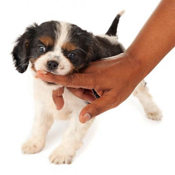
Anal sacs are two glands on either side of and slightly below the dog's anus, which contain secretions. When the dog defecates, the passing feces squeezes some of the secretions out, in normal cases. The sacs may change their positions if the dog is overweight. Soft bowel movements may prevent the sacs from secreting properly. If the glands do not secrete normally, the contents will thicken and cause the glands to enlarge, becoming "impacted".
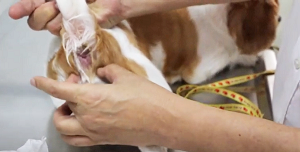 Anal disorders have been found to be common among dogs. The most
common one is called anal sacculitis, which is
inflammation and possible infection of the anal glands. Symptoms include scooting, localized skin irriation, painful
defecation (dyschezia), and the continuous sensation of the need to pass
stools (tenesmus). Initial treatment involves periodic
manual "expression" of the glands (see photo at right), which
involves applying pressure to the outside of a gland with the forefinger
of one hand and simultaneously applying pressure to the outside of the
other gland with the thumb of the other hand. Other management includes cleaning
of the sac, antibiotics if they are infected, and dietary changes, such
as adding fiber or psyllium powder. For those cases which do
not respond favorably to such treatments, removal of the anal sacs (anal
sacculectomy) is the next option. Removal of the anal sacs also is the
prescribed treatment for cases which include cancerous growths, such as
anal sac tumors (apocrine cell carcinoma). (See
this section of our
Cancer webpage for more details.)
Anal disorders have been found to be common among dogs. The most
common one is called anal sacculitis, which is
inflammation and possible infection of the anal glands. Symptoms include scooting, localized skin irriation, painful
defecation (dyschezia), and the continuous sensation of the need to pass
stools (tenesmus). Initial treatment involves periodic
manual "expression" of the glands (see photo at right), which
involves applying pressure to the outside of a gland with the forefinger
of one hand and simultaneously applying pressure to the outside of the
other gland with the thumb of the other hand. Other management includes cleaning
of the sac, antibiotics if they are infected, and dietary changes, such
as adding fiber or psyllium powder. For those cases which do
not respond favorably to such treatments, removal of the anal sacs (anal
sacculectomy) is the next option. Removal of the anal sacs also is the
prescribed treatment for cases which include cancerous growths, such as
anal sac tumors (apocrine cell carcinoma). (See
this section of our
Cancer webpage for more details.)
The cavalier King Charles spaniel is considered "at increased risk of requiring anal sacculectomy", according to three UK studies which reviewed medical records of dogs treated for anal sac disorders. In a September 2006 article examining the medical records of British dogs diagnosed with anal sac gland carcinoma (ASGC), cavaliers were ranked third (behind English cocker spaniels and springer spaniels) as having a breed pre-disposition to developing ASGC.
In a July 2014 article, in which the records of dogs undergoing anal sacculetomies between 2003 and 2013, the author found that:
"Cavalier King Charles spaniels and Labrador-type dogs were over-represented within this study population. ... the CKCS and Labrador/Labrador crosses were likely to be truly over-represented and may be at increased risk of requiring anal sacculectomy."
In a July 2021 article, investigators examined the VetCompass records of 104,212 dogs treated in 110 UK veterinary practices in 2013 and found that the cavalier was the most at-risk breed for anal sac disorders (ASDs). Anal disorders were over 3 times more prevalant in the cavalier as in the average mixed breed dog, with a prevalence of 14.83%. The King Charles spaniel (English toy spaniel) was close behind in second place, with a prevalence of 13.78%. The breed in third place, the Bichon Frise, had a prevalence of only 7.29% .The most common treatments consisted of antimicrobials and analgesics. Anal sacculectomy (surgery) was performed in fewer than 1% of cases. See our Veterinary Resources section below for the list of articles in which this disorder is discussed.
RETURN TO TOP
Artery fistulas
While reports of diagnosis and treatment of artery fistulas -- abnormal connections to the artery -- are rare, in two recent articles, cavaliers have been the predominant affected breed, with their ages ranging from 4 months to 18 months. All dogs underwent surgery for the insertion of shunts within the affected arteries.
In a December 2018 article, Italian cardiologists reviewed the records of 13 dogs of 8 breeds -- including 4 cavaliers (30+%) -- which had aortic-to-pulmonary arterial shunts inserted due to systemic-to-pulmonary arteriovenous fistulas originating from the descending aorta and connect with pulmonary arteries. None of the procedures were due solely to patent ductus arteriosus (PDA). The four cavaliers varied in age from 8 months to 18 months.
In a March 2019 article, a team of veterinary clinicians from three USA universities (R.L. Winter, J.A. Horton, D.K. Newhard, M. Holland) diagnosed and repaired two artery fistulas (abnormal connections to the artery) by transcatheter embolization to block the flow of blood through the fistulas. The dog was a 4-month-old cavalier King Charles spaniel (CKCS) which had grade 2 murmur at the base of the heart (basilar). X-rays showed mild enlargement of the left atrium. Echocardiography "suggested anomalous vessels around the main pulmonary artery". The pulse of the femoral arterial pulse was "hyperdynamic". Computed tomography angiography (CTA) showed two systemic-to-pulmonary artery fistulas (SPAFs). Fistulas are abnormal shunts leading from the artery and by-passing it. But for the dog's breed's history of mitral valve disease, corrective surgery may not have been performed. However, since the dog was a CKCS, her owner elected to have transcatheter embolization surgery performed. "Transcatheter" means that the surgical devices were delivered to the sites of the fistulas through the artery by inserting a tubular device. "Embolization" means to lodge an object in the blood vessel to obstruct the offending blood flow. The surgeons inserted embolization coils and silk suture threads through the left femoral artery to the fistulas, guided by CTA. The surgical procedure was successful; complee blockage into the fistulas was observed; vertebral heart size decreased; the left atrium size decreased; there no longer was a murmur.
RETURN TO TOP
Arthritis
 Arthritis
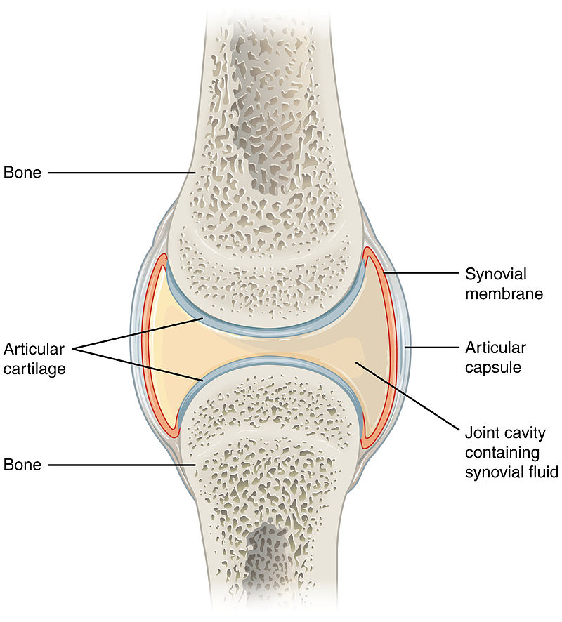
(osteoarthritis -- OA) is the most commonly diagnosed joint disease in dogs.
Arthritis usually is a progressive degeneration of what are known as
synovial joints, which are joints with a capsule filled with synovial
fluid which surrounds the bones' surfaces. The degeneration leads to
impaired function and pain. Arthritis is a multifactorial disease,
primarily genetic but often exacerbated by the dog's diet and exercise
levels. Arthritis often is secondary to a joint abnormality such as a
ligament rupture or patellar luxation.
Arthritis
(osteoarthritis -- OA) is the most commonly diagnosed joint disease in dogs.
Arthritis usually is a progressive degeneration of what are known as
synovial joints, which are joints with a capsule filled with synovial
fluid which surrounds the bones' surfaces. The degeneration leads to
impaired function and pain. Arthritis is a multifactorial disease,
primarily genetic but often exacerbated by the dog's diet and exercise
levels. Arthritis often is secondary to a joint abnormality such as a
ligament rupture or patellar luxation.
An idiopathic form of arthritis, immune-mediated polyarthritis (IMPA), causing recurring episodes of lameness and joint swelling, has been reported in cavalier King Charles spaniels. See our Immune-Mediated Polyarthritis (IMPA) webpage.
In an April 2018 article, UK researchers examining all reported cases of arthritis in a UK dog population under primary veterinary care during 2013 found that 2.42% were treated for arthritis. This made the CKCS the 11th most frequently affected breed in the study. Of 10,143 cavaliers in the study, 245 were diagnosed with osteoarthritis. The most frequently affected breeds were: (1) golden retriever, (2) Labrador retriever, (3) Rottweiler, (4) German shepherd, and (5) border collie. The overall average of all dogs diagnosed with osteoarthritis was 2.5%.
In a November 2016 article, a team of Wisconsin and Michigan researchers studied the clinical records of 79 dogs diagnosed with immune-mediated polyarthritis (IMPA). Of those 79 dogs, 13 had erosive IMPA, of which, two were cavalier King Charles spaniels. The 13 affected dogs had erosive lesions in their carpal joints. The estimated median synovial fluid lymphocyte count for dogs with erosive IMPA was significantly greater than that for dogs with nonerosive IMPA. Results indicated erosive IMPA most commonly affected the carpal joints of middle-aged small-breed dogs.
Medications which may be prescribed include NSAIDs (nonsteroidal anti-inflammatory drugs), which are designed to relieve pain and reduce inflammation. An NSAID growing in popularity for treating arthritis in dogs is grapiprant (Galliprant). A COX-2 selective NSAID, enflicoxib, has been found in a September 2021 study to be effective when administered orally. Other NSAIDs include Loxicom (Meloxicam).
Polysulfated glycosaminoglycan (PSGAG) (Adequan) is injected into the muscles to relieve pain and inflammation and improve range of motion due to arthritis in dogs. Studies have found that it prevents catabolic enzymes from degrading cartilage and bone, the causes of arthritis. PSGAG has been approved by the US Food & Drug Administration (FDA) for the management of OA in dogs.
Bedinvetmab (Librela, Beransa) is a injectable solution to alleviate pain associated with osteoarthritis (OA) in dogs. It is administered monthly. It is a monoclonal antibody which binds to and targets Nerve Growth Factors (NGF), which have key involvement in OA pain. It is manufactured by Zoetis. In May 2023, Zoetis announced that the USA Food & Drug Administration approved Librela (bedinvetmab injection) for the control of pain associated with osteoarthritis (OA) in dogs. Zoetis stated in its press release that Librela is the first and only once-monthly, anti-NGF monoclonal antibody treatment for canine OA pain and is approved as safe and effective in providing long-term control of OA pain symptoms in dogs, which can improve their mobility and overall quality of life.
Nevertheless, bedinvetmab is the subject of controversy due to reports of unanticipated adverse reactions, including ataxia, hind-end weakness, inability to walk, new or worsened seizures, and organ damage. Among expressed concerns are that blocking NGF may prevent its known protective and reparative roles elsewhere in the body. See this May 2015 article and this October 2023 article.
Synovetin OA is a "conversion electron therapeutic veterinary device" which is injected into the dog's elbow joints which are afflicted with osteoarthritis to reduce pain and inflammation of the synovial membrane which lines the elbow (synovitis).
Palmitoylethanolamide (PEA) is a
N-acylethanolamine molecule in a family of long-chain fatty acid
 amides
called ALIAmides. PEA has been found in rat and mice studies to limit
hyperactvity in immune cells and thereby control inflammatory responses
and resulting tissue damage. PEA is produced by the animal's body as
needed in response to certain types of injuries. PEA is a product of
normal fatty acid synthesis from palmitic acid. It is found in many
common foods, particularly palm oil, soy beans, egg yolks, and peanuts.
The commercial version is most commonly manufactured from palm oil*.
No
objective, unbiased clinical studies of the
effect of PEA on treating arthritis have been published.
amides
called ALIAmides. PEA has been found in rat and mice studies to limit
hyperactvity in immune cells and thereby control inflammatory responses
and resulting tissue damage. PEA is produced by the animal's body as
needed in response to certain types of injuries. PEA is a product of
normal fatty acid synthesis from palmitic acid. It is found in many
common foods, particularly palm oil, soy beans, egg yolks, and peanuts.
The commercial version is most commonly manufactured from palm oil*.
No
objective, unbiased clinical studies of the
effect of PEA on treating arthritis have been published.
Not all PEA is alike. There are three types of PEA.
• Basic PEA is insoluble in water and therefore the oral intake of it (rather than being injected directly into the abdomen) has very poor bioavailability, meaning that it does not get absorbed well in the dog's gut.
• Micronized PEA (m-PEA) is a patented technique that reduces the diameter of PEA particles, making them absorbable in the intestine, which has been found to be more effective than ordinary naive PEA in activating PEA levels in blood plasma in dogs. See this August 2014 article.
• Ultra-micronized PEA (um-PEA), also patented, reduces the PEA particle size further, to enable it to cross the blood-brain barrier, likewise has been found to be much more effective than naive PEA. See this August 2014 article.
 If a PEA product is not advertised as being micronized or
ultra-micronized, then
Dr. Clare Rusbridge advises that
"You
probably are wasting your money."
A variety of brands of
micronized and ultra-micronized PEA are offered on-line.
If a PEA product is not advertised as being micronized or
ultra-micronized, then
Dr. Clare Rusbridge advises that
"You
probably are wasting your money."
A variety of brands of
micronized and ultra-micronized PEA are offered on-line.
As for dosages, in a study using micronized PEA, the range was up to 24 mg/kg for osteoarthritis.
* Palm oil: The palm oil cultivation industry has been destroying rainforests in Sumatra and Borneo in Indonesia and Malaysia, the only habitats of orangutans. If you are going to obtain PEA, we suggest that you do so only from vendors whose PEA has been manufactured with palm oil from sustainable sources and not the deforestation of rainforests. This link connects to a "PalmOil Scan Mobile App" which will enable you to determine if the PEA vendors you select obtain their palm oil from sustainable sources.
Nutraceuticals are nutrients necessary for supporting or improving normal structure and function of the weight-bearing joints. They have been found to:
• Support or enhance metabolism of cartilage cells (chondrocyte) and joint membranes (synoviocyte) -- an anabolic effect;
• Inhibit damaging enzymes within synovial fluid and cartilage -- a catabolic effect;
• Inhibit formation of thrombi in small blood vessels supplying the joint (antithrombolic).
The most common nutraceuticals are glucosamine and
chondroitin.
Glucosamine regulates the synthesis of
 collagen in cartilage, and may provide mild anit-inflammatory
effects. Crystalline glucosamine sulphate has the greatest efficacy and
bioavailablity for osteoarthritis. Chondroitin sulfate inhibits
destructive enzymes in joint fluid and cartilage. Both of them also
contribute to the building blocks synthesis of glycoaminoglycans and
proteoglycans) for the formation and repair of cartilage.
collagen in cartilage, and may provide mild anit-inflammatory
effects. Crystalline glucosamine sulphate has the greatest efficacy and
bioavailablity for osteoarthritis. Chondroitin sulfate inhibits
destructive enzymes in joint fluid and cartilage. Both of them also
contribute to the building blocks synthesis of glycoaminoglycans and
proteoglycans) for the formation and repair of cartilage.
Undenatured type-II collagen (UC-II), a patented form of collagen extracted from the cartilage of the chicken’s sternum, has been found effective in reducing pain and discomfort experienced by dogs suffering from (osteoarthritis.
Microlactin (Duralactin) is a nutraceutical consisting of a protein concentrate derived from the milk of hyper-immunized cows, called hyper-immune milk factor (HIMF). It reportedly impedes inflammation and can be used safely in dogs. It is slow to respond to pain, taking a week to two weeks for maximum effectiveness.
 Cannabinol (CBD) is
is a cannabinoid compound produced from hemp and marijuana (Cannabis
Sativa) plants. CBD oil mimics the endocannabinoid molecules which the
dog’s (and our) body produces in several different organs. They play
roles in reducing pain, regulating inflammation, and affecting the
immune system, by initially binding to receptors in the brain.
Cannabinol (CBD) is
is a cannabinoid compound produced from hemp and marijuana (Cannabis
Sativa) plants. CBD oil mimics the endocannabinoid molecules which the
dog’s (and our) body produces in several different organs. They play
roles in reducing pain, regulating inflammation, and affecting the
immune system, by initially binding to receptors in the brain.
CBD is non-psychoactive, unlike tetrahydrocannabinol (THC), another cannabinoid compound from marijuana, which is considered psychoactive by altering the mental state, and can be highly toxic to dogs.
Varieties of CBD: Cannabidiol-based veterinary products are derived mainly from hemp (Cannabis sativa) and must contain less than 0.3% tetrahydrocannabinol (THC). This form of CBD can be processed into “full spectrum” or “broad spectrum” and also may be in the form of a “distillate”, in which all THC has been removed, or in the form of CBD “isolate”, which is a purifed powder.
• Full Spectrum: Full spectrum CBD contains other extracts found in the cannabis plant, including terpenes, and up to 0.3% THC.
• Broad Spectrum: Broad spectrum CBD also contains some other cannabis compounds but no more than trace amounts of THC.
• CBD Isolate: CBD isolate is pure CBD and contains no other cannabis plant compounds.
• Naked CBD: Naked CBD describes CBD oil by itself, as opposed to being capsultated or microcapsulated or combined with any other substance, such as deoxycholic acid (DCA).
• Liposomal CBD: This is an orally administered encapsultated CBD which is packaged within liposomes, small fatty cellular sacs which improve bioavailability of the CBD by enabling it to be withstand digesstion in the stomach and degradation in the liver. Lipsomal CBD was tested on dogs in this September 2020 article.
• Cannabidiolic acid (CBDA) is an acid precursor of CBD. It forms CBD when heated. It has been shown in some studies to be more potent that CBD for treating rats. It has been found to be more readily absorbed into the human bloodstream than CBD. Aa theory is that adding CBDA to doses of CBD may make the CBD more absorbable. In this September 2020 article, the investigators found that CBDA is absorbed at least twice as well as CBD in dogs within a 24 hour period, with some differences depending upon the medium used to deliver the oral treatment.
 The most canine studies of CBD thus far have been regarding
osteoarthritis (OA). The results have been fairly favorable, with the
clear exception of one study, the one reported in the
March 2021 article. Each of them is summarized below.
The most canine studies of CBD thus far have been regarding
osteoarthritis (OA). The results have been fairly favorable, with the
clear exception of one study, the one reported in the
March 2021 article. Each of them is summarized below.
Interestingly, only one of the studies thus far has included any cavalier King Charles spaniels. And in the March 2020 article below, the two CKCSs were singled out as requiring the highest dose of CBD to achieve any measurable relief.
In a July 2018 article, researchers report on a study of 16 dogs to determine the safety of CBD extract and its efficacy in allevieating pain in dogs diagnosed with osteoarthritis (OA). The equal mix of CBD and carboxylic acid of CBD (CBDA), was administered at 2mg/kg every 12 hours for 4 weeks. The treated dogs were:
"... perceived to be more comfortable and active. There appear to be no observed side effects of the treatment ... dogs undergoing OA treatment for a month duration. There were some dogs with incidental rises in alkaline phosphatase that could be related to the treatment. Further long-term studies with larger populations are needed to identify long-term effects of CBD rich industrial hemp treatment, however short term effects appear to be positive."
In a March 2020 article about a pilot study of 32 osteoarthritic dogs, in which two cavalier King Charles spaniels were included, hemp-derived CBD oil administered to the dogs reportedly "appears to positively affect dogs with chronic maladaptive pain by decreasing their pain, thereby improving their mobility and quality of life." The specific ingredients of the CBD oil and its dosages were:
"At the initial evaluation and enrollment, qualified dogs received a CBD oil product at a dose of 0.25 mg/kg delivered on food QD for 3 days and then morning and night (approximately every 12 hours). The product given was a certified organic, cold-pressed hemp seed oil infused with 1,000 mg of full-spectrum hemp extract derived from organically grown hemp plants, cultivated in Colorado. Full-spectrum extract includes cannabinoids (such as cannabidiolic acid, CBD, cannabigerol, cannabichromene), flavonoids, terpenes, and other constituents within the cannabis plant."
Most interestingly, the investigators singled out the two CKCSs:
"Among these 30 dogs, the dose of CBD needed to achieve a positive effect ranged from 0.3 up to 4.12 mg/kg BID. The 2 dogs in the study requiring the highest dose of the CBD product were both Cavalier King Charles spaniels (not related to one another), and neither of these dogs experienced any changes/elevations in liver enzymes."
In an August 2020 article, 9 dogs being treated for chronic osteoarthritis-related pain with conventional medications were dosed with oral transmucosal (OTM) cannabidiol (CBD) (2 mg/kg) every 12 hours for 12 weeks. The investigators report that the Pain Severity Score and the Pain Interference Score were significantly lower in CBD than in the control group of 12 other dogs, and the Quality of Life Index was significantly higher in the CBD group. They concluded:
"The addition of OTM CBD showed promising results. Further pharmacokinetics and long-term studies in larger populations are needed to encourage its inclusion into a multimodal pharmacological approach for canine osteoarthritis-related pain."
In a September 2020 article, 15 dogs diagnosed with osteoarthritis were divided into 3 groups and administered (a) 20 mg/day (0.5 mg/kg) naked CBD, (b) 50 mg/day (1.2 mg/kg) naked CBD, or (c) 20 mg/day liposomal CBD for 30 days. The dogs were tested on the first and last days for four different movements: sitting to standing, lying to standing, walking, and running. The investigators report that, among the 5 dogs receiving 20 mg/day of naked CBD, there generally were no improvements noted among all four movement categories. As for the 5 dogs being dosed 50 mg/day, there were "significant improvements" among all four assessment categories, as also were the 5 dogs receiving 20 mg/day liposomal CBD.
In a March 2021 article, 23 dogs with naturally occurring osteoarthritis of appendicular joints received 2.5 mg/kg of a CBD isolate in hempseed oil for six weeks. The investigators measured outcome objective gait analysis, activity counts (via accelerometry) and clinical metrology instruments. They report finding no differences noted between the CBD group and the placebo group at any time point for any of the recorded outcome measures. As adverse events, then noted elevation in liver enzymes in a majority of the dogs, and vomiting in two. They concluded that, "The pilot data from this study do not support the use of CBD as a symptom-relieving agent for canine OA."
See our Cannabis webpage for additional details about CBD, including delivery methods, bioavailability, dosages, and adverse reactions.
 Laser therapy, called "photobiomodulation"
(PBM), which is continuous wave laser treatments at
acupuncture
sites, is a treatment option, either with or without medication.
Laser therapy, called "photobiomodulation"
(PBM), which is continuous wave laser treatments at
acupuncture
sites, is a treatment option, either with or without medication.
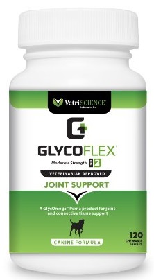
Owners may provide their affected dogs with supplements such as glucoamine HCI, methylsulfonylmethane (MSM), N,N-Dimethylglycine HCI (DMG), and manganese. Retail combinations of these supplements include Vetri-Science GlycoFlex Joint Support.
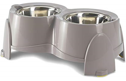
An aid to cavaliers suffering from arthritis is an elevated dog food
bowl. The
Ergo Feeder
(above right) is an
 example.
Read more about canine arthritis on the Canine Arthritis Management website.
RETURN TO TOP
Blood pressure
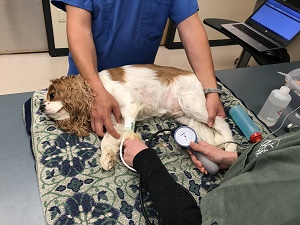 The
usual reading of the blood pressure of a dog is its systolic pressure,
as it is the most potentially variable and most dependent on the
patient's conditions (breed, age, sex, temperament, disease status,
exercise conditioning, diet). The systolic pressure reading is taken
when, as the heart beat, it measures the amount of force put on the
arteries. The diastolic blood pressure reading measures the amount of
force on the arteries when the heart is resting.
The
usual reading of the blood pressure of a dog is its systolic pressure,
as it is the most potentially variable and most dependent on the
patient's conditions (breed, age, sex, temperament, disease status,
exercise conditioning, diet). The systolic pressure reading is taken
when, as the heart beat, it measures the amount of force put on the
arteries. The diastolic blood pressure reading measures the amount of
force on the arteries when the heart is resting.
The normal range of systolic blood pressure (BP) values in dogs are breed-specific and also depend upon the other conditions of the patient described above. As examples: (a) Breed: normal BP varies among breeds, with larger breeds tending to lower BP and terriers, beagles, and sight hounds higher BP; (b) Age: normal BP tends to increase with the dog's age; (c) Sex: entire females have the lowest BP and intact males the highest and neutered dogs in between; (d) Temperament: BP is higher in more nervous dogs; (e) Diseases: diseased dogs tend to have higher BP than healthy ones; (f) Exercise: the more exercise a dog gets, the lower its BP; (g) Obesity: overweight dogs have higher BP than normal weight dogs; (h) Diet: dogs on home-made diets have lower BP than those fed commercial foods. (Source: this March 1996 article.)
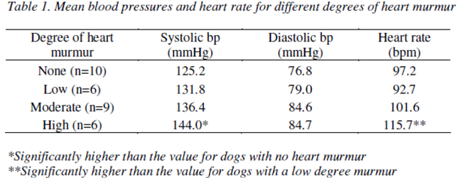
Cavalier King Charles spaniel: In a 2007 study of the BP of 29 healthy cavaliers, ranging in age from 1 to 11 years, their normal systolic BP ranged from 108 mmHg to 144 mmHg; their diastolic BP ranged from 58 to 84 mmHg, and their normal heart rate ranged from 65 to 147 beats-per-minute. This systolic range was slightly lower thn the average values for dogs and spaniels in general. The diastolic pressure results, however, were very similar to the species-wide and spaniel ranges.
In the same 2007 study, the BP of 31 CKCSs aged from 5 to 14 years were measured to determine the effect of MVD murmurs on BP. See "Table 1" below.
The so-called "gold standard" direct method for accurately measuring BP is invasive, in that it calls for insertion of a "cannula", a narrow tube, into an artery and which also is connected to a transducer in a surgical environment. An alternative indirect methods, using either ultrasound Doppler or oscillometric techniques, are the methods of choice by most cardiologists. They are not 100% as accurate as the invasive direct method in the best of environments, but they are accurate enough, particularly on a periodic timeframe, to be acceptable. However, human BP cuffs are not at all reliable for measuring the BP of dogs.
RETURN TO TOP
Blood transfusions
 In a
September 2017 article by a team of Italian veterinary blood
specialists, they studied 7,414 dogs, including 103 cavalier King
Charles spaniels, to determine the potential sensitization risk to dogs
receiving blood transfusions consisting of the Dog Erythrocyte Antigen
(DEA) 1 blood group.
In a
September 2017 article by a team of Italian veterinary blood
specialists, they studied 7,414 dogs, including 103 cavalier King
Charles spaniels, to determine the potential sensitization risk to dogs
receiving blood transfusions consisting of the Dog Erythrocyte Antigen
(DEA) 1 blood group.
Dogs have natural occuring antibodies against the DEA 1 antigen in red blood cells (erythrocytes or RBCs). In a dog with DEA 1 negative RBCs and with an immune system which is highly sensitized to DEA 1 positive RBCs, the system will produce alloantibodies following the first transfusion of DEA 1 positive blood. Following a second transfusion of DEA 1 positive blood, a dog which is highly sensitized to DEA 1 positive may develop an acute reaction, including fever, pigmenturia, and lethargy, and its packed cell volume (PCV) will not rise as expected. Death may result from such a mis-matched second transfusion. See this May 1995 article.
In the 2017 Italian study, they found that CKCSs were among the few breeds having the highest percentage likelihood to become sensitized (25.0%) following the first mis-matched transfusion, and that cavaliers also were among the breeds having the highest risk (6.2%) of an acute transfusional reaction following a second such transfusion. They therefore recommend:
""Blood typing to identify the presence of DEA 1 and the cross-match to establish full compatibility should be performed before each transfusion in order to reduce the risk of sensitization or immunological reaction between donor and recipient dogs."
RETURN TO TOP
Bromide toxicity
Excessively high levels of bromide in the dog's blood stream can produce symptoms such as head twitching (similar to myoclonus), body twitching, severe lack of muscle control, particularly in the legs, and overall depression. Potassium bromide is a standard medical treatment for idiopathic epilepsy, and so when a dog is being treated with that drug, bromide toxicity should be suspected. In an April 2014 report, a spayed female cavalier diagnosed with idiopathic epilepsy was treated with potassium bromide and phenobarbital. Eight days later, the veterinarians found her suffering from a lack of muscle control and repetitive twitching of the limbs. They concluded she was overdosed with the bromide solution. She had no similar episodes following a reduction in the dosing.
In a September 2009 article, another cavalier suffering from similar symptoms was diagnosed with hyperchloremia -- a high quantity of chlorine in the blood system. The clinicians reported that the dog was being prescribed potassium citrate for a urinary tract disorder. The drug was prepared at a local pharmacy, and it was discovered that, instead of the pharmacy's inadvertent use of citrate in the compounding formula, it used bromide, resulting in excessive intake of bromide on a daily basis. Once the bromide was discontinued, the dog's hyperchloremia and symptoms ceased.
RETURN TO TOP
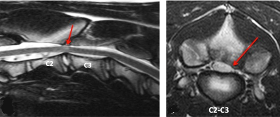
Cervical spondylomyelopathy
 Cervical
spondylomyelopathy (CSM), also known as Wobbler syndrome, is a disease
of compression of the cervical spine in the dog's neck. The dog's spinal
cord becomes compressed, resulting in neck pain and nervous system
disorders, including head tilt, loss of cordination (ataxia), facial
twitching, and head shaking. It may lead to some paralysis in severe
cases. While CSM is most common among giant breeds and other large
breeds, it has been reported in at least four case studies of cavalier
King Charles spaniels, at the junction of the C2 and C3 discs, high in
the neck.
Cervical
spondylomyelopathy (CSM), also known as Wobbler syndrome, is a disease
of compression of the cervical spine in the dog's neck. The dog's spinal
cord becomes compressed, resulting in neck pain and nervous system
disorders, including head tilt, loss of cordination (ataxia), facial
twitching, and head shaking. It may lead to some paralysis in severe
cases. While CSM is most common among giant breeds and other large
breeds, it has been reported in at least four case studies of cavalier
King Charles spaniels, at the junction of the C2 and C3 discs, high in
the neck.
 In one of the CKCS cases, bony (osseous) lesions or cysts were
identified during a
magnetic
resonance imaging (MRI) scan, which appear to have caused
narrowing of the spinal cord (stenosis). (See this
October 2011 article.) In the other three CKCS cases, the stenosis
appeared to have been caused by degeneration of the joint between the C2
and C3 vertebrae, compressing the spinal cord. (See this
October 2011 article and this
November 2015 article.) All cavaliers also were diagnosed with
Chiari-like malformation (CM) but not with syringomyelia. The CM was
discounted as being a cause of the disorder.
In one of the CKCS cases, bony (osseous) lesions or cysts were
identified during a
magnetic
resonance imaging (MRI) scan, which appear to have caused
narrowing of the spinal cord (stenosis). (See this
October 2011 article.) In the other three CKCS cases, the stenosis
appeared to have been caused by degeneration of the joint between the C2
and C3 vertebrae, compressing the spinal cord. (See this
October 2011 article and this
November 2015 article.) All cavaliers also were diagnosed with
Chiari-like malformation (CM) but not with syringomyelia. The CM was
discounted as being a cause of the disorder.
 Treatment normally consists of either medical management or surgery,
depending upon severity of symptoms and the owner's preference.
Medicines could include gabapentin, carprofen, tramadol,
paracetamol, robenacoxib,an NSAID, and/or prednisolone. Surgery options
would be decompression of the spinal cord by either a haemilaminectomy
or dorsal laminectomy. See also this
June 2023 article describing "instrumented cervical fusion" using
end-plate conforming interbody devices with a micro-porus structure.
Treatment normally consists of either medical management or surgery,
depending upon severity of symptoms and the owner's preference.
Medicines could include gabapentin, carprofen, tramadol,
paracetamol, robenacoxib,an NSAID, and/or prednisolone. Surgery options
would be decompression of the spinal cord by either a haemilaminectomy
or dorsal laminectomy. See also this
June 2023 article describing "instrumented cervical fusion" using
end-plate conforming interbody devices with a micro-porus structure.
RETURN TO TOP
Coronavirus
In one case study, a cavalier diagnosed with injury to its heart (myocardial injury) was found to be infected with coronavirus disease (COVID-19). In this December 2021 article, Italian clinicians reported that a six-year-old CKCS had a two-month history of severe exercise intolerance and syncope. Cardiac auscultation revealed a grade 2 murmur. Chest x-rays showed mild generalised enlargement of heart with a vertebral heart scale (VHS) 11.5. Echocardiography showed that the mitral valve leaflets were normal but that a mild mitral regurgitation with central jet was present. No other echocardiographic abnormalities were identified. Clinical signs had developed during a local wave of coronavirus disease (COVID-19) two weeks after the dogs family members had become infected with coronavirus 2 (SARS-CoV-2). Serological and molecular tests aimed at diagnosing SARS-CoV-2 infection were subsequently performed, especially in light of the dog’s peculiar history. Results of such tests supported a presumptive diagnosis of COVID-19-associated myocardial injury.
RETURN TO TOP
Dewclaws
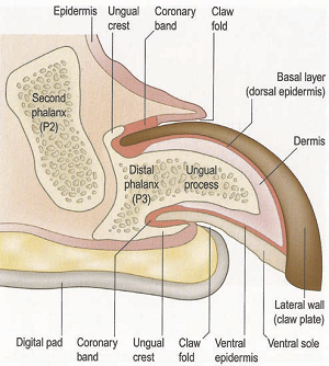 Dewclaws
(or dew claws) on the front legs of cavaliers are in fact toes, the
first digit of each foot, equivalent to human thumbs. All dogs are born
with a dewclaw on each front leg. Few dogs, however, will be born with
rear leg dewclaws, and they are very rarely found on any CKCSs. Dogs'
foreleg dewclaws are not "rudimentary" or "vestigial". Dewclaws have
their own sets of muscles, tendons, nerves, and blood supply. (See
diagram at right.)
Dewclaws
(or dew claws) on the front legs of cavaliers are in fact toes, the
first digit of each foot, equivalent to human thumbs. All dogs are born
with a dewclaw on each front leg. Few dogs, however, will be born with
rear leg dewclaws, and they are very rarely found on any CKCSs. Dogs'
foreleg dewclaws are not "rudimentary" or "vestigial". Dewclaws have
their own sets of muscles, tendons, nerves, and blood supply. (See
diagram at right.)
The foreleg dewclaws aid in stabilizing the dog's ankles (carpus). decrease rotational forces on the other digits, and help prevent age-related arthritis in the carpal joints. Veterinary researchers recommend that healthy foreleg dewclaws not be amputated, and that the nails be periodcically trimmed short.
RETURN TO TOP
Dilated cardiomyopathy
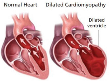 Dilated cardiomyopathy (DCM) is a disease in which the heart muscle
weakens and does not function properly in order to to pump blood
adequately, leading to poor circulation. This is called a loss of
"myocardial contractility". Other consequences are an irregular heart
beat and, ultimately, heart failure. Symptoms include difficulty
breathing, weakness, and collapse.
Dilated cardiomyopathy (DCM) is a disease in which the heart muscle
weakens and does not function properly in order to to pump blood
adequately, leading to poor circulation. This is called a loss of
"myocardial contractility". Other consequences are an irregular heart
beat and, ultimately, heart failure. Symptoms include difficulty
breathing, weakness, and collapse.
Thus far, fortunately, DCM is rare in the cavalier King Charles spaniel. In a January 2009 review of DCM in 369 dogs at clinics in the United Kingdom between 1993 and 2006, only two were cavaliers. It is much more common in some larger breeds, particularly the Doberman pinscher and the boxer, and ususally at middle-age. In some cases, DCM can be diagnosed early by detecting a heart murmur, which raises a conflict for CKCSs since their most common disorder, mitral valve disease, also is first detected by hearing a murmur. However, the DCM murmur is a different sound and located at a different part of the dog's heart. So the examining veterinarian must be adept at distinguishing between the two differing murmurs. DCM can be confirmed by x-ray showing significant enlargement of the heart, and electrocardiogram (EKG) and echocardiogram.
DCM has been associated with taurine deficiency in the blood, so in such cases, additional taurine likely will be prescribed. Medications such as diuretics, ACE-inhibitors, and pimobendan has been shown to be effective improving the quality of life of dogs affected by DCM, but there are no medications which cure this disease.
RETURN TO TOP
 Ectrodactyly
Ectrodactyly* is an extremely rare congenital malformation in which the development of the dog's paw bone (mesenchymal) cells are interrupted during gestation. Causes could include genetic mutations, environmental factors such as maternal disease or diet, drugs, vaccines, or radiation. Ectrodactyly often is displayed as a cleft between metacarpal bones, usually the first and second metacarpal bones. They may be abnormally formed or missing. In a 2017 case of a cavalier King Charles spaniel puppy in New Zealand, both forelegs are affected, with one shorter than the other and resembling a lobster claw and the other with a split between the toes. (See photo at right.)
* Also known as split-hand deformity or lobster syndrome.
RETURN TO TOP
Encephalitis
Encephalitis is inflammation of the brain, and autoimmune
encephalitis occurs when the dog's immune system mistakenly attacks
healthy brain cells, leading to that inflammation. GABA is the main
inhibitory neurotransmitter in the brain, which enables the regulation
of neuronal activity. GABAAR are ion channels in
 the
brain control the rapid transmission of nerve impulses between neurons.
the
brain control the rapid transmission of nerve impulses between neurons.
In a June 2022 article, German veterinary neurologists described the first reported case of a dog -- a 1-year-old cavalier -- diagnosed with anti-GABAAR (γ-aminobutyric acid-A receptor) autoimmune encephalitis. The cavalier (right) had multiple generalized seizures and showed alternating stupor and hyperexcitability, ataxia, repetitive muscle contractions, and circling to the left side. Auto-antibody encephalitis, with antibody-mediated dysfunction of receptors was suspected. Despite treatment with multiple antiseizure medications (diazepam followed by phenobarbital) the seizures and behavior abnormalities continued, and the dog alternated between stupors and hyperexcitability. Immunotherapy with dexamethasone, a corticosteroid, led to rapid improvement of the clinical signs and the CKCS improved continuously. A month later, GABAAR auto-antibodies had decreased significantly. An antibody search in the cerebral-spinal fluid (CSF) and serum located a neuropil staining pattern of GABAAR antibodies. At the examination four weeks after the start of immunotherapy, the dog was clinically normal; the GABAAR antibody titer in serum had strongly decreased; and the antibodies were no longer detectable in the CSF. The clinicians report that this is "the first veterinary patient with an anti-GABAAR encephalitis with a good outcome following ASM and corticosteroid treatment." They concluded:
"Based on the clinical signs and presentation (epileptic seizures lacking any response to ASMs, erratic behavioral changes) and the results of the initial diagnostics (lack of abnormal findings on conventional MRI and CSF examination), an autoimmune encephalitis was suspected, proven, and successfully treated. Clinicians should consider to test for autoantibodies and start immunotherapy in cases with a similar clinical presentation and lack of response to anti-seizure medication even if an inflammatory/infectious or neoplastic cause was clinically excluded on MRI and CSF."
RETURN TO TOP
Epiglottic retroversion
Epiglottic retroversion (ER) is an intermittent obstruction of the upper respiratory tract in some small breed dogs, including the cavalier King Charles spaniel. The epiglottis is a leaf-shaped flap in the throat that prevents food from entering the windpipe and the lungs; it is open during breathing, allowing air into the larynx, but during swallowing, it closes to prevent aspiration of food into the lungs. The most common symptoms are chronic intermittent inspiratory high-pitched, wheezing sound (stridor) caused by disrupted airflow, exercise intolerance, shortness of breath (dyspnea), coughing and gagging, reverse sneezing, bluish gums (cyanosis), rapid breathing (tachypnea), and sneezing. ER could be related to other concurrent upper airway disorders, including brachycephalic airway obstruction syndrome (BAOS), laryngeal paralysis, and tracheal collapse. ER is diagnosed using video-laryngosopy. Diagnosis of ER is confirmed when the epiglottis is not positioned against the base of the tongue at inspiration. Surgical options include a laser-surgical treatment called temporary epiglottopexy, and a procedure called unilateral cricoarytenoid lateralisation which involves tying the windpipe to the wall of the trachea. See these reported cases.
In a June 2020 article, diagnosis and treatment of two cavaliers was reported, in which both had concurrent BAOS and one also had laryngeal paralysis. One was treated with a temporary epiglottopexy and the other with a unilateral cricoarytenoid lateralisation. Both dogs had excellent long-term outcomes.
RETURN TO TOP
Facial nerve paralysis -- Bell's Palsy
 Idiopathic facial nerve paralysis
or facial palsy is reportedly common in the cavalier
King Charles spaniel. It is described as a sudden onset of either
unilateral or bilateral facial nerve paralysis with no other abnormal
signs. They may include drooping or inability to blink or move lips,
ears, or other muscles of the face. Onset of the
signs usually are sudden and do not worsen over time. The signs
typically occur in dogs aged 2 years or older.
Idiopathic facial nerve paralysis
or facial palsy is reportedly common in the cavalier
King Charles spaniel. It is described as a sudden onset of either
unilateral or bilateral facial nerve paralysis with no other abnormal
signs. They may include drooping or inability to blink or move lips,
ears, or other muscles of the face. Onset of the
signs usually are sudden and do not worsen over time. The signs
typically occur in dogs aged 2 years or older.
The cause of facial nerve paralysis is said to be idiopathic facial neuropathy in 75% of the cases, which essentially means the cause has not been determined. It may also be diagnosed as Horner's Syndrome. In 40% to 70% of the cases, it has been combined with vestibular syndrome. The cause of that combination likewise is unknown. In fact, that combination condition has been named "facial and vestibular neuropathy of unknown origin" (FVNUO). Seriously(!).
Recovery may be within weeks, but in the most cases the abnormality is permanent. There is an increased risk of exposure corneal ulcers in dogs with protruding eyes, such as cavaliers. Facial nerve paralysis in the CKCS may also be associated with primary secretory otitis media (PSOM), masticatory muscle myositis (MMM), hypothyroidism, and/or vestibular syndrome.
In a November 2016 abstract, French researchers studied 69 dogs with facial nerve paralysis. The report does not provide a count of cavalier King Charles spaniels included in the study, but the researchers concluded that the CKCS and the French bulldog should be added to the list of predisposed breeds to the disorder. Additionally, they found:
"Idiopathic facial paralysis was diagnosed in 48% of dogs. Vestibular signs were the most common additional clinical signs and were observed in 36% of dogs with idiopathic facial paralysis. Peripheral nervous system disease was diagnosed in 19% of dogs, and central nervous system disease occurred in 30% of dogs. ... Improved diagnostic methods enabled the diagnosis of a higher percentage of inflammatory/infectious diseases, which were absent in the central nervous system aetiologies of a previous similar study, and revealed metabolic (hypothyroidism), inflammatory and neoplastic aetiologies for peripheral nervous system disease."
In an April 2017 article, a cavalier suffered facial nerve paralysis which caused sleep apnea and an inability to blink her eyes. Specialists at the Animal Health Trust performed surgery on her eyelids to help protect her eyes while awaiting nerve function to slowly return. The author states:
"Fortunately most dogs cope very well with such nerve paralysis, which can be permanent, as long as we can keep their eyes healthy."
In an October 2017 abstract, Dr. Dustin Dees of Austin, Texas reports successfully treating a cavalier suffering from idiopathic facial nerve paralysis by using continuous wave laser treatments at four acupuncture sites corresponding to a branch of the facial nerve. The method is called "photobiomodulation". Treatments were performed twice a week for five weeks. The results were successful. The dog's blinking ability substantially improved, and corneal dryness resolved with marked improvement of the dog's Schirmer testing.
In a December 2019 article by Univeristy of Sydney, Australia researchers, they reviewed the records of 122 dogs presented at their hospital from 2001 to 2016 which had symptoms of facial nerve paralysis (FNP). FNP can be a symptom of any of a variety of disorders, including primary secretory otitis media (PSOM), direct injury, hypothyroidism, and drug sensitivity. Idiopathic FNP (IFNP) applies to FNP symptoms due to unknown causes. It is not considered to be effectively treatable. Of the 36 purebred dogs in the entire study, 15 (42%) were cavaliers, the highest number of purebreds by far. Of the 15 purebreds diagnosed with IFNP, cavaliers numbered 9 (60%). The authors concluded:
"This study found differences in the incidence of FNP causes compared with the only previous study, which are presumed to be a result of geographic and temporal differences. Breeds and age groups are significant risk factors correlated to acquisition of FNP and IFNP. Vestibular dysfunction, hypersalivation and keratoconjunctivitis sicca were common findings in dogs with IFNP. Dogs with IFNP have guarded prognosis for recovery, and often require life-long eye lubrication. There is no evidence to date that treatment alters the outcome in dogs with IFNP."
Bell's palsy is an idiopathic facial nerve paralysis, which occurs without evidence of infection, reduced thryroid function, or injury. It has been identified in one veterinary report of three cavalier King Charles spaniels. It may be the same disorder as facial nerve paralysis.
RETURN TO TOP
Fear avoidance in puppies
 It generally is accepted that early life experiences are of prime
importance in shaping later behaviors. This appears to be as true with
canines as with humans and other mammal species. We call this process
“socialization” (or “socialisation” in UK parlance) – the learning process
of interacting acceptably with others.
It generally is accepted that early life experiences are of prime
importance in shaping later behaviors. This appears to be as true with
canines as with humans and other mammal species. We call this process
“socialization” (or “socialisation” in UK parlance) – the learning process
of interacting acceptably with others.
While a study of the level of fear avoidance among puppies of different breeds is not a "disorder", it has been shown in a July 2015 study that cavalier puppies from 4 to 10 weeks of age exhibited a later onset of fear-related avoidance behavior when compared with German shepherd dog puppies and Yorkshire terrier puppies. In the same study, CKCSs demonstrated the highest incidence of crouching in response to a loud noise, followed by the Yorkshire terriers. Breed differences in puppy mobility were observed beginning at 6 weeks of age, with German shepherd dogs demonstrating the most mobility and cavaliers the least. The authors concluded that the results of their study support the hypothesis that emotional and behavioral development, as well as the onset of fear-related avoidance behavior, varies among breeds of domestic dogs.
Puppy socialization consists of a variety of sub-sets. They include interacting (a) with the puppy’s mother, (b) with its fellow siblings and other dogs, preferably in the breeder’s home or kennel, (c) and with humans. A lack of proper and timely socialization with other dogs has been found to result in higher levels of fear of other dogs. Similarly, the same is the case with lack of timely, positive contacts with humans – fear of people.
Puppies are said to have a sensitive period for this dog and human socialization in their early post-natal life, during which their neurological and emotional development is immature and most receptive for novel external stimuli of all of their senses. (See this August 2020 article.) This early process of learning behavioral patterns sometimes is called “imprinting”. This time period, found to occur especially from the third week to the fourteenth week of age, can significantly affect a dog’s behavior throughout its life. (See this March 1961 article and this July 2015 article.)
It is during the third week that most puppies’ eyes and ears
become functional, and they become more mobile. (See
this
November 2022
article.) This is followed by an initial fear response exhibited by puppies
 to
sudden sounds and other startling novel events, which has been found to
develop around the sixth or seventh week, give or take up to a couple of
weeks, depending upon the breed and particular dog. The puppy’s typical
reaction to fearful events is avoidance. Recent research has found that this
fear of novelty – leading to increased avoidance – continues to increase in
intensity until the twelfth to fourteenth week. (See
this
November 2022
article.)
to
sudden sounds and other startling novel events, which has been found to
develop around the sixth or seventh week, give or take up to a couple of
weeks, depending upon the breed and particular dog. The puppy’s typical
reaction to fearful events is avoidance. Recent research has found that this
fear of novelty – leading to increased avoidance – continues to increase in
intensity until the twelfth to fourteenth week. (See
this
November 2022
article.)
And so, an important window of opportunity exists for both puppy-to-other-dogs socialization and puppy-to-humans socialization during those weeks from the third to up to fourteenth. It would appear that the best source for providing these new sensory stimulations to other dogs and humans on a consistent and continuing knowledgeable basis, would be the puppy’s breeder.
All of this brings us to the uniqueness of the cavalier King Charles spaniel as a breed. Published studies have found these characteristics about the development of the cavalier’s nervous system:
• Cavalier puppies tend to have their eyes open and begin weaning and exploration at a later age than many other breeds. (See this July 2015 article.)
• Unlike other breeds (e.g., including German shepherd dogs and Yorkshire terriers in the Morrow, July 2015 study) cavalier puppies failed to respond to any stimuli until five weeks of age, rather than the usual three weeks. (See this July 2015 article.)
• Cavalier puppies demonstrated a significantly later onset of fear-related avoidance behavior (compared with both the Yorkshire terrier and German shepherd dog puppies in the 2015 study) (See this July 2015 article.)
• Cavaliers appear to require a longer socialization period than other breeds. (See this July 2015 article.)
Notwithstanding this evidence, CKCS breed clubs adhere to a species-wide policy of recommending that cavalier puppies be transferred from breeder to buyer as early as the eighth week.
In the United Kingdom (UK), the Cavalier King Charles Spaniel Club’s Code of Ethics provides that “Members who breed or exhibit should: ... Not part with a puppy under the age of eight weeks to a new owner.” That club also requires that its members “Will abide by all aspects of the Animal Welfare Act.’
Interestingly, the UK’s “Animal Welfare (Licensing of Activities Involving Animals) (England) Regulations (2018 ed.)” states at Schedule 6, Section 1(5):
“No puppy aged under 8 weeks may be sold or permanently separated from its biological
mother.”
So, separating puppy from mother, litter, and breeder is condoned ethically and legally in the UK at eight weeks of age.
The American Kennel Club (AKC) recommends that:
“You should not expect to bring a puppy home until it is between eight to 12 weeks of age. Puppies need ample time to mature and socialize with their mother and littermates to set them up for success in their future homes.”
This, of course, is a species-wide recommendation – in other words, for all puppies of all breeds – and therefore is not based upon any scientific basis, because all breeds are not alike.
The American Cavalier King Charles Spaniel Club (ACKCSC – the AKC’s
“parent” club for cavaliers) goes one step
 further by stating in its
“ethical guidelines” that:
further by stating in its
“ethical guidelines” that:
“I [meaning, the cavalier puppy’s breeder] will not allow any puppy to leave for its new home before the age of eight weeks although twelve weeks is suggested.”
The Cavalier King Charles Spaniel Club, USA, (CKCSC,USA) has a code of ethics which includes this statement:
“I [the cavalier puppy’s breeder] will not allow any puppy to leave for its new home before the age of eight weeks. The CKCSC recommends ten to twelve weeks as the appropriate age for transfer.”
Contrary to all of the above regulations, cavalier club ethical codes, and recommendations, it is quite clear that separating pup from mother, litter, and breeder prior to the fourteenth week is not in the best interests of the long-term healthy development of cavaliers. Cavaliers in general have their peculiarities, and among them is their overly slow development from birth to their fourteenth week.
Cavaliers need to be socialized longer than other breeds, and that socialization should be with mother, siblings and other dogs, and humans, on a consistent and continuing basis from their weaning until at least their fourteenth week.
RETURN TO TOP
Gallbladder disorders
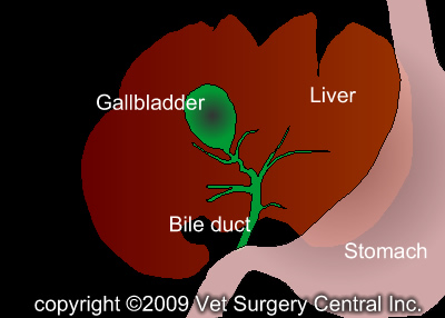 The gallbladder is a
pear-shaped sac located between the two lobes of the liver,
which stores bile, a yellow fluid which includes fat and cholesterol and
which is secreted through its bile duct by the liver into the small
intestine to help breakdown fats in the dog's ingested food. The bile
enables the vitamins and other nutrients to be absorbed into the
bloodstream and also removes certain wastes.
The gallbladder is a
pear-shaped sac located between the two lobes of the liver,
which stores bile, a yellow fluid which includes fat and cholesterol and
which is secreted through its bile duct by the liver into the small
intestine to help breakdown fats in the dog's ingested food. The bile
enables the vitamins and other nutrients to be absorbed into the
bloodstream and also removes certain wastes.
The primary disorders of the dog's gallbladder and biliary tract include gallstones (cholelithiasis), swelling (mucoceles), inflammation (cholecystitis). Others include the absence of the gallbladder (agenesis) and undeveloped biliary tract (biliary atresia). The cavalier is the only breed reported in veterinary literature to be "over-represented" in developing cholecystitis. See this August 2020 article, and this October 2021 article.
• Gallstones (cholelithiasis)
A gallstone (cholelith) is a solid mass formed from bile precipitates (usually consisting of calcium carbonate, cholesterol, and bilirubin) in the gallbladder or in the intra-hepatic or extra-hepatic ductal system. Cholelithiasis describes gallstone(s) in the gallbladder.
While cholelithiasis is very common in humans, affecting up to 25%, symptomatic cholelithiasis is much rarer, affecting only 10% of those diagnosed with gallstones. The overall prevalence of canine cholelithiasis has been estimated to be between 0.03% and 15%. These stones usually remain in the gallbladder or pass through the common bile duct unobstructed. Complications of cholelithiasis in dogs include extrahepatic biliary duct obstruction, chholangitis / cholecystitis, gallbladder perforation, and cholecystocutaneous fistula. Choledocholithiasis (also called bile duct stones or gallstones in the bile duct) is the presence of a gallstone in the bile duct. See citations of all reported cases here.
In a March 2020 article, UK researchers identified 68 dogs diagnosed with cholelithiasis on ultrasound between January 2010 and June 2018 at the Small Animal Hospital at the University of Glasgow. Of those, the records of 61 dogs out of 31 breeds were reviewable. Cholelithiasis was classified as an incidental finding in 53 (86.9%) dogs, including 4 cavaliers, with 8 (13.1%) dogs being classified as symptomatic, inlcuding 2 cavaliers, having complications of cholelithiasis that included biliary duct obstruction (1 CKCS), biliary peritonitis, emphysematous cholecystitis, and acute cholecystitis (1 CKCS). The breed rankings were, mixed breeds (11 dogs), Labrador retrievers (7 dogs), border collies (5 dogs), Cocker spaniels (4 dogs), and cavaliers (4 dogs).
The two cavaliers diagnosed with symptomatic cholelithiasis were (a) a 4 year old spayed female exhibiting anorexia and jaundice and diagnosed with acute cholelithiasis due to a single stone within the gallbladder, and (b) a 5 year old castrated male exhibiting vomiting, anorexia, and painful abdomen, diagnosed with obstructive cholelithiasis due to a single stone within the bile duct.
In an August 2020 article, a 6-year-old cavalier with diarrhea, vomiting, and anorexia was diagnosed with partially obstructive cholelithlasis by abdominal ultrasound and blood work. The dog was treated with ursodeoxycholic acid (UDCA) (Ursodiol) and a low-fat diet for 8 months, resulting in the nearly complete dissolution of the cholelith at that point.
In an October 2021 article, University of Cambridge (UK) researchers reviewed the medical records of 38 dogs diagnosed with cholelithiasis by abdominal ultrasound (AUS) between 2010 and 2019 at The Queen's Veterinary School Hospital, University of Cambridge. Cavalier King Charles spaniels was the most common breed, comprising 4 (10.5%) of the patients. The investigators confirmed a "high prevalence" of cholelithiasis in CKCSs. Of the 18 (47.4%) dogs classified as symptomatic, the clinical signs of cholelithiasis included:
• vomiting
• decreased appetite
• lethargy
• diarrhea
• jaundice (icterus)
• abdominal pain
• weight loss
• excessive urination (polyuria)
• frequent urination (pollakiuria)
• slow, difficult, painful urinary flow (stranguria)
• blood in the urine (hematuria)
• excessive thirst (polydipsia)
• thinning of haircoat
• deafness
• shifting lameness
Symptomatic cases had higher alkaline phosphatase, gamma-glutamyl transferase, and alanine transferase activities than did non-symptomatic cases, and 44.4% of symptomatic cases also had choledocholithiasis (gallbladder stones in the bile duct).
Treatment categories consisted of those 13 dogs (34.2%) medically
treated, 10 surgically treated (26.3%), and 15 not treated (29.5%). They
found that medical treatment with ursodeoxycholic acid (UDCA) resolved
or improved cholelithiasis in several dogs. They therefore concluded
that, "with careful case selection, UDCA may be a safe and effective
tool for management of cholelithiasis [without choledocholithiasis] in
dogs." They also recommended that dogs "with marked increases in ALP,
ALT, and GGT activity with or without ultrasonographic evidence of
choledocholiths and with or without symptomatic cholelithiasis can be
considered for surgical intervention." They also recommended that
"development of evidence-based guidelines for management of
cholelithiasis in dogs would enable improved outcomes for patients and
offer more reliable prognoses for clinicians, especially given the lack
of consensus regarding medical management of
 cholelithiasis in dogs."
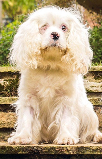
• Swelling (mucoceles)
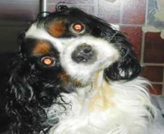
Gallbladder mucoceles (GBM) is the leading gallbladder and bile duct disease in dogs. GBM is the accumulation of immobile bile and mucus that causes the gallbladder to swell and obstructs the bile duct. Some cavaliers have been reported to have been diagnosed with GBM. For example, this all-white CKCS named Snoopy (right) has been diagnosed with GBM (and a variety of other internal disorders). See the reported GBM veterinary journal articles here.
RETURN TO TOP
Growth plate (physeal) fractures
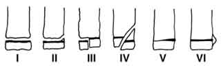 Fractures
of the bones of immature dogs can include a growth plate (physis),
called a physeal fracture. The growth plate is composed mostly of
cartilage, meaning that it is weaker than adjoining ossified bone. A
hazard of damaging and/or repairing a growth plate is that it could
cause premature closure of the plate. Degrees of growth plate fractures
are classified by the Salter-Harris classification system (see
diagram). In a
February 2016 article, UK orthopedists discuss a growth plate
(Salter-Harris type one) fracture of the humerus of a cavalier King
Charles spaniel. The radiograph photos in the linked article show the
CKCS at left with the fracture and at right how it has been stabilized
with parallel K-wires through the greater tubercle into the humeral
diaphysis.
Fractures
of the bones of immature dogs can include a growth plate (physis),
called a physeal fracture. The growth plate is composed mostly of
cartilage, meaning that it is weaker than adjoining ossified bone. A
hazard of damaging and/or repairing a growth plate is that it could
cause premature closure of the plate. Degrees of growth plate fractures
are classified by the Salter-Harris classification system (see
diagram). In a
February 2016 article, UK orthopedists discuss a growth plate
(Salter-Harris type one) fracture of the humerus of a cavalier King
Charles spaniel. The radiograph photos in the linked article show the
CKCS at left with the fracture and at right how it has been stabilized
with parallel K-wires through the greater tubercle into the humeral
diaphysis.
RETURN TO TOP
Heatstroke
BOTTOM LINE: Cool the heatstroke victim first, before rushing to the veterinarian. Cool with cold-water immersion for young or healthy dogs. Evaporative cooling (applying cool water plus air movement -- fan, breeze, air conditioning) for any dog is the next best option.
 If the body temperature reaches 41.1°C (106°F) or above, the dog is
said to have heatstroke. In a
June 2020 article, UK researchers reviewed veterinary records of
over 900,000 dogs in the UK in 2016, finding reports of 395 heat-related
(heatstroke) illnesses, with an incidence rate of 0.04%. Clinical signs
of heatstroke included:
If the body temperature reaches 41.1°C (106°F) or above, the dog is
said to have heatstroke. In a
June 2020 article, UK researchers reviewed veterinary records of
over 900,000 dogs in the UK in 2016, finding reports of 395 heat-related
(heatstroke) illnesses, with an incidence rate of 0.04%. Clinical signs
of heatstroke included:
• panting excessively or continuously despite removal from heat/cessation of exercise,
• collapse not subsequently attributed to another cause (e.g. heart failure, Addison’s),
• stiffness, lethargy or reluctance to move,
• gastrointestinal disturbance including hypersalivation, vomiting or diarrhea,
• neurological dysfunction including ataxia, seizures, coma or death,
• haematological disturbances including petechiae or purpura.
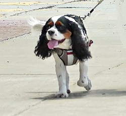
The three main risk factors for heatstroke were found to be breed
type, high bodyweight, and older age. Cavalier King Charles spaniels
ranked 6th among nine breeds most likely to develop heatstroke, when
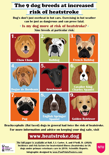 compared to Labrador retrievers, which were the least likely. The
cavalier followed the Chow Chow, bulldog, French bulldog, Dogue de
Bordeaux, and greyhound in breed risk for heatstroke. The rest of the
top nine breeds were the pug, English springer spaniel, and golden
retriever.
compared to Labrador retrievers, which were the least likely. The
cavalier followed the Chow Chow, bulldog, French bulldog, Dogue de
Bordeaux, and greyhound in breed risk for heatstroke. The rest of the
top nine breeds were the pug, English springer spaniel, and golden
retriever.
To treat headstroke, the first step is to lower the dog's body temperature. Then oxygen supplementation, fluids, and treatments of any complications. The veterinary website Heatstroke.dog advises:
"Our current understanding is that if your dog has overheated, but is still conscious, the most effective methods of cooling are to either immerse them in water (cold tap water is perfect), or wet them (with whatever water you have available) and fan them – air conditioning is perfect if you’re transporting them to the vet. If your dog has overheated and has lost consciousness, it is ESSENTIAL that you protect their airway, and don’t let them inhale any water. Dogs that have lost consciousness will cool far more slowly, so it is even more important to use effective cooling methods, such as spraying with water plus air movement."
After pouring cold water over the dog or immersing it in cold water,
immediately take it to the nearest veterinary clinic. The dog should not
be totally submerged in water, and its nose and mouth always should be above the
water level. The body temperature needs to be reduced slowly to avoid
stress. A sudden drop in body temperature can cause more complications.
Avoid ice cold water baths, because sudden cold can cause the
 arteries and veins to constrict and force the blood back to the organs.
Leaving wet towels on the dog will prevent heat from escaping, so do not
do that. Constantly pouring cold or cool water over the dog's body, while the dog is on
in cool metal sink or metal table, is preferrable. The cooling process
should take about a half hour to an hour. An intravenous catheter will
allow fluids to be given to help support cardiac output; however, fluids
should be overloaded. See this
July 2019 article for more details.
arteries and veins to constrict and force the blood back to the organs.
Leaving wet towels on the dog will prevent heat from escaping, so do not
do that. Constantly pouring cold or cool water over the dog's body, while the dog is on
in cool metal sink or metal table, is preferrable. The cooling process
should take about a half hour to an hour. An intravenous catheter will
allow fluids to be given to help support cardiac output; however, fluids
should be overloaded. See this
July 2019 article for more details.
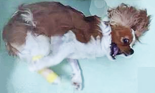
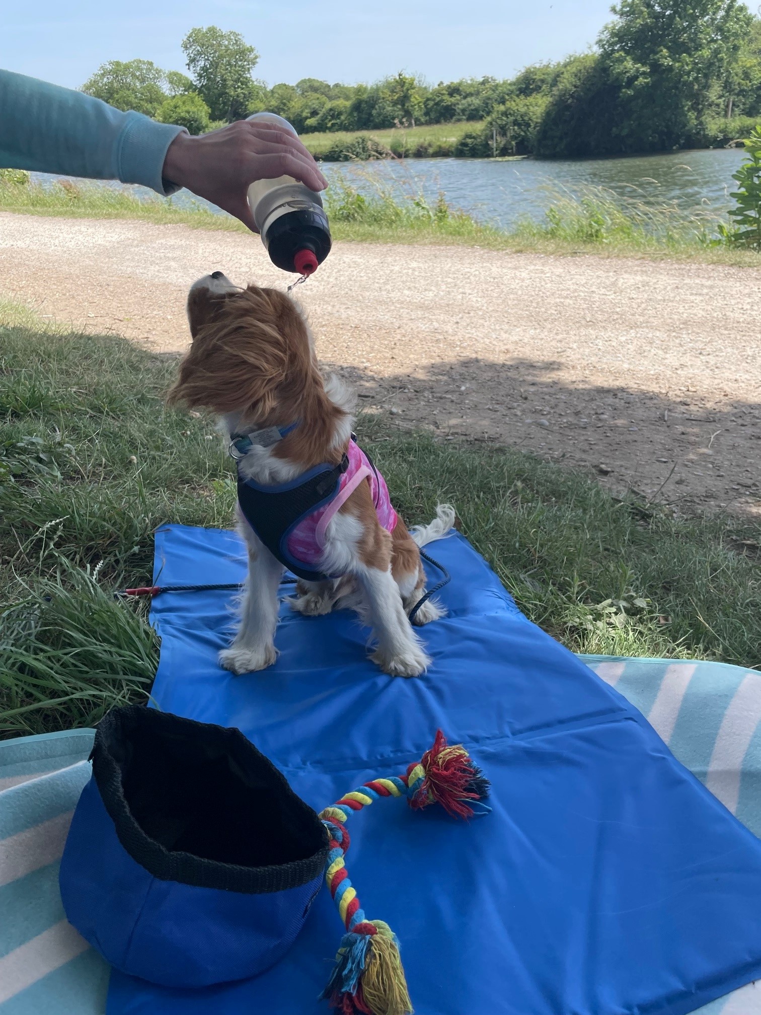
To prevent heatstroke in dogs which must remain outdoors in a very hot environment, consider having the dog wear a cooling vest or wet towels, but they must be kept wet. Once they dry out, they will hold the heat in because air cannot circulate around the dog's skin. In this photo, a cavalier is in the shade, wearing a cooling vest which is being doused with additional water to keep it wet.
RETURN TO TOP
Icterus (jaundice)
• xylitol poisoning
In an October 2009 article, a clinician reported that a cavalier which had eaten 2 or 3 pieces of chewing gum containing xylitol subsequently developed jaundice, severe lethary, anorexiz, and vomiting over a 5-day period. Laboratory testing indicated several adnormalities indicating liver damage. Helatic lymphadenopahty was detected on abdominal ultrasound. Xylitol toxicosis was presumed. LIver support therapy was started, and the dog recovered following several weeks of treatment.
• zinc poisoning
Icterus is a yellowing, such as jaundice. In a July 2010 article, a cavalier had an episode of collapse and weakness. The dog had icteric sclera and mucous membranes, and was diagnosed with probable zinc toxicity. Abdominal radiography revealed metallic foreign bodies in the stomach. Multiple coins, including some zinc containing pennies minted after 1982, and a medallion were removed endoscopically.
See, also, this July 2013 article in which a cavalier was diagnosed with acute zinc toxicity following ingesting a metallic object which also resulted in hemolytic anemia and acute pancreatitis.
RETURN TO TOP
Insecticide poisoning reactions
• Fipronil
Fipronil (Frontline, Fiproguard, Flevox, PetArmor)
has been found to cause epiletic seizures in at least on cavalier King
Charles spaniel. Fipronil is a phenylpyrazole antiparasitic agent primarily used to kill fleas,
ticks, lice, sarcoptic mange, and cheyletiellosis on dogs. It is applied
to the skin (topically)
 rather than by mouth (ingested). It works by
interfering with the brain and spinal cord of insects, resulting in
their death. Frontline Plus contains 9.8% fipronil.
rather than by mouth (ingested). It works by
interfering with the brain and spinal cord of insects, resulting in
their death. Frontline Plus contains 9.8% fipronil.
Fipronil is a prescription drug and can only be obtained by prescription from veterinarians. Its makers claim that it is not absorbed into the body and does not circulate through the blood stream. However, studies have found fipronil, in the form of fipronil sulfone, in the bloodstream of humans.
Fipronil has been reported to cause thyroid disruption in humans. It has been found to disrupt GABAergic signaling activity,, and the GABA system closely interacts with thyroid hormones Fipronil sulfone concentrations were negatively correlated with serum thyroid-stimulating hormone (TSH) concentrations in employees at a fipronil production facility, suggesting that fipronil has a central inhibitory effect on TSH secretion in humans. The U.S. Environmental Protection Agency (EPA) has classified fipronil as a possible human carcinogen based on data showing an increase in thyroid follicular cell tumors in rats after long term exposure.
In a January 2019 article, the investigators reported that fipronil sulfone in the blood of pregnant women transfers to the fetuses and adversely affects the infants’ health, including measurements of thyroid function.
In an October 2018 article, Turkish veterinarians reported on the diagnosis and treatment of a 3-year-old neutered female cavalier diagnosed with mitral valve disease which began to experience epileptic seizures. The epilepsy first was treated with levetiracetam, which resulted in the epileptic seizures disappearing. Then, three months later, the cavalier was treated for fleas and ticks with fipronil. The epileptic attacks "dramatically increased and became more severe just after the fipronil treatment (~7 times per day)." Gabapentin was added to the levetiracetam treatment protocol, and the epileptic seizures. The researchers concluded:
"Fipronil, phenylpyrazole origin and other antiparasitic drugs should be used very carefully and under veterinary consultant supervision due to their side effects especially in epileptic dogs."
RETURN TO TOP
• Pyrethrins
Pyrethrins
(pyrethroid, permethrin) has been found to be highly
toxic in cavalier King Charles spaniels. Pyrethrins are the active ingredients of certain insecticides,
including topical ointments applied to dogs' skin. We report three
separate published case studies of cavaliers diagnosed with pyrethrin
poisoning.
In a September 2014 article, Israeli researchers reported on the diagnosis and treatment of a cavalier suffering from generalized body tremors, facial twitching, and salivation. Biospotix spray (geraniol essential oils) and Advantage spot on had been applied on the dog’s skin, and the owner’s household was sprayed against insects using a commercial pyrethroid preparation (Admiral) containing 7.9% bifenthrin, 24 hours before clinical signs appeared. The dog was otherwise healthy, fully vaccinated, lived in an apartment and leashwalked. They diagnosed bifenthrin toxicosis. They treated with diazepam, methocarbamol and IV fluids, followed by general anesthesia with isofluran and diazepam CRI. After twenty-four hours, the dog was no longer under general anesthesia. Seventy two hours after admission the dog was discharged, was alert and responsive when stimulated, and walked and ate normally.
In an November 2014 article, a 17-month-old cavalier "exhibited clinical signs compatible with pyrethrin toxicity (swaying, hypersalivation, seizures, tremors, twitching of the facial muscles, four limb ataxia and paddling) consistent with the TS-syndrome [tremor associated with salivation]." The dog had been exposed to two different compounds of the pyrethrins/pyrethroids group and imidacloprid in a product named Admiral. He was treated with diazepam, metacarbamol and IV fluids, followed by general anesthesia with isofloran and diazepam CRI. The dog was discharged afer 72 hours, fully recovered.
In an April 2020 case report, a seven-month-old cavalier King Charles spaniel displayed coarse muscle tremors that progressed to seizures, after licking a cleaning product containing permethrin. At the time of the event, the puppu was being treated with prednisone for masticatory muscle myositis (MMM). The dog was treated with midazolam, dexmedetomidine, and intravenous lipid emulsion (ILE). This is the first reported use of ILE as therapy in the successful management of canine permethrin toxicosis. No further tremors or seizure activity, nor any adverse effects were observed following administration of ILE therapy.
RETURN TO TOP
Meningoencephalitis
Meningoencephalitis is inflammation of the brain and its protective membranes, usually caused by a bacteria. Meningoencephalomyelitis [MEM] is a form of meningoencephaliits which also affects the spinal cord.
At least three veterinary studies have found cavaliers to be more susceptible to this disorder than the average dog. In a May 2013 study, 5% of the affected dogs were cavaliers. In an October 2018 study, 5.25% were CKCSs.
In a June 2018 article, a team of PennVet clinicians diagnosed meningoencephalomyelitis caused by the bacteria Pasteurella multocida in a 5-year-old cavalier King Charles spaniel. (Meningoencephalomyelitis [MEM] is inflammation of the brain, including its protective membranes, and of the spinal cord, caused by a bacteria.) The dog had severe periodontal disease, with contact ulceration and possible necrotizing stomatitis. Her symptoms included fever, lethargy, inappetence, and multifocal neurologic signs, mainly dull mentation. Based upon examination of her cerebrospinal fluid (CSF) and blood culture, and her response to therapy (anti-emetics and gastroprotectants, an opioid for analgesia, and dexamethasone sodium phosphate as an antiinflammatory), P. multocida meningoencephalomyelitis was diagnosed. They opine that the severe periodontal disease led to a bacteremia causing hematogenous seeding of a bacterial meningitis originating at the disrupted blood–spinal cord barrier.They concluded:
"In the future, be aware that a fever with multifocal neurologic signs and severe periodontal disease in a canine patient may suggest a P. multocida infection and both CSF and blood cultures can be submitted for confirmation."
RETURN TO TOP
Myoclonus
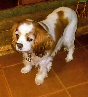 Myoclonus
is an involuntary movement syndrome displaying tremors, symptoms of spasmodic jerky
contractions of groups of muscles, mainly of the head (such as, rapid
eye movements, nodding, or shuddering) and forelimbs when the dog is
standing or sitting. The movements can be "positive" (caused by
contraction of the muscles) or, rarely, "negative" (loss of muscular
control). The syndrome can be progressive with affected dogs
suffering frequent jerks which may cause the dog to fall or stumble.
Myoclonus
is an involuntary movement syndrome displaying tremors, symptoms of spasmodic jerky
contractions of groups of muscles, mainly of the head (such as, rapid
eye movements, nodding, or shuddering) and forelimbs when the dog is
standing or sitting. The movements can be "positive" (caused by
contraction of the muscles) or, rarely, "negative" (loss of muscular
control). The syndrome can be progressive with affected dogs
suffering frequent jerks which may cause the dog to fall or stumble.
 Click
on the photo of the Blenheim at right to watch a video of its episode,
and click on the photo of the ruby below to watch a video of its
episode. See, also, these veterinary reports for
details about this disorder in CKCSs. Se, also,
this YouTube video.
Click
on the photo of the Blenheim at right to watch a video of its episode,
and click on the photo of the ruby below to watch a video of its
episode. See, also, these veterinary reports for
details about this disorder in CKCSs. Se, also,
this YouTube video.
In a March 2014 article, UK clinicians report that a cavalier developed generalized appendicular repetitive myoclonus as a result of a toxicity reaction to dosages of potassium bromide, which had been prescribed to treat idiopathic epilepsy. The CKCS improved after the bromide dosage was reduced, and no similar episodes were reported at follow ups of 26 months.
In a September 2015 article, the International Veterinary Epilepsy Task Force stated:
"Older Cavalier King Charles spaniels (>5 years old) have a high prevalence of myoclonus, which manifests most commonly as a brief jerking of the head and forelimbs when the dog is standing or sitting. Initially the syndrome is relatively benign but can be progressive with affected dogs suffering frequent jerks which may interfere with function, for example cause the dog to fall or stumble. The syndrome can be confused with focal epileptic seizures but generally does not respond to AEDs [antiepileptic drugs] licenced for dogs although may respond to levetiracetam."
In an abstract presented at the 2017 symposium of the European College of Veterinary Neurology (ECVN), a team of UK veterinary neurologists examined the records of 40 cavalier King Charles spaniels displaying myoclonic jerks. In these cases, the signs included rapid eyelid blinking, head nodding, shuddering, and exensions down the limbs resulting in falling or stumbling. The movements lasted for seconds, usually when the dog was stationary or sitting and did not lose consciousness. Multiple daily episodes were reported and increased in frequency and intensity over time. The median age of onset was 9 years, with a main range from 7 to 10 years. They found that the condition improved with treatment with levetiracetum and possibly corticosteroids.
In a March 2022 article, UK researchers reviewed the medical records of 39 cavaliers diagnosed with myocluonus. In this study, the researchers described the symptoms as:
"The myoclonic event was a mixture of positive and negative myoclonus and described as rapid eyelid blinking with head nodding/shuddering and variable extension down the thoracic limbs, that could result in falling or stumbling of the thoracic limbs. The movement lasted seconds had no obvious trigger and occurred mostly when stationary in a sternal or sitting position. It did not seem to be associated with loss of consciousness (see Video S1 and Video S2). Multiple daily episodes were seen and increased in frequency and intensity over time. Owners whose dogs would frequently fall, often disrupting them while eating, reported the myoclonus to interfere with the dog's quality of life. The mean age of onset of myoclonus was 8.38 years (SD ±1.96)."
Videos which accompany this March 2022 article are linked here: (1) ruby cavalier; (2) Blenheim cavalier. In addition to classic signs of myoclonus, 13 of the dogs (33%) had epileptic seizures or similar events. All of the dogs had Chiari-like malformation (CM), and 20 of them showed signs of CM-associated pain. Twenty of 30 dogs also had syringomylia (SM), and 13 dogs had primary secretory otitis media (PSOM). Twenty-three dogs were being treated for CM/SM with gabapentin or pregabalin. The researchers concluded that the cavaliers had a "higher prevalence" of myoclonus after age 8 years, and that:
"Myoclonus is characterized by spontaneous onset, rapid eyelid blinking and head nodding, a jerking movement of the head sometimes extending down the thoracic limbs, occasionally involving the whole body and might cause the dog to stumble or fall. There was no apparent loss of consciousness. Myoclonus was not associated with a trigger or sleep but did occur when standing, sitting, or lying in a sternal position. Multiple episodes in a day were often reported. Clinical course was slowly progressive with the episodes becoming more frequent and more intense over time. The myoclonus observed in our cohort was not action-related nor stimulus sensitive. ... Based on the presenting features of myoclonus in our reported cohort a cortical or cortico-subcortical etiology is suspected."
Other possible causes of these jerky movements include (1) steroid-responsive tremor syndrome, (2) cerebellitis (inflammation of the cerebellum), (3) exposure to toxins, including pesticides, amphetamines, narcotics, macadamia nuts, marijuana, (4) endocrine disease, (5) infections such as rabies or distemper, (6) bromide toxicity.
In other breeds, similar symptoms, when rapid and repetitive, have been described as canine idiopathic head tremor syndrome (IHTS). See this April 2015 article.
Medical treatment typically would include levetiracetam (Keppra), an anticonvulsant. It appears to be relatively safe for dogs, and reportedly rarely has any adverse side effects and does not appear to affect the liver or liver enzymes. In an April 2024 artcle, the researchers reported finding that "Levetiracetam can be used effectively to rapidly stop myoclonic seizures and to decrease the frequency of myoclonic seizures."
RETURN TO TOP
Necrotizing fasciitis (NF)
Necrotizing fasciitis (NF) is a spreading bacterial infection of the soft tissues beneath the dog's skin, particularly the fascia (see diagram). "Necrotizing" means that the bacteria destroys the cells of the affected tissues. NF may be caused by a bacterial infection with Streptococcus canis, B-hemolytic streptococcus, Staphylococcus pseudintermedius, or E. Coli. The bacteria progresses so rapidly that unless immediate surgical treatment is not provided the likelihood of death has been 100%. NF does not respond to medical care.
In a March 2021 article, two New York clinicians reported on the diagnosis and treatment for bacterial infections of necrotizing fasciitis (NF) in three dogs. One of the dogs was a 4.5 year old female cavalier King Charles spaniel, with symptoms which included inappetence, vomiting, diarrhea, dehydration, high temperature, periodic rapid heartbeat, an indication on x-rays of soft tissue wisps within the regions of fat beneath the skin, an painful soft-tissue swelling. The dog underwent three surgeries within two days, which included removal of the dead latissimus dorsi, additional muscular necrosis which required "debridement" (removal of dead tissue). By the sixth day of hospitalization, the dog had improved.
RETURN TO TOP
Neutering & Spaying
Gonadectomy is the veterinary term describing removal of either the
male dog's testes (also called neutering) and the females ovaries (also
called spaying or ovariectomy). Neutering cavalier males and spaying
cavalier
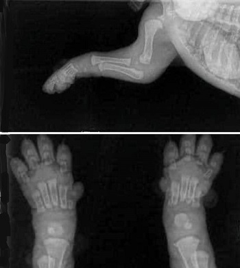 females are controversial, and doing so before they reach
physical maturity -- 18 to 20 months -- is especially controversial.
Neutering males involves castration, and spaying females involves even
more invasive surgery to remove both the ovaries and uteris.
females are controversial, and doing so before they reach
physical maturity -- 18 to 20 months -- is especially controversial.
Neutering males involves castration, and spaying females involves even
more invasive surgery to remove both the ovaries and uteris.
The most controversial aspects of both procedures are the consequences they have to the cavaliers' physical growth, behavioral issues, and future obesity and urinary incontinence. Spaying and neurtering do not just remove the dogs' sexually abilities, but also hormones essential to their growth. As an example, cavalier puppies are born without connective tissues -- joints and cartilage -- between their weight-bearing bones. (See x-rays at right.) As they mature, the bones continue to grow, and the cartilage and joints form and connect to those bones. Hormones in the dogs' sexual organs are instrumental in regulating that growth, and the premature removal of the sources of those hormones have been found to disrupt growth-plate closures (see chart below) and result in joint disorders and failure to appropriately end bone growth cycles.
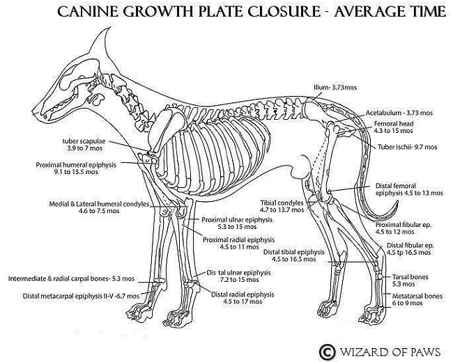
In a January 2022 article, Dr. Karen M. Tobias reviewed the current veterinary literature regarding gonadectomies in dogs and concluded that:
"Dogs should be allowed to reach musculoskeletal maturity (ie, be fully grown, with a fully functional urethral sphincter) before being neutered. Breed type and temperament should also be considered."
In a December 2016 article, a team of University of California-Davis reasearchers examined the medical records from 1995 to 2010 of 90,090 dogs of all AKC-recognized breeds treated at their veterinary hospital, to determine the relation between neuter status and autoimmune diseases. They report finding that neutered dogs had a significantly greater risk of:
• atopic dermatitis (ATOP)
• autoimmune hemolytic anemia (AIHA)
• hypoadrenocorticism (ADD)
• hypothyroidism (HYPO)
• immune-mediated thrombocytopenia (ITP)
• inflammatory bowel disease (IBD)
than intact dogs with neutered females being at greater risk than neutered males for all but AIHA and ADD. Neutered females, but not males, had a significantly greater risk of lupus erythematosus (LUP) than intact females. Pyometra was a greater risk for intact females.
In a January 2019 study by Italian researchers of 21,281 dogs of 31 different breeds, cavaliers ranked third, behind standard Schnauzers and fox terriers in being at risk of developing Cushing's disease (spontaneous hypercortisolism), and that neutered female dogs were at the highest risk for spontaneous hypercortisolism.
In a July 2020 article, University of California-Davis researchers reviewed the school's hospital records of over 50,000 dogs to analyze the increased risks of specific joint disorders and cancers associated with neutering dogs at various ages. A total of 35 breeds, including cavalier King Charles spaniels, were included in the study. The joint disorders were: hip dysplasia (HD), cranial cruciate ligament tears or ruptures (CCL), and elbow dysplasia (ED); the cancers were lymphoma/lymphosarcoma (LSA), hemangiosarcoma (HSA), mast cell tumors (MCT), and osteosarcoma (OSA). The CKCS results were:
"The study population was 51 intact males, 72 neutered males, 87 intact females, and 76 spayed females, for a sample size of 286 cases. Joint disorders: hip dysplasia (HD), cranial cruciate ligament rupture (CCL), and elbow dysplasia (ED). For males and females left intact, the occurrences of one or more joint disorders were just 1-4 percent. For males and females neutered at any age, there was no noteworthy increase of joint disorder occurrence, undoubtedly reflecting the small body size. Cancers: lymphoma (LSA), mast cell tumor (MCT), hemangiosarcoma (HSA), and osteosarcoma (OSA). In intact males, the occurrence of cancers was 2 percent and zero in intact females. For both sexes, neutering was not associated with any increase in this measure. Mammary Cancer (MC), Pyometra (PYO) and Urinary Incontinence (UI) in Females. The occurrence of MC in females left intact was zero. There was no occurrence of UI in spayed females. PYO was diagnosed in 2 percent of intact females. Bottom line. Lacking a noticeable occurrence of increased joint disorders or cancers with neutering males or females, those wishing to neuter should decide on the appropriate age."
The study included intervertebral disc disorders (IDD) in only two breeds, the Corgi and Dachshund, even though the cavalier also is widely regarded as being prone to a form of IDD known as Hansen type I disc extrusions. The authors reported that for male Corgis neutered before age 6 months, 18% developed IDD. For the Dachsund, no increase in IDD cases was noted in neutered dogs. This study was limited to certain specified joint disorders and cancers. It did not include any other known consequences of neutering prior to the dog reaching physical maturity.
In a January 2023 article, the health and behavior outcomes were compared among neutered dogs (castrated – full gonadectomy), sexually intact dogs, and dogs which underwent vasectomy (males) or ovary-sparing hysterectomy (females) by a team of USA researchers. Information was gathered by answers to questionnaires submitted by the dogs’ owners. Data was acquired about 6,018 dogs, including 2,786 males (1,672 castrated, 1,056 intact, and 58 vasectomized) and 3,232 females (2,281 spayed, 792 intact, and 159 ovary-sparing spayed). The breeds of dogs were not specified. The health issues in the questionnaire included the orthopedic problems of hip dysplasia, elbow dysplasia, cranial cruciate ligament injury (knees – patellas), osteochondrosis dissecans (cartilage separation from bone at joints), panosteitis (inflammation of leg bones), and patellas luxation (knee separation). Various types of cancers were listed. Other health conditions listed were prostate enlargement or infection, Addison’s disease, Cushing’s disease, diabetes mellitus, hypothyroidism, and obesity. The investigators report:
“Our most important finding was that longer duration that gonads were present, regardless of reproductive status, was associated with fewer general health problems and both problematic and nuisance behaviors. It was also associated with an increased lifespan.”
“Being sexually intact or having an OSS [ovary-sparing spay] were associated with lower odds of orthopedic problems, consistent with other studies showing lower risks of a variety of orthopedic problems in sexually intact dogs as compared to gonadectomized dogs.”
“Our results were also consistent with previous studies indicating that the associated odds of cancer is significantly lower for sexually intact dogs than gonadectomized dogs. Although gonadectomized dogs had a later cancer diagnosis, significantly more gonadectomized dogs were diagnosed with cancer compared to sexually intact dogs.”
“Gonadectomized dogs and dogs that underwent OSS were more likely to be obese than sexually intact dogs, but there was no relationship between duration that gonads were present and obesity. This suggests that the causes of obesity are more complex than just the absence of gonadal hormones.”
“Our data also indicated that the associated prevalence of both problematic and nuisance behaviors decreased with increased exposure to gonadal hormones. Male dogs of all reproductive groups and dogs with an OSS were reportedly more likely to mark or mount than sexually intact and SF dogs. Further, there was no difference in marking or mounting between gonadectomized and IM dogs.
“Taken together, these data suggest that gonadal hormone exposure time may be a more sensitive way to examine the effects of reproductive surgery on health and behavior than categorizing dogs into groups by type of reproductive surgery. This is likely because there may be significant overlap in the length of exposure to gonadal hormones between the 6 groups examined in this study.”
They concluded that: “ Our findings emphasize the importance of gonadal hormone exposure to canine health and behavior.”
In a June 2023 letter in the British VeterinaryRecord, 30+ veterinarians called upon their colleagues to stop recommending "gonadectomy as a blanket policy". They stated:
"Despite the limitations of a survey-based and retrospective study, the findings are clear: longer exposure to gonadal hormones is associated with fewer general health problems, longer lifespan, lower odds of orthopaedic problems, cancer and obesity, and fewer problematic and nuisance behaviours. ... Numerous peer-reviewed research papers now describe how routine neutering of dogs has been linked to increased incidence of health problems such as cruciate ligament rupture, hip dysplasia, osetoarthritis, hypothyroidim and many cancers (including haemangiosarcoma, mast cell tumours, lymphosarcoma, osteosarcoma, transitional cell carcinoma of the bladder), and the assertino that gondadectomised dogs live longer has largely been discredited. ... Kutzler concludes: 'Gonads should no longer be considered mere gamete-producing or ancillary sex organs but rather necessary endocrine glands for normal metabolic, endocrinologic, musculoskeletal, and anti-neoplastic health. Reproductive sterillisation with gonad removal should not be considered a routine procedure.'"
RETURN TO TOP
Orofacial clefts
Orofacial clefts -- cleft lip (CL), cleft palate (CP) and cleft lip and palate (CLP) -- are abnormal fissures of the mouth or face which occur due to incomplete fusion of tissues during embryonic development. In a November 2019 article, Cornell University veterinary researchers reseached the incidence of orofacial clefts in 7,429 live births of purebred puppies of 78 AKC-recognized breeds in the USA during a 12 month period. In those litters, 228 puppies had orofacial clefts (3%), of which there were 59 CLS (26%), 134 CPs (59%) and 35 CLFs (15%). The cavalier King Charles spaniel ranked third behind the Boston terrier and French bulldog in having significantly increased odds of CL and of CLP. Of 134 live births of cavaliers, there was a total of 10 CLs and CLPs, amounting to 7.5% of the puppies.
RETURN TO TOP
Parasites
Parasitic insects have been reported to invade the bodies of cavalier King Charles spaniels as well as other canines and also humans. In some instances, cavaliers are singled out in veterinary research literature as case studies for such parasitic invasions. In particular, the lungworm Angiostrongylus vasorum, also called the French heartworm and the small heartworm, has been reported frequently in the CKCS. See our Lungworm webpage for more information about the A. vasorum.
 Another parasite reported to be found in the cavalier is the larvae of
flies. In a
February 2023 article, Belgium veterinary clinicians report
finding the rat-tailed larvae of the common drone fly (Eristalis
tenax) being expelled in the feces of a cavalier. (Images shown
at right are the drone fly and its larva.) The dog's owner
observed several living larvae in fresh feces. For seven weeks before
these larvae were found, the dog’s feces had been dark black, tarry
feces that usually are associated with upper gastrointestinal bleeding.
The dog showed signs of abdominal discomfort. The dog was treated with
Milbemax and Panacur, following which the dog’s feces tested negative
for occult blood; no more larvae were found, and the symptoms
disappeared. Nevertheless, the investigators suspect that the dog's
signs of discomfort and the blood-darkened feces were not due to the
larvae, because they are known to inhabit only the rectum. They
concluded:
Another parasite reported to be found in the cavalier is the larvae of
flies. In a
February 2023 article, Belgium veterinary clinicians report
finding the rat-tailed larvae of the common drone fly (Eristalis
tenax) being expelled in the feces of a cavalier. (Images shown
at right are the drone fly and its larva.) The dog's owner
observed several living larvae in fresh feces. For seven weeks before
these larvae were found, the dog’s feces had been dark black, tarry
feces that usually are associated with upper gastrointestinal bleeding.
The dog showed signs of abdominal discomfort. The dog was treated with
Milbemax and Panacur, following which the dog’s feces tested negative
for occult blood; no more larvae were found, and the symptoms
disappeared. Nevertheless, the investigators suspect that the dog's
signs of discomfort and the blood-darkened feces were not due to the
larvae, because they are known to inhabit only the rectum. They
concluded:
"In conclusion, discovery of living drone fly larvae in pet’s feces may be frightening for the owner, but in most cases, facultative myiasis does not involve pathology."
RETURN TO TOP
 Plants
-- toxic, poisonous
Plants
-- toxic, poisonous
Some varieties of plants, including common decorative ones, are toxic to dogs, capable of causing serious illness and death from the consumption of relatively small amounts of leaves, berries, or seeds. The most toxic plants for dogs include: lilies, castor bean, cycad palms (such as sago), yews, autumn crocuses, foxglove, lily of the valley, oleander, and "yesterday, today, and tomorrow". See this March 2005 article for details.
In this May 2017 article, 14 Texas dogs (including two cavaliers), suffered toxicosis from eating the seeds or other parts of the cycad (sago) palm tree (see example at right). Nine of the 14 dogs died as a direct result of cycad palm intoxication. Three of the five survivors had persistently elevated liver enzymes, indicating liver damage. Serum albumin levels and nadir platelet counts were significantly lower in nonsurvivors compared to survivors. The article does not indicate in which category the CKCSs fell.
RETURN TO TOP
Polyglandular deficiency syndrome
In a March 2021 article, Japanese veterinarians reported diangnosing and treating a cavalier King Charles spaniel in the first reported case of a dog with a combination of polyglandular deficiency syndrome (PDS), diabetes mellitus, and hypoadrenocorticism (Addison's disease). PDS is defined as a multiple endocrine organ failure, including the gonads, the endocrine pancreas, and the parathyroid glands. They reported:
"The dog was diagnosed with diabetic ketoacidosis based on hyperglycemia and renal glucose and ketone body loss. The dog’s condition improved on intensive treatment of diabetes mellitus; daily subcutaneous insulin detemir injection maintained an appropriate blood glucose level over half a year. However, the dog’s body weight gradually decreased from day 207, and on day 501, it presented with a decreased appetite; the precise cause could not be determined. Based on mild hyponatremia and hyperkalemia, hypoadrenocorticism was suggested; the diagnosis was made using an adrenocorticotropic hormone stimulation test. Daily fludrocortisone with low-dose prednisolone oral administration resulted in poor recovery of the blood chemistry abnormalities; however, monthly desoxycorticosterone pivalate (DOCP) subcutaneous injection with daily low-dose prednisolone oral administration helped in the significant recovery of the abnormalities. Therefore, clinicians should consider the possibility of coexistence of hypoadrenocorticism in dogs with diabetes mellitus presenting with undifferentiated weight loss. Additionally, DOCP (not fludrocortisone) may be useful in treating dogs with diabetes mellitus complicated with hypoadrenocorticism."
RETURN TO TOP
Porencephaly
Porencephaly is the congenital cerebral defect and a rare
malformation with the main clinical sign of seizure symptoms.
![]() In a
July 2015 article, Japanese researchers reported
discovering porencephaly -- a congenital cerebral cavity, filled with
cerebrospinal fluid (CSF) -- in a 9 month old female cavalier. The dog exhibited symptoms of chewing and excitement
before secondary generalized seizures and fly-biting after the seizures
for 5–6 min. (At right, see MRI scan of CKCS' lesion at arrow.)
They examined a total of two affected dogs and one affected
cat. Their aim of the study was to find if there was any hippocampal
atrophy in cases of porencephaly, and they found in all three cases,
less hippocampal volume or hippocampal loss at the lesion side or the
larger defect side. They also noted that the severity of seizure
symptoms was attributed to cyst ratio and asymmetric ratio. Both the
cyst ratio and asymmetric ratio had correlation with the seizure
symptoms. They concluded that porencephaly may coexist with hippocampal
atrophy, and that clinicians should evaluate carefully the hippocampal
volume and asymmetry in MRI, because the atrophy may have relationships
with porencephaly-related seizures.
In a
July 2015 article, Japanese researchers reported
discovering porencephaly -- a congenital cerebral cavity, filled with
cerebrospinal fluid (CSF) -- in a 9 month old female cavalier. The dog exhibited symptoms of chewing and excitement
before secondary generalized seizures and fly-biting after the seizures
for 5–6 min. (At right, see MRI scan of CKCS' lesion at arrow.)
They examined a total of two affected dogs and one affected
cat. Their aim of the study was to find if there was any hippocampal
atrophy in cases of porencephaly, and they found in all three cases,
less hippocampal volume or hippocampal loss at the lesion side or the
larger defect side. They also noted that the severity of seizure
symptoms was attributed to cyst ratio and asymmetric ratio. Both the
cyst ratio and asymmetric ratio had correlation with the seizure
symptoms. They concluded that porencephaly may coexist with hippocampal
atrophy, and that clinicians should evaluate carefully the hippocampal
volume and asymmetry in MRI, because the atrophy may have relationships
with porencephaly-related seizures.
RETURN TO TOP
Pregnancy
 Cavalier
King Charles spaniel females have been found to come into season
(estrus) beginning at between 6 and 9 months, averaging at 7 months, and
each estrus lasting 3 weeks, with repeating cycles at 6 month intervals
on average.
Cavalier
King Charles spaniel females have been found to come into season
(estrus) beginning at between 6 and 9 months, averaging at 7 months, and
each estrus lasting 3 weeks, with repeating cycles at 6 month intervals
on average.
Recent studies comparing the pregnancies of cavalier's to other breeds have found that CKCS' pregnancies tend to have shorter terms than those of the other breeds. The gestation terms for cavaliers in these studies has ranged from 59.5 days to 66 days, with the consensus average term being 62.8 days, while the species average has been 65 days.
In a December 2011 article by a team of French veterinarians, they studied 25 cavalier pregnancies compared to 137 pregnancies of 52 other breeds of dogs. They reported that the CKCS had a shorter duration of gestation (61 ± 1.5 days) than the mean length of 63.1 ± 2.1 days.
In a December 2020 article by USA researchers, the duration of 17 pregnancies of 10 cavalier bitches from central North Carolina was compared to those of 17 other breeds. They report finding that the CKCS duration was shorter (62.8 ± 2.0 days, with a range of 60 to 66 days) than in the control group (64.5 ± 1.4 days, range 62 to 68 days). They concluded that their observation had clinical implications for pregnancy management of cavlaiers, including recommendations for scheduling a timed caesarean section and approaches to managing late-erm complications. See also this July 2016 poster abstract of the same study.
In a September 2023 article, reproductive specialist Cheryl Lopate stated: "In the author's experience, Cavalier King Charles Spaniels also have shorter gestation length."
A June 2016 veterinary journal article examined the normal pregnancies of six CKCSs. The researchers were comparing progesterone concentrations (P4) at various stages of normal pregnancies. They found:
• The six cavaliers delivered a range of one to five puppies, between 62 and 65 days after ovulation.
• The P4 concentrations continuously decreased from the first to the last sampling during pregnancy.
In an October 2016 report by a team of French reproduction specialists, they studied 2,561 heats of 1,668 cavalier King Charles spaniels in France. They found:
Number of puppies born per litter: 4.4 ± 2.0
Stillbirth rate: 8.8%
Post-natal mortality rate: 7.5%
Number of puppies surviving per whelping: 3.7 ± 2.0
RETURN TO TOP
Pulmonic stenosis
Pulmonic stenosis (pulmonary valve stenosis) is a common congenital cardiac defect. It is characterized by the narrowing and obstruction of blood through the heart's pulmonary valve, which connects the pulmonary artery to the right ventricle chamber.
Cavaliers are among the breeds at increased risk for this disorder. It
can be classified as sub-valvular, valvular
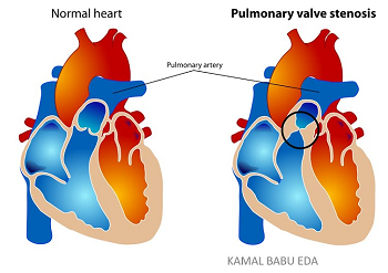 or supra-valvular according
to the location of the lesion. Valvular stenosis is the most common
form. There are two main types – A and B. Type A is typified by fusion
of the peripheral edges of the semilunar valves (commissural fusion) and
poststenotic dilatation of the pulmonary trunk. Type B may involve
hypoplasia of the pulmonary annulus and pulmonary trunk, with the cusps
appearing thickened and immobile. Type A is more prevalent. See
these veterinary reports for details about this disorder in cavaliers.
or supra-valvular according
to the location of the lesion. Valvular stenosis is the most common
form. There are two main types – A and B. Type A is typified by fusion
of the peripheral edges of the semilunar valves (commissural fusion) and
poststenotic dilatation of the pulmonary trunk. Type B may involve
hypoplasia of the pulmonary annulus and pulmonary trunk, with the cusps
appearing thickened and immobile. Type A is more prevalent. See
these veterinary reports for details about this disorder in cavaliers.
In a June 1991 article, two 5-month-old cavalier puppies were diagnosed with pulmonic stenosis, having murmurs in the pulmonic area. But were treated with balloon valvuloplasty.
In an August 2000 report, researchers found wrinkling and irregularity of the surface of the main pulmonary artery and its branches. The pulmonary artery wall showed sever intimal thickening due to subendothelial fibrosis and lesions.
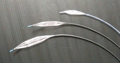 In
this
July 2016 article, a team of Thailand veterinary surgeons and other
specialists report a successful insertion of a balloon in the pulmonary
valves of three dogs, including a cavalier (#2 in the report), affected
by pulmonic stenosis. In this study, the balloon was inserted by a
catheter and then inflated at the location of the narrowing of the
pulmonary valve. (See examples of balloon catheters at right.)
Once the balloon was inflated, the narrowing of the valve disappeared
and the pressure through the valve greatly decreased in the CKCS.
In
this
July 2016 article, a team of Thailand veterinary surgeons and other
specialists report a successful insertion of a balloon in the pulmonary
valves of three dogs, including a cavalier (#2 in the report), affected
by pulmonic stenosis. In this study, the balloon was inserted by a
catheter and then inflated at the location of the narrowing of the
pulmonary valve. (See examples of balloon catheters at right.)
Once the balloon was inflated, the narrowing of the valve disappeared
and the pressure through the valve greatly decreased in the CKCS.
In a
July 2016 newspaper article, veterinary cardiologist Simon Swift of
the University of Florida's veterinary
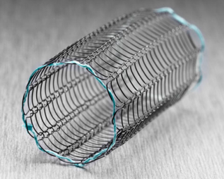 college
reported a case of a Havanese spaniel with pulmonic stenosis which for
which a valvuloplasty procedure was unsuccessful because the dog had an
"unusually thick right ventricle and abnormal valve". Dr. Swift and a
surgical team, which included the university's pediatric cardiology
specialists, put a stent, a metal tube made of wire mesh (see photo
at left), on a balloon and entered the chest straight into the
heart. Then, they expanded the balloon to open the stent and relieve the
blockage in his artery, Swift said.
college
reported a case of a Havanese spaniel with pulmonic stenosis which for
which a valvuloplasty procedure was unsuccessful because the dog had an
"unusually thick right ventricle and abnormal valve". Dr. Swift and a
surgical team, which included the university's pediatric cardiology
specialists, put a stent, a metal tube made of wire mesh (see photo
at left), on a balloon and entered the chest straight into the
heart. Then, they expanded the balloon to open the stent and relieve the
blockage in his artery, Swift said.
In an August 2019 article, 26 cavaliers from seven different clinics worldwide were diagnosed with pulmonic stenosis, most of which had severe cases of the disease. The authors concluded that CKCSs "might be predisposed to pulmonic stenosis".
RETURN TO TOP
Quadricuspid aortic valve
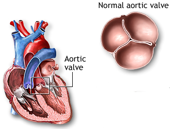 The
aortic valve is the gateway for blood to exit out of the ventricle and
pass through the aorta artery for the body to receive oxygenated blood.
The aortic valve is composed of (1) the aortic annulus, (2) the aortic
root, (3) cusps (leaflets), and (4) commissures. Normal aortic valves
have three equally sized leaflets, called (1) the right coronary cusp,
(2) the left coronary cusp, and (3) the non-coronary cusp. (See
diagram at left.) Quadrisupid aortic valve (QAV) is a rare
congenital heart disease in which a fourth cusp develops (see
diagram at right), resulting in
The
aortic valve is the gateway for blood to exit out of the ventricle and
pass through the aorta artery for the body to receive oxygenated blood.
The aortic valve is composed of (1) the aortic annulus, (2) the aortic
root, (3) cusps (leaflets), and (4) commissures. Normal aortic valves
have three equally sized leaflets, called (1) the right coronary cusp,
(2) the left coronary cusp, and (3) the non-coronary cusp. (See
diagram at left.) Quadrisupid aortic valve (QAV) is a rare
congenital heart disease in which a fourth cusp develops (see
diagram at right), resulting in
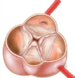 regurgitation
through the aortic valve, causing the sound of a murmur detected by
auscultation.
regurgitation
through the aortic valve, causing the sound of a murmur detected by
auscultation.
In this June 2008 article, French researchers reported six cases of dogs having QAV. Two of the dogs (33.3%) were cavalier King Charles spaniels. They both had developed a fourth cusp, smaller than the other three equally sized larger cusps. The two cavaliers also had an abnormally dilated left coronary artery ostium (an opening in the aortic sinuses). One of them also had MVD with a ruptured chordae, and the other has a ventricular septal defect. Neither of these dogs, nor any of the other four in this study, displayed any clinical signs of the disorder.
RETURN TO TOP
Salivary glands disorders
Dogs have several salivary glands, located in pairs on either side of
the dog's head, from near the eyes to the lower jaw.
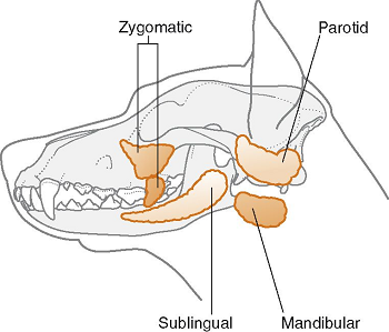 The
four major pairs are known as the parotid, zygomatic (orbital),
mandibular, and sublingual glands. There also are minor glands,
including the palatine and lacrimal. Each gland is connected by a duct
which delivers the saliva to areas of the dog's mouth, providing
lubrication to aid in the chewing process, soften the food, release
digestive enzymes, keep the mouth moist, flush particles of food, and
cool the dog when overheated and panting.
The
four major pairs are known as the parotid, zygomatic (orbital),
mandibular, and sublingual glands. There also are minor glands,
including the palatine and lacrimal. Each gland is connected by a duct
which delivers the saliva to areas of the dog's mouth, providing
lubrication to aid in the chewing process, soften the food, release
digestive enzymes, keep the mouth moist, flush particles of food, and
cool the dog when overheated and panting.
These glands may become subject to any of a variety of disorders which could be due to infection, trauma, inflammation, stone obstructions in their ducts, cancer, and/or immune-mediated disease. Disorders of the salivary glands found to be particularly problemlatic for cavalier King Charles spaniels are listed below.
• Hypersialism
In this November 1992 article, two cavaliers were found to salivate excessively due to their swollen mandibular glands producing more saliva than they were capable of swallowing, a disorder which they discribed as hypersialism associated with non-painful, symmetrical enlargement of both mandibular salivary glands. The clinicians were unable to determine the cause of the hypersialism, but they suspected from the lack of any structural or functional abnormalities that the cause was not an inability to swallow but rather a reluctance to do so. They treated the two cavaliers with the anticonvulsant medication phenobarbitone, which completely resolved the condition.
• Sialadenitis
Sialadenitis is inflammation of the mandibular and/or zygomatic salivary glands. It is the principle disorder which affects dogs' salivary glands. Sialadenitis has been linked to a peripheral form of epilepsy. Studies have reported complete clinical recoveries following oral administration of phenobarbital with no recurrence of disease once medication was tapered off.
In this November 1992 article, two cavaliers were found to salivate excessively due to their swollen mandibular glands producing more saliva than they were capable of swallowing, a disorder called hypersialism. The clinicians were unable to determine the cause of the hypersialism, but they suspected from the lack of any structural or functional abnormalities that the cause was not an inability to swallow but rather a reluctance to do so. They treated the two cavaliers with the anticonvulsant medication phenobarbitone, which completely resolved the condition.
In this August 2018 article, a cavalier was diagnosed with zygomatic sialadenitis which had been diagnosed by computed tomography (CT scan). The dog's symptoms had been trismus (inability to normally open the mouth -- lockjaw), exophthalmos (abnormal protrusion of the eyeballs, and bilateral blindness over the previous three days. The symptoms occurred following abdominal surgery to remove a foreign body from the gastrointestinal region. Phenobarbital was added to to antibiotic treatment, and two days later, the exophthalmos and trismus cleared up. The dog returned to normal behavior but blindness remained in both eyes.
• Sialoceles
Sialoceles is the accumulation of saliva in the tissues surrounding the affected salivary gland due to leakage from the gland or salivary duct. The leakage causes an inflammatory reaction. A fluid-filled pocket develops, resulting in swelling, which is the most typical sign. Most such swellings are not painful but may become quite large.
The most commonly affected gland is the monostomatic portion of the sublingual gland, resulting in neck swelling and formation of a saliva-filled cyst (ranula) on the floor of the mouth. Depending upon which gland is affected, the region of swelling will surround it. Sialoceles of the zygomatic gland may cause exopthalmos (abnormal protrusion of the eyeball from its socket).
In a May 2022 article, 22 dogs, including one cavalier, were diagnosed with sialoceles of various glands. The cavalier had a swollen jaw due to sialoceles of the parotid glands. Transverse CT scanning was able to detect this condition.
Apart from visual examination, aspiration of the swelling is a form of diagnosis, resulting in removal of a thick, vicouse fluid that usually is yellowish. The usual treatment of sialoceles is surgical removal of the affected salivary gland. Temporary alternatives to surgery are drainage and anti-inflammatories.
• Sialolithiasis
Sialolithiasis is the formation of a stone, which blocks a duct from a salivary gland to the dog's mouth. They mostly are composed of calcium and develop in any of the dog's major salivary glands, the parotid, submandibular (uner the jaw), and sublingual (under the tongue), or a minor salivary gland. Each of the the two parotid glands are located in front of each ear. The glands attach to a duct which transports saliva from the gland to the mouth. The duct attached to the parotid gland is called Wharton's duct.
While sialolithiasis of the parotid duct in dogs has been reported infrequently, there have been at least two reorted cases of parotid duct sialolithiasis in cavaliers. See this June 1996 article and this July 1997 article, both reporting case studies of cavaliers. In both cases, surgery was conducted to remove the stones and repair the parotid glands and their ducts.
RETURN TO TOP
Sand impaction
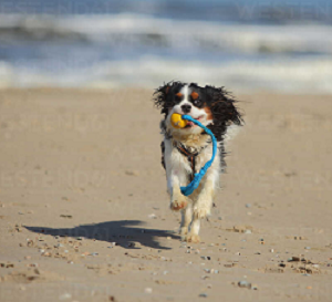 Sand
impaction results from dogs swallowing too much sand, typically during
trips to a beach. It can occur accidentally while playing on the sand, if the dog repeatedly picks up wet play objects, like toys or balls with
sand encoating them. Sand impaction is an intestinal blockage, such as
the white, sausage-shaped one visible in the x-ray of a cavalier, below.
Sand
impaction results from dogs swallowing too much sand, typically during
trips to a beach. It can occur accidentally while playing on the sand, if the dog repeatedly picks up wet play objects, like toys or balls with
sand encoating them. Sand impaction is an intestinal blockage, such as
the white, sausage-shaped one visible in the x-ray of a cavalier, below.
Symptoms, which may not appear until hours or even days later, include vomiting, loss of appetite, lethargy, and abdominal pain. X-rays can confirm the obstruction, usually located in the small intestine. In mild cases, treatment may be limited to an enema to oral paraffin oil to medication (ampicillin, metronidazole, panteprazole, ranitidine, dextrans 70). In more severe cases, surgery also will be necessary.
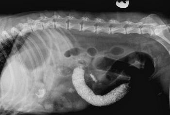 Survival
is not assured. In this
December
2009 article, clinicians reported that an 11 year old cavalier
(see x-ray at left) had experienced clinical signs for 18 days. It
had a prior history of mitral valve disease (MVD), including congestive heart
failure, as well as gastrointestinal disease. It was treated with an
enema, gastric lavage, pentastarch (a blood volume expander to manage
shock), and plasma, followed by surgery. The CKCS died after the surgery.
The authors attributed the death to the combination of MVD, acidosis,
gastrointestinal disease, and the surgery.
Survival
is not assured. In this
December
2009 article, clinicians reported that an 11 year old cavalier
(see x-ray at left) had experienced clinical signs for 18 days. It
had a prior history of mitral valve disease (MVD), including congestive heart
failure, as well as gastrointestinal disease. It was treated with an
enema, gastric lavage, pentastarch (a blood volume expander to manage
shock), and plasma, followed by surgery. The CKCS died after the surgery.
The authors attributed the death to the combination of MVD, acidosis,
gastrointestinal disease, and the surgery.
RETURN TO TOP
Shadow chasing
 Shadow chasing is a repetitive and seemingly uncontrollable behavior
-- a neurological disorder -- in a small minority of dogs, similar to
"obsessive compulsive disorder" (OCD) in humans. It is categorized as a
canine compulsive disorder (CCD). It is attributed to an alteration in
the dog's dopaminergic neurotransmitter (DAT) system in the brain.
Shadow chasing is a repetitive and seemingly uncontrollable behavior
-- a neurological disorder -- in a small minority of dogs, similar to
"obsessive compulsive disorder" (OCD) in humans. It is categorized as a
canine compulsive disorder (CCD). It is attributed to an alteration in
the dog's dopaminergic neurotransmitter (DAT) system in the brain.
In this November 2010 case-study, a female cavalier King Charles spaniel's shadow chasing was getting progressively frequent and long term. Examination revealed a significantly higher dopamine transporter (DAT) striatal-to-brain ratio. The Belgian veterinarians treated the disorder with the tricyclic antidepressant clomipramine. Noticeable improvement in the cavalier's condition began after seven days of treatment. Shadow chasing disappeared almost completely after three weeks and, if performed, was stopped by calling the dog’s name. Clomipramine treatment was continued at the same dose for two months, at which point the researchers noted the DAT binding regained normal values. Nevertheless, the shadow chasing reocurred after discontinuing the medication for two days. The most significant side effect was gluttony (e.g., persistent searching and opening garbage pails) after six weeks of treatment, which the vets treated by reducing the dosage of clomipramine every other day thereafter. The researchers concluded that the successful treatment with clomipramine indicated the role of dopamine in CCD.
CAUTION: Clomipramine (Anafranil), a tricyclic antidepressant (TCA), inhibits both norepinephrine and serotonin reuptake. It reportedly can lead to serotonin accumulation and serotonin syndrome (symptomatically, twitching, tremor, tachycardia, myoclonic movements, and hyperthermia) in humans when used in combination with monoamine oxidase inhibitors (MAOIs, which decrease the breakdown of serotonin) or serotonin reuptake inhibitors, which increase synaptic serotonin concentrations. Deaths have been reported in humans given clomipramine plus MAOIs—a well-established interaction. See this June 2013 report. Also, high levels of serotonin in cavaliers' blood platelets and mitral valve tissues have been associated with mitral valve disease. For more information, see this discussion on our MVD webpage.
See, also, our related discussion of fly catching.
RETURN TO TOP
Temporomandibular joint morphology (TMJ)
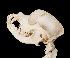 The temporomandibular joint (TMJ) is the hinge of the jaw that
connects the jaw to the temporal bones of the skull.
Temporo-mandibular joint dysplasia refers to
pain and dysfunction of the muscles that move the temporomandibular
joints and the jaw. Unfortunately, the typical shape of the cavalier's
temporomandibular joint is uniquely odd. In a
May 2001 report, the researchers compared the morphology of the
temporomandibular joints of numerous breeds of dogs, from a radiographic
positioning standpoint. They noted that:
The temporomandibular joint (TMJ) is the hinge of the jaw that
connects the jaw to the temporal bones of the skull.
Temporo-mandibular joint dysplasia refers to
pain and dysfunction of the muscles that move the temporomandibular
joints and the jaw. Unfortunately, the typical shape of the cavalier's
temporomandibular joint is uniquely odd. In a
May 2001 report, the researchers compared the morphology of the
temporomandibular joints of numerous breeds of dogs, from a radiographic
positioning standpoint. They noted that:
"The temporomandibular joints of the cavalier king Charles spaniel and Pekingese skulls in the anatomical collection were considered to be grossly abnormal. They were therefore excluded from further consideration in the present study and their rotational angles omitted from Table 2."
In a May 2002 article, veterinary radiologists reported on temporomandibular joint dysplasia in the skulls of 33 cavaliers, finding evidence of bilateral temporomandibular joint dysplasia. They concluded:
"This finding suggests that temporomandibular joint dysplasia is a widespread asymptomatic condition in the CKCS and should be regarded as a normal morphologic variation rather than a pathologic anomaly."
In an August 2016 study, eleven CKCSs were included in a review of TMJ in 48 dogs, examining the morphologic and morphometric features of the TMJ using computed tomography (CT). They found:
"The TMJs of Cavalier King Charles spaniels showed remarkably lower values associated with shallow mandibular fossae and incongruent articular heads.
"Cavalier King Charles spaniels had the shallowest mandibular fossae of all breeds. The retroarticular process appeared as a bony extension on the medioventral aspect of the mandibular fossa, articulating with the caudomedial aspect of the condylar process. This bony extension was prominent in Labrador retrievers, German shepherds, boxers, and English bulldogs. The retroarticular process was less developed in cocker spaniels, shih tzus, and pug. In Cavalier King Charles spaniels, it was small or absent.
"The wide angles documented in Labrador retrievers, German shepherds, boxers, and English bulldogs were consistent with long and well-defined retroarticular processes, whereas the narrow angles found in cocker spaniels, shih tzus, Cavalier King Charles spaniels, and a pug were due to less developed or absent retroarticular processes. A dramatically reduced ventral extension of the retroarticular process, as observed in cocker spaniels and Cavaliers King Charles spaniels in this study, may permit slight caudal dislocation of the head of the condylar process and predispose to TMJ instability.
"German shepherds, boxers, and English bulldogswere consistent with deeper (more concave) mandibular fossae and more prominent retroarticular processes, whereas the narrow angles found in cocker spaniels, shih tzus, CavalierKing Charles spaniels, and the pug were due to more shallower (less concave) mandibular fossae and less prominent or absent retroarticular processes.
"The present study agrees with the asymptomatic dysplastic condition of the TMJ previously reported in the Cavalier King Charles spaniel and suggests that this morphological features may exist in certain dog breeds such as cocker spaniels and small breeds such as shih tzus and pugs."
Diagram C of Figure 11 below shows the severe shallowness of the CKCS's mandibular fossa, compared to those of the Labrador retriever, German shepherd boxer, and English bulldog (diagram A) and those of the Cocker spaniel, pug, and Shih Tzus (diagram C).
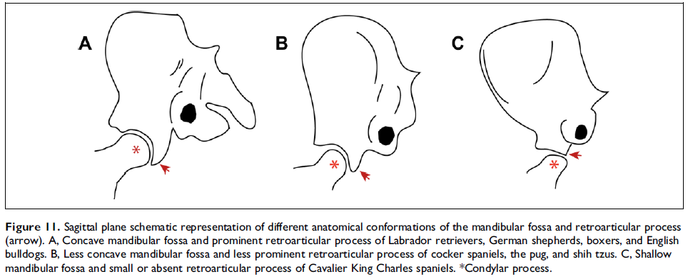
Beginning in 2021, a classification system of five degrees of severity of TMJ was established for humans. The system is based upon MRI and CT scanning observations. The five stages, along with their sub-grades, are:
Stage 0: Normal MRI/CT study.
Stage 1A: A normal condyle-disk-fossa relationship associated with pathologic changes of the LPM + /joint effusion.
Stage 1B: A normal condyle-disk-fossa relationship associated with pathologic changes of the disk + /Bone degenerative process of the condyle.
Stage 2A: Anterior disk displacement in the closed mouth position with reduction to the normal position in the open mouth position associated with pathologic changes of the LPM + /joint effusion.
Stage 2B: Anterior disk displacement in the closed mouth position with reduction to the normal position in the open mouth position associated with pathologic changes of the disk + /Bone degenerative process of the condyle.
Stage 2C: Anterior disk displacement in the closed mouth position with reduction to the normal position in the open mouth position with condylar hypertranslation.
Stage 3A: Anterior disk displacement in the closed mouth position without reduction to the normal position in the open mouth position associated with pathologic changes of the LPM + /joint effusion.
Stage 3B: Anterior disk displacement in the closed mouth position without reduction to the normal position in the open mouth position associated with pathologic changes of the disk + /Bone degenerative process of the condyle.
Stage 3C: Anterior disk displacement in the closed mouth position without reduction to the normal position in the open mouth position associated with normal translation movement of the mandibular condyle (no limitation of the mouth opening).
Stage 4: Posterior disk displacement.
In an April 2023 article, UK and USA researchers assessed the temporomandibular joint (TMJ) by computed tomography (CT) in several breeds of brachycephalic dogs, including cavalier King Charles spaniels. The other breeds among 153 dogs were French bulldogs, English bulldogs, boxers, chihuahuas, Lhasa Apsos, pugs, Shih Tzus, and Staffordshire bull terriers. None of the dogs displayed any symptoms of TMJ disorders. The severity of the TMJ morphological changes was determined using their modification of the 5-grade classification system for humans. They report finding widespread variations in the shape of the head of the condylar process of the mandible (see diagram above), the mandibular fossa, and other areas, ranging from a rounded concave TMJ with a long retroarticular process to a flattened TMJ without a process. Variations in the articular surface of the head of the condyle ranged from flat, through curved and trapezoid to sigmoid. They found that the prevalence of severe TMJ dysplasia (grades B3 and C) in the cavaliers was high (69.2% of the CKCSs in the study). They concluded that the "marked changes" in the CKCS appear to be "highly prevalent" and "should be considered a breed variation."
RETURN TO TOP
Tonsillitis
We know of no veterinary journal articles about cavaliers having tonsillitis. However, it is a known disorder in purebred dogs, especially in smaller breeds. See this article on the general nature of the disorder. Since tonsillitis is an infection, it usually is successfully treated with antibiotics. Removal of the tonsils as treatment -- tonsillectomy -- reportedly is quite rare. As the linked article notes, tonsils play a "vital role in fighting infection of the oropharyngeal cavity (mouth and throat)." As a result, tonsillectomy may become necessary only if "there is poor response to treatment or if tonsillitis becomes a recurring condition. Recurrent tonsillitis is more likely to occur in small breeds of dogs."
Nevertheless, we know of one reported case of a cavalier undergoing a tonsillectomy. It was performed on Rex, the CKCS owned by President and Mrs. Ronald Reagan, and was performed in January 1986, only a month after the Reagans acquired Rex in December 1985 at age one year. See this brief New York Times article.
RETURN TO TOP
Vasculitis
Vasculitis is the inflammation of blood vessel walls. In a June 2015 article, researchers reported on vasculitis in six cavaliers (14+%), along with 36 dogs of other breeds.
RETURN TO TOP
Ventricular septal defect
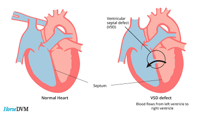 Ventricular septal defect (VSD) is a hole between the interior wall
(the muscular septum) of the heart separating the right ventricle and
the left ventricle. Blood may flow through the hole (shunting) from the
left ventricle (LV) to the righ ventricle (RV), called
left-to-right (L-R) shunting, or from the RV to the LV, called
right-to-left (R-L) shunting, or even
by-directional during the heart cycle. R-L shunting is called
reversed VSD. It is a congenital defect in the sense
that it is an opening that exists during the dog's fetal stage but which
fails to close as the fetus continues to develop. VSD is most common
among English bulldogs, English springer spaniels, French bulldogs, and
Germn shepherd dogs, but it has been diagnosed periodically in cavalier
King Charles spaniels.
Ventricular septal defect (VSD) is a hole between the interior wall
(the muscular septum) of the heart separating the right ventricle and
the left ventricle. Blood may flow through the hole (shunting) from the
left ventricle (LV) to the righ ventricle (RV), called
left-to-right (L-R) shunting, or from the RV to the LV, called
right-to-left (R-L) shunting, or even
by-directional during the heart cycle. R-L shunting is called
reversed VSD. It is a congenital defect in the sense
that it is an opening that exists during the dog's fetal stage but which
fails to close as the fetus continues to develop. VSD is most common
among English bulldogs, English springer spaniels, French bulldogs, and
Germn shepherd dogs, but it has been diagnosed periodically in cavalier
King Charles spaniels.
There are various types of VSDs, depending upon the location of the hole and the structures adjacent to it. VSD can progress so rapidly to the stage of congestive heart failure (CHF) that the puppy dies before its first visit to a veterinarian. Most puppies which are examined and diagnosed with VSD have such a mild case that they do not exhibit any clinical symptoms. L-R VSD first may be detected by asucultation, with the sound of a murmur loudest on the right side of the heart. VSD may be expected to cause enlargement of the LV and then the left atrium, and thereby lead to CHF. RV enlargement and pulmonary hypertension also occur if the hole is very large. X-rays and echocardiography will show the heart enlargement, and an echo scan can detect the shunting of blood through the defect.
Small VSDs may close on their own within the first two years. Dogs with small to moderate shunting of blood may be expected to have normal life spans. Affected dogs which develop left chamber enlargement and/or CHF are treated as MVD-affected dogs in Stages B2 and C and D are. There are various forms of surgical treatments, including cardio-pulmonary bypass, hypothermia and intracardiac surgery and a transcatheter-delivered device. See this October 2022 article.
R-L VSD has different symptoms and consequences. Poor stamina, a rapid respiratory rate, cyanosis (bluish discoloration of the skin and mucous membranes resulting from inadequate oxygenation of the blood), collapse, and seizures are common signs. X-rays and echocardiographic scans will diagnose this form of VSD. Surgery may be more risky for dogs with R-L VSE, and shortened life spans may be expected.
In this June 2008 article, French researchers reported a case of a cavalier having a ventricular septal defect. Thd CKCS did not display any clinical signs of the disorder.
RETURN TO TOP
Zoonotic diseases (zoonosis)
Zoonotic disease (zoonosis) is defined as a disease which is transmitted between an animal or insect and a human. Customarily, the transmision is from the animal to the human, but the definition allows for the opposite of that direction. In a June 2020 article, Italian veterinary researchers diagnosed a ten year old male cavalier King Charles spaniel with severe symptoms of infection in its ears due to the bacteria Acinetobacter baumannii. A. baumannii is an infection typically acquired by hospitalized humans. Both the cavalier and its primary owner, who had a very long hospital stay before the dog exhibited any symptoms, were found to to have A. baumannii. The dog exhibited symptoms of severe, chronic discharges of pus from its ear canals. The owner had no outward signs of the disease, but nasal swab tests confirmed the owner's bacterial infection. The investigators concluded:
"A. baumannii ... can be considered a zoonotic pathogen and this clinical case demonstrates a possible transmission between human and dog. As the person with an asymptomatic nasal colonization of A. baumannii was hospitalized for a long period before the onset of the symptoms in the dog, we hypothesize that the pet owner could have been the potential reservoir for the canine infection. Therefore, further experimental studies are needed in order to understand if this was an isolated case or if A. baumannii can successfully infect both pets and owners."
RETURN TO TOP
Research News
April 2023:
Cavaliers have the highest prevalence (69.2%) of
temporomandibular joint (TMJ) dysplasia in a study of 153 asymptomatic
brachycephalic dogs.
 In
an
April 2023 article, UK and USA researchers (Emilie Paran [right],
Sarah Bouyssou, Alison King) assessed the temporomandibular joint (TMJ)
by computed tomography (CT) in several breeds of brachycephalic dogs,
including cavalier King Charles spaniels. The other breeds among 153
dogs were French bulldogs, English bulldogs, boxers, chihuahuas, Lhasa
Apsos, pugs, Shih Tzus, and Staffordshire bull terriers. None of the
dogs displayed any symptoms of TMJ disorders. The severity of the TMJ
morphological changes was determined using their modification of the
5-grade classification system (linked here).
They report finding widespread variations in the shape of the head of
the condylar process of the mandible (see diagram), the mandibular
fossa, and other areas, ranging from a rounded concave TMJ with a long
retroarticular process to a flattened TMJ without a process. Variations
in the articular surface of the head of the condyle ranged from flat,
through curved and trapezoid to sigmoid. They found that the prevalence
of severe TMJ dysplasia (grades B3 and C) in the cavaliers was high
(69.2% of the CKCSs in the study). They concluded that the "marked
changes" in the CKCS appear to be "highly prevalent" and "should be
considered a breed variation."
In
an
April 2023 article, UK and USA researchers (Emilie Paran [right],
Sarah Bouyssou, Alison King) assessed the temporomandibular joint (TMJ)
by computed tomography (CT) in several breeds of brachycephalic dogs,
including cavalier King Charles spaniels. The other breeds among 153
dogs were French bulldogs, English bulldogs, boxers, chihuahuas, Lhasa
Apsos, pugs, Shih Tzus, and Staffordshire bull terriers. None of the
dogs displayed any symptoms of TMJ disorders. The severity of the TMJ
morphological changes was determined using their modification of the
5-grade classification system (linked here).
They report finding widespread variations in the shape of the head of
the condylar process of the mandible (see diagram), the mandibular
fossa, and other areas, ranging from a rounded concave TMJ with a long
retroarticular process to a flattened TMJ without a process. Variations
in the articular surface of the head of the condyle ranged from flat,
through curved and trapezoid to sigmoid. They found that the prevalence
of severe TMJ dysplasia (grades B3 and C) in the cavaliers was high
(69.2% of the CKCSs in the study). They concluded that the "marked
changes" in the CKCS appear to be "highly prevalent" and "should be
considered a breed variation."
February 2023:
Fly larvae
found in a cavalier in Belgium.
 In
a
February 2023 article, Belgium veterinary clinicians (Hendrik De
Bosschere [right], A.-S. Platteeuw, A. Verstraeten) report
finding the rat-tailed larvae of the common drone fly (Eristalis tenax)
being expelled in the feces of a cavalier King Charles spaniel. The
dog's owner observed several living larvae in fresh feces. For seven
weeks before these larvae were found, the dog’s feces had been dark
black, tarry feces that usually are associated with upper
gastrointestinal bleeding. The dog showed signs of abdominal discomfort.
The dog was treated with Milbemax and Panacur, following which the dog’s
feces tested negative for occult blood; no more larvae were found, and
the symptoms disappeared. Nevertheless, the investigators suspect that
the dog's signs of discomfort and the blood-darkened feces were not due
to the larvae, because they are known to inhabit only the rectum. They
concluded:
In
a
February 2023 article, Belgium veterinary clinicians (Hendrik De
Bosschere [right], A.-S. Platteeuw, A. Verstraeten) report
finding the rat-tailed larvae of the common drone fly (Eristalis tenax)
being expelled in the feces of a cavalier King Charles spaniel. The
dog's owner observed several living larvae in fresh feces. For seven
weeks before these larvae were found, the dog’s feces had been dark
black, tarry feces that usually are associated with upper
gastrointestinal bleeding. The dog showed signs of abdominal discomfort.
The dog was treated with Milbemax and Panacur, following which the dog’s
feces tested negative for occult blood; no more larvae were found, and
the symptoms disappeared. Nevertheless, the investigators suspect that
the dog's signs of discomfort and the blood-darkened feces were not due
to the larvae, because they are known to inhabit only the rectum. They
concluded:
"In conclusion, discovery of living drone fly larvae in pet’s feces may be frightening for the owner, but in most cases, facultative myiasis does not involve pathology."
January 2023:
USA study finds that castration and spaying are associated with
greater health problems and nuisance behaviors in dogs.
 In a
January 2023 article, the health and behavior outcomes were compared
among neutered dogs (castrated – full gonadectomy), sexually intact
dogs, and dogs which underwent vasectomy (males) or ovary-sparing
hysterectomy (females) by a team of USA researchers (Chris Zink
[right], Mikel M. Delgado, Judith L. Stella). Information was gathered by answers to
questionnaires submitted by the dogs’ owners. Data was acquired about
6,018 dogs, including 2,786 males (1,672 castrated, 1,056 intact, and 58
vasectomized) and 3,232 females (2,281 spayed, 792 intact, and 159
ovary-sparing spayed). The breeds of dogs were not specified. The health
issues in the questionnaire included the orthopedic problems of hip
dysplasia, elbow dysplasia, cranial cruciate ligament injury (knees –
patellas), osteochondrosis dissecans (cartilage separation from bone at
joints), panosteitis (inflammation of leg bones), and patellas luxation
(knee separation). Various types of cancers were listed. Other health
conditions listed were prostate enlargement or infection, Addison’s
disease, Cushing’s disease, diabetes mellitus, hypothyroidism, and
obesity. The investigators report:
In a
January 2023 article, the health and behavior outcomes were compared
among neutered dogs (castrated – full gonadectomy), sexually intact
dogs, and dogs which underwent vasectomy (males) or ovary-sparing
hysterectomy (females) by a team of USA researchers (Chris Zink
[right], Mikel M. Delgado, Judith L. Stella). Information was gathered by answers to
questionnaires submitted by the dogs’ owners. Data was acquired about
6,018 dogs, including 2,786 males (1,672 castrated, 1,056 intact, and 58
vasectomized) and 3,232 females (2,281 spayed, 792 intact, and 159
ovary-sparing spayed). The breeds of dogs were not specified. The health
issues in the questionnaire included the orthopedic problems of hip
dysplasia, elbow dysplasia, cranial cruciate ligament injury (knees –
patellas), osteochondrosis dissecans (cartilage separation from bone at
joints), panosteitis (inflammation of leg bones), and patellas luxation
(knee separation). Various types of cancers were listed. Other health
conditions listed were prostate enlargement or infection, Addison’s
disease, Cushing’s disease, diabetes mellitus, hypothyroidism, and
obesity. The investigators report:
“Our most important finding was that longer duration that gonads were present, regardless of reproductive status, was associated with fewer general health problems and both problematic and nuisance behaviors. It was also associated with an increased lifespan.”
“Being sexually intact or having an OSS [ovary-sparing spay] were associated with lower odds of orthopedic problems, consistent with other studies showing lower risks of a variety of orthopedic problems in sexually intact dogs as compared to gonadectomized dogs.”
“Our results were also consistent with previous studies indicating that the associated odds of cancer is significantly lower for sexually intact dogs than gonadectomized dogs. Although gonadectomized dogs had a later cancer diagnosis, significantly more gonadectomized dogs were diagnosed with cancer compared to sexually intact dogs.”
“Gonadectomized dogs and dogs that underwent OSS were more likely to be obese than sexually intact dogs, but there was no relationship between duration that gonads were present and obesity. This suggests that the causes of obesity are more complex than just the absence of gonadal hormones.”
“Our data also indicated that the associated prevalence of both problematic and nuisance behaviors decreased with increased exposure to gonadal hormones. Male dogs of all reproductive groups and dogs with an OSS were reportedly more likely to mark or mount than sexually intact and SF dogs. Further, there was no difference in marking or mounting between gonadectomized and IM dogs.
“Taken together, these data suggest that gonadal hormone exposure time may be a more sensitive way to examine the effects of reproductive surgery on health and behavior than categorizing dogs into groups by type of reproductive surgery. This is likely because there may be significant overlap in the length of exposure to gonadal hormones between the 6 groups examined in this study.”
They concluded that: “ Our findings emphasize the importance of gonadal hormone exposure to canine health and behavior.”
September 2022:
UK neurologists are seeking cavaliers with myoclonus.
 In a September 2022 article in the Veterinary
Record, UK neurologists
Matthew Green (right), Mark Lowrie, and Clare Rusbridge have issued a "call for
cases" of cavalier King Charles spaniels which are suspected of having
myoclonus. They describe the condition as "recurrent and uncontrollable
episodes of abnormal jerk-like movements that may commonly involve the
head and face but also legs and body." They report "seeing increasing
numbers of Cavalier King Charles Spaniels with this condition." They
request:
In a September 2022 article in the Veterinary
Record, UK neurologists
Matthew Green (right), Mark Lowrie, and Clare Rusbridge have issued a "call for
cases" of cavalier King Charles spaniels which are suspected of having
myoclonus. They describe the condition as "recurrent and uncontrollable
episodes of abnormal jerk-like movements that may commonly involve the
head and face but also legs and body." They report "seeing increasing
numbers of Cavalier King Charles Spaniels with this condition." They
request:
"We are hoping to find people that own, or have previously owned, Cavalier King Charles Spaniels with suspected myoclonus. We would ask you to provide a short video of the abnormal episodes for review byour neurology team, if possible, followed by a short online questionnaire to further study this condition. It is hoped that we can better understand the ways in which dogs are affected, which may then lead to improved recognition and management of the condition. If you think your dog may be affected, or you would like further information, please contact Matthew Green at Dovecote Veterinary Hospital, via e-mail: matthew.green@dovecoteveterinaryhospital.co.uk"
Click on the photo of the cavalier above to view a video of its episode of myoclonus, or click here.
June 2022:
Cavalier is first reported case of anti-GABAAR autoimmune
encephalitis.
 In a
June
2022 article, German veterinary neurologists (Enrice I. Huenerfauth,
Christian G. Bien, Corinna Bien, Holger A. Volk [right], Nina
Meyerhoff) describe the first
reported case of a dog -- a 1-year-old cavalier King Charles spaniel --
diagnosed with anti-GABAAR (γ-aminobutyric acid-A receptor) autoimmune
encephalitis. Encephalitis is inflammation of the brain, and autoimmune
encephalitis occurs when the dog's immune system mistakenly attacks
healthy brain cells, leading to that inflammation. GABA is the main
inhibitory neurotransmitter in the brain, which enables the regulation
of neuronal activity. GABAAR are ion channels in the brain control the
rapid transmission of nerve impulses between neurons. The cavalier had
multiple generalized seizures and showed alternating stupor and
hyperexcitability, ataxia, repetitive muscle contractions, and circling
to the left side. Auto-antibody encephalitis, with antibody-mediated
dysfunction of receptors was suspected. Despite treatment with multiple
antiseizure medications (diazepam followed by phenobarbital) the
seizures and behavior abnormalities continued, and the dog alternated
between stupors and hyperexcitability. Immunotherapy with dexamethasone,
a corticosteroid, led to rapid improvement of the clinical signs and the
CKCS improved continuously. A month later, GABAAR auto-antibodies had
decreased significantly. An antibody search in the cerebral-spinal fluid
(CSF) and serum located a neuropil staining pattern of GABAAR
antibodies. At the examination four weeks after the start of
immunotherapy, the dog was clinically normal; the GABAAR antibody titer
in serum had strongly decreased; and the antibodies were no longer
detectable in the CSF. The clinicians report that this is "the first
veterinary patient with an anti-GABAAR encephalitis with a good outcome
following ASM and corticosteroid treatment." They concluded:
In a
June
2022 article, German veterinary neurologists (Enrice I. Huenerfauth,
Christian G. Bien, Corinna Bien, Holger A. Volk [right], Nina
Meyerhoff) describe the first
reported case of a dog -- a 1-year-old cavalier King Charles spaniel --
diagnosed with anti-GABAAR (γ-aminobutyric acid-A receptor) autoimmune
encephalitis. Encephalitis is inflammation of the brain, and autoimmune
encephalitis occurs when the dog's immune system mistakenly attacks
healthy brain cells, leading to that inflammation. GABA is the main
inhibitory neurotransmitter in the brain, which enables the regulation
of neuronal activity. GABAAR are ion channels in the brain control the
rapid transmission of nerve impulses between neurons. The cavalier had
multiple generalized seizures and showed alternating stupor and
hyperexcitability, ataxia, repetitive muscle contractions, and circling
to the left side. Auto-antibody encephalitis, with antibody-mediated
dysfunction of receptors was suspected. Despite treatment with multiple
antiseizure medications (diazepam followed by phenobarbital) the
seizures and behavior abnormalities continued, and the dog alternated
between stupors and hyperexcitability. Immunotherapy with dexamethasone,
a corticosteroid, led to rapid improvement of the clinical signs and the
CKCS improved continuously. A month later, GABAAR auto-antibodies had
decreased significantly. An antibody search in the cerebral-spinal fluid
(CSF) and serum located a neuropil staining pattern of GABAAR
antibodies. At the examination four weeks after the start of
immunotherapy, the dog was clinically normal; the GABAAR antibody titer
in serum had strongly decreased; and the antibodies were no longer
detectable in the CSF. The clinicians report that this is "the first
veterinary patient with an anti-GABAAR encephalitis with a good outcome
following ASM and corticosteroid treatment." They concluded:
"Based on the clinical signs and presentation (epileptic seizures lacking any response to ASMs, erratic behavioral changes) and the results of the initial diagnostics (lack of abnormal findings on conventional MRI and CSF examination), an autoimmune encephalitis was suspected, proven, and successfully treated. Clinicians should consider to test for autoantibodies and start immunotherapy in cases with a similar clinical presentation and lack of response to anti-seizure medication even if an inflammatory/infectious or neoplastic cause was clinically excluded on MRI and CSF."
March 2022:
Myoclonus (spasmodic jerky contractions of head and
forelimbs muscles) is more prevalent in older cavaliers, UK study
finds.
 In
a March
2022 article, UK researchers Carina Rotter (right),
Danielle Whittaker, and Clare Rusbridge reviewed the medical records of
39 cavalier King Charles spaniels diagnosed with myocluonus, a syndrome
displaying symptoms of spasmodic jerky contractions of groups of
muscles, mainly of the head and forelimbs when the dog is standing or
sitting. In this study, the researchers described the symptoms as:
In
a March
2022 article, UK researchers Carina Rotter (right),
Danielle Whittaker, and Clare Rusbridge reviewed the medical records of
39 cavalier King Charles spaniels diagnosed with myocluonus, a syndrome
displaying symptoms of spasmodic jerky contractions of groups of
muscles, mainly of the head and forelimbs when the dog is standing or
sitting. In this study, the researchers described the symptoms as:
"The myoclonic event was a mixture of positive and negative myoclonus and described as rapid eyelid blinking with head nodding/shuddering and variable extension down the thoracic limbs, that could result in falling or stumbling of the thoracic limbs. The movement lasted seconds had no obvious trigger and occurred mostly when stationary in a sternal or sitting position. It did not seem to be associated with loss of consciousness (see Video S1 and Video S2). Multiple daily episodes were seen and increased in frequency and intensity over time. Owners whose dogs would frequently fall, often disrupting them while eating, reported the myoclonus to interfere with the dog's quality of life. The mean age of onset of myoclonus was 8.38 years (SD ±1.96)."
Click
on the photos of the two cavaliers here to view their videos, or click
this link for the ruby and
this link for the Blenheim.
In addition to classic signs of myoclonus, 13 of the dogs (33%) had epileptic seizures or similar events. All of the dogs had Chiari-like malformation (CM), and 20 of them showed signs of CM-associated pain. Twenty of 30 dogs also had syringomyelia (SM), and 13 dogs had primary secretory otitis media (PSOM). Twenty-three of 30 had lateral ventriculomegaly, which was mild in 9/23 dogs, moderate in 11 and marked in 3 dogs. One dog had hydrocephalus which was managed with placement of a ventriculo-peritoneal shunt. Twenty-three dogs were being treated for CM/SM with gabapentin or pregabalin. The researchers concluded that the cavaliers had a "higher prevalence" of myoclonus after age 8 years, and that:
December 2021:
COVID-19 virus causes heart muscle damage in a cavalier King
Charles spaniel.
 In
a
December 2021 article, a team of Italian veterinary clinicians and
researchers (Giovanni Romito [right], Teresa Bertaglia, Luigi
Bertaglia, Nicola Decaro, Annamaria Uva, Gianluca Rugna, Ana Moreno,
Giacomo Vincifori, Francesco Dondi, Alessia Diana, Mario Cipone)
diagnosed a 6-year old cavalier King Charles spaniel as being
COVID-19-positive and suffering from myocardial (heart muscle) injury
(MI). While the dog had a mild mitral valve murmur, mild mitral
regurgitation, and possibly a slightly enlarged left ventricle, the
researchers dismissed MVD as being the cause of the myocardial injury.
They also determined that no bacteria or parasites or non-infectious
diseases known to cause MI were present in the dog's systems. They
concluded that SARS-CoV-2, the causative agent of COVID-19 (coronavirus
disease 2019) was the cause of this dog's myocardial injury.
In
a
December 2021 article, a team of Italian veterinary clinicians and
researchers (Giovanni Romito [right], Teresa Bertaglia, Luigi
Bertaglia, Nicola Decaro, Annamaria Uva, Gianluca Rugna, Ana Moreno,
Giacomo Vincifori, Francesco Dondi, Alessia Diana, Mario Cipone)
diagnosed a 6-year old cavalier King Charles spaniel as being
COVID-19-positive and suffering from myocardial (heart muscle) injury
(MI). While the dog had a mild mitral valve murmur, mild mitral
regurgitation, and possibly a slightly enlarged left ventricle, the
researchers dismissed MVD as being the cause of the myocardial injury.
They also determined that no bacteria or parasites or non-infectious
diseases known to cause MI were present in the dog's systems. They
concluded that SARS-CoV-2, the causative agent of COVID-19 (coronavirus
disease 2019) was the cause of this dog's myocardial injury.
October 2021:
UK study finds cavaliers having a high prevalence of gallbladder
stones (cholelithiasis).
 In
an
October 2021 article, University of Cambridge (UK) researchers
(Frederik Allan, Penny J. Watson, Katie E. McCallum [right])
reviewed the medical records of 38 dogs diagnosed with cholelithiasis by
abdominal ultrasound (AUS) between 2010 and 2019 at The Queen's
Veterinary School Hospital, University of Cambridge. Cavalier King
Charles spaniels was the most common breed, comprising 4 (10.5%) of the
patients. The investigators confirmed a "high prevalence" of
cholelithiasis in CKCSs. Of the 18 (47.4%) dogs classified as
symptomatic, the clinical signs of cholelithiasis included:
In
an
October 2021 article, University of Cambridge (UK) researchers
(Frederik Allan, Penny J. Watson, Katie E. McCallum [right])
reviewed the medical records of 38 dogs diagnosed with cholelithiasis by
abdominal ultrasound (AUS) between 2010 and 2019 at The Queen's
Veterinary School Hospital, University of Cambridge. Cavalier King
Charles spaniels was the most common breed, comprising 4 (10.5%) of the
patients. The investigators confirmed a "high prevalence" of
cholelithiasis in CKCSs. Of the 18 (47.4%) dogs classified as
symptomatic, the clinical signs of cholelithiasis included:
• vomiting
• decreased appetite
• lethargy
• diarrhea
• jaundice (icterus)
• abdominal pain
• weight loss
• excessive urination (polyuria)
• frequent urination (pollakiuria)
• slow, difficult, painful urinary flow (stranguria)
• blood in the urine (hematuria)
• excessive thirst (polydipsia)
• thinning of haircoat
• deafness
• shifting lameness
Symptomatic cases had higher alkaline phosphatase, gamma-glutamyl transferase, and alanine transferase activities than did non-symptomatic cases, and 44.4% of symptomatic cases also had choledocholithiasis (gallbladder stones in the bile duct).
Treatment categories consisted of those 13 dogs (34.2%) medically treated, 10 surgically treated (26.3%), and 15 not treated (29.5%). They found that medical treatment with ursodeoxycholic acid (UDCA) resolved or improved cholelithiasis in several dogs. They therefore concluded that, "with careful case selection, UDCA may be a safe and effective tool for management of cholelithiasis [without choledocholithiasis] in dogs." They also recommended that dogs "with marked increases in ALP, ALT, and GGT activity with or without ultrasonographic evidence of choledocholiths and with or without symptomatic cholelithiasis can be considered for surgical intervention." They also recommended that "development of evidence-based guidelines for management of cholelithiasis in dogs would enable improved outcomes for patients and offer more reliable prognoses for clinicians, especially given the lack of consensus regarding medical management of cholelithiasis in dogs."
July 2021:
Cavaliers were the most at-risk breed for anal sac disorders in
2013 in the UK.
 In
a
July 2021 article, investigators (Dan G. O'Neill [right],
Anke Hendricks, Jennifer A. Phillips, Dave C. Brodbelt, David B. Church,
Anette Loeffler) examined the VetCompass records of 104,212 dogs treated
in 110 UK veterinary practices in 2013 and found that the cavalier King Charles spaniel
was the most at-risk breed for anal sac disorders (ASDs). Anal disorders were
over 3 times more prevalant in the cavalier as in the average mixed
breed dog, with a prevalence of 14.83%. The King Charles spaniel (English toy spaniel) was close
behind in second place, with a prevalence of 13.78%. The breed in third
place, the Bichon Frise, had a prevalence of only 7.29% .The most common
treatments consisted of antimicrobials and analgesics. Anal sacculectomy
(surgery) was performed in fewer than 1% of cases.
In
a
July 2021 article, investigators (Dan G. O'Neill [right],
Anke Hendricks, Jennifer A. Phillips, Dave C. Brodbelt, David B. Church,
Anette Loeffler) examined the VetCompass records of 104,212 dogs treated
in 110 UK veterinary practices in 2013 and found that the cavalier King Charles spaniel
was the most at-risk breed for anal sac disorders (ASDs). Anal disorders were
over 3 times more prevalant in the cavalier as in the average mixed
breed dog, with a prevalence of 14.83%. The King Charles spaniel (English toy spaniel) was close
behind in second place, with a prevalence of 13.78%. The breed in third
place, the Bichon Frise, had a prevalence of only 7.29% .The most common
treatments consisted of antimicrobials and analgesics. Anal sacculectomy
(surgery) was performed in fewer than 1% of cases.
April 2021:
Cavalier with sleep disordered breathing (SDB) is successfully
fitted with a ventilation mask every night.
 In an
April 2021 article, a team of Australian veterinary specialists
(Waylon Wiseman [right], A. Rosenblatt, M. Kunga)
report a case of a 4-year-old female cavalier King Charles spaniel
experiencing sleep apnea episodes every 10 to 15 minutes during sleep.
They diagnosed "dynamic pharyngeal collapse" (DPC), which involves
partial or complete collapse of the pharynx. They performed a series of
surgical procedures -- bilateral tonsillectomy, bilateral laryngeal
sacculectomy, and unilateral cricoarytenoid lateralisation -- with
minimal improvement. The cavalier's breathing pattern quality improved
while awake, but she continued to suffer from SBD at night, with apnea
episodes every 30 minutes. The dog was fitted with a "continuous
positive airway pressure" (CPAP) device, which included a mask connected
to an air hose and pump, to create a mild positive pressure in the dog's
airway. (See Figures 1 & 2.) A muzzle was fitted over the mask to secure
it to the dog's head. The cavalier gradually got accustomed to the
device, with the owner using positive reinforcement with treats and
removing the apparatus before the dog displayed signs of distress.
Eventually, after continuing to suffer SDB episodes without the device,
the dog sought out the mask and muzzle. With the CPAP device in place,
the cavalier was able to sleep between 4 to 5 hours each night. Four
years later, when the cavalier was 8 years old, she was examined with a
dynamic fluoroscope to determine if the SDB was due to DPC. The
fluoroscopy exam confirmed partial pharyngeal collapse.
In an
April 2021 article, a team of Australian veterinary specialists
(Waylon Wiseman [right], A. Rosenblatt, M. Kunga)
report a case of a 4-year-old female cavalier King Charles spaniel
experiencing sleep apnea episodes every 10 to 15 minutes during sleep.
They diagnosed "dynamic pharyngeal collapse" (DPC), which involves
partial or complete collapse of the pharynx. They performed a series of
surgical procedures -- bilateral tonsillectomy, bilateral laryngeal
sacculectomy, and unilateral cricoarytenoid lateralisation -- with
minimal improvement. The cavalier's breathing pattern quality improved
while awake, but she continued to suffer from SBD at night, with apnea
episodes every 30 minutes. The dog was fitted with a "continuous
positive airway pressure" (CPAP) device, which included a mask connected
to an air hose and pump, to create a mild positive pressure in the dog's
airway. (See Figures 1 & 2.) A muzzle was fitted over the mask to secure
it to the dog's head. The cavalier gradually got accustomed to the
device, with the owner using positive reinforcement with treats and
removing the apparatus before the dog displayed signs of distress.
Eventually, after continuing to suffer SDB episodes without the device,
the dog sought out the mask and muzzle. With the CPAP device in place,
the cavalier was able to sleep between 4 to 5 hours each night. Four
years later, when the cavalier was 8 years old, she was examined with a
dynamic fluoroscope to determine if the SDB was due to DPC. The
fluoroscopy exam confirmed partial pharyngeal collapse.
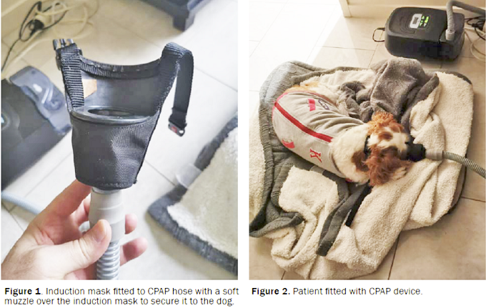
March 2021:
Cavalier is diagnosed with first canine case of
combination of polyglandular deficiency plus diabetes and Addison's
disease.
 In a
March 2021 article, Japanese veterinarians (Sho Furukawa,
Natsuko Meguri, Kazue Koura, Hiroyuki Koura, Akira Matsuda [right]) report
diangnosing and treating a cavalier King Charles spaniel in the first
reported case of a dog with a combination of
polyglandular deficiency
syndrome (PDS), diabetes mellitus, and
hypoadrenocorticism (Addison's
disease). PDS is defined as a multiple endocrine organ failure,
including the gonads, the endocrine pancreas, and the parathyroid
glands. They report:
In a
March 2021 article, Japanese veterinarians (Sho Furukawa,
Natsuko Meguri, Kazue Koura, Hiroyuki Koura, Akira Matsuda [right]) report
diangnosing and treating a cavalier King Charles spaniel in the first
reported case of a dog with a combination of
polyglandular deficiency
syndrome (PDS), diabetes mellitus, and
hypoadrenocorticism (Addison's
disease). PDS is defined as a multiple endocrine organ failure,
including the gonads, the endocrine pancreas, and the parathyroid
glands. They report:
"The dog was diagnosed with diabetic ketoacidosis based on hyperglycemia and renal glucose and ketone body loss. The dog’s condition improved on intensive treatment of diabetes mellitus; daily subcutaneous insulin detemir injection maintained an appropriate blood glucose level over half a year. However, the dog’s body weight gradually decreased from day 207, and on day 501, it presented with a decreased appetite; the precise cause could not be determined. Based on mild hyponatremia and hyperkalemia, hypoadrenocorticism was suggested; the diagnosis was made using an adrenocorticotropic hormone stimulation test. Daily fludrocortisone with low-dose prednisolone oral administration resulted in poor recovery of the blood chemistry abnormalities; however, monthly desoxycorticosterone pivalate (DOCP) subcutaneous injection with daily low-dose prednisolone oral administration helped in the significant recovery of the abnormalities. Therefore, clinicians should consider the possibility of coexistence of hypoadrenocorticism in dogs with diabetes mellitus presenting with undifferentiated weight loss. Additionally, DOCP (not fludrocortisone) may be useful in treating dogs with diabetes mellitus complicated with hypoadrenocorticism."
March 2021:
Cavalier is successfully surgically treated for necrotizing
fasciitis.
 In
a
March 2021 article, two New York clinicians (Alicia Mastrocco,
Jennifer Prittie) reported on the diagnosis and treatment for bacterial
infections of necrotizing fasciitis (NF) in three dogs. One of the dogs
was a 4.5 year old female cavalier King Charles spaniel, with symptoms
which included inappetence, vomiting, diarrhea, dehydration, high
temperature, periodic rapid heartbeat, an indication on x-rays of soft
tissue wisps within the regions of fat beneath the skin, an painful
soft-tissue swelling. The dog underwent three surgeries within two days,
which included removal of the dead latissimus dorsi, additional muscular
necrosis which required "debridement" (removal of dead tissue). By the
sixth day of hospitalization, the dog had improved.
In
a
March 2021 article, two New York clinicians (Alicia Mastrocco,
Jennifer Prittie) reported on the diagnosis and treatment for bacterial
infections of necrotizing fasciitis (NF) in three dogs. One of the dogs
was a 4.5 year old female cavalier King Charles spaniel, with symptoms
which included inappetence, vomiting, diarrhea, dehydration, high
temperature, periodic rapid heartbeat, an indication on x-rays of soft
tissue wisps within the regions of fat beneath the skin, an painful
soft-tissue swelling. The dog underwent three surgeries within two days,
which included removal of the dead latissimus dorsi, additional muscular
necrosis which required "debridement" (removal of dead tissue). By the
sixth day of hospitalization, the dog had improved.
December 2020:
Duration
of pregnancy is shorter in cavaliers.
 In a
December 2020 article by
North Carolina State University researchers (Kristina Baltutis,
Katherine Settle, Theresa Beachler [right], Sara Lyle, C. Scott
Bailey), the durations of 17 pregnancies of 10 cavalier King Charles
spaniel bitches from central North Caolina were compared to those of 17 other
breeds. They report finding that the average CKCS duration was shorter (62.8 ±
2.0 days, with a range of 60 to 66 days) than in the control group (64.5
± 1.4 days, range 62 to 68 days). They concluded that their observation
had clinical implications for pregnancy management of cavlaiers,
including recommendations for scheduling a timed caesarean section and
approaches to managing late-erm complications.
In a
December 2020 article by
North Carolina State University researchers (Kristina Baltutis,
Katherine Settle, Theresa Beachler [right], Sara Lyle, C. Scott
Bailey), the durations of 17 pregnancies of 10 cavalier King Charles
spaniel bitches from central North Caolina were compared to those of 17 other
breeds. They report finding that the average CKCS duration was shorter (62.8 ±
2.0 days, with a range of 60 to 66 days) than in the control group (64.5
± 1.4 days, range 62 to 68 days). They concluded that their observation
had clinical implications for pregnancy management of cavlaiers,
including recommendations for scheduling a timed caesarean section and
approaches to managing late-erm complications.
July 2020:
20-year study of Italian dogs with congenital heart disease
ranks cavaliers 12th among 92 breeds.
 In a
July 2020 article, a team of Italian veterinary researchers (Paola
Giuseppina Brambilla [right], Michele Polli, Danitza Pradelli, Melissa Papa,
Rita Rizzi, Mara Bagardi, Claudio Bussadori) examined the records of
1,377 dogs of 92 breeds (and 191 crossbreeds) which were diagnosed with
congenital heart diseases (CHDs) over the 20-year period from 1997 to
2017. The two most common CHDs were pulmonic stenosis (PS) and patent
ductus arteriosus (PDA). The cavalier King Charles spaniel ranked 12th
among all breeds with CHDs, with 60% of cavaliers diagnosed with PDA and
the remaining 40% with PS. They found that the probability of detecting
a CHD at the first examination was highest among French bulldogs and
cavaliers. Specifically regarding CHDs in cavaliers, they warned that
signs of cavalier puppies' CHDs may be overlooked because murmurs due to
mitral valve disease (which is not a CHD) may be regarded as normal for
the breed. They stated:
In a
July 2020 article, a team of Italian veterinary researchers (Paola
Giuseppina Brambilla [right], Michele Polli, Danitza Pradelli, Melissa Papa,
Rita Rizzi, Mara Bagardi, Claudio Bussadori) examined the records of
1,377 dogs of 92 breeds (and 191 crossbreeds) which were diagnosed with
congenital heart diseases (CHDs) over the 20-year period from 1997 to
2017. The two most common CHDs were pulmonic stenosis (PS) and patent
ductus arteriosus (PDA). The cavalier King Charles spaniel ranked 12th
among all breeds with CHDs, with 60% of cavaliers diagnosed with PDA and
the remaining 40% with PS. They found that the probability of detecting
a CHD at the first examination was highest among French bulldogs and
cavaliers. Specifically regarding CHDs in cavaliers, they warned that
signs of cavalier puppies' CHDs may be overlooked because murmurs due to
mitral valve disease (which is not a CHD) may be regarded as normal for
the breed. They stated:
"It is ... possible that owners do not perceive the clinical signs of some inherited cardiac disorders as problems, but rather as normal, breed-specific characteristics (e.g., murmur in CKCS)."
They concluded that:
"In this context, the importance of creating a network of veterinary cardiology centers that monitor the distribution of a breed and treat the problem of CHDs using the same clinical approach and diagnostic procedures is clear. This approach could be a useful instrument to provide breeders with effective support in implementing the breeding program in order to control the diffusion of CHDs, without impoverishing the genetic pool."
July 2020:
Study of 286 cavaliers finds neutering at any age does not
affect increased likelihood of certain joint disorders or cancers.
 In a
July 2020 article, University of California-Davis researchers
(Benjamin L. Hart [right], Lynette A. Hart, Abigail P. Thigpen,
Neil H. Willits)
reviewed the school's hospital records of over 50,000 dogs to analyze
the increased risks of specific joint disorders and cancers associated
with neutering dogs at various ages. A total of 35 breeds, including
cavalier King Charles spaniels, were included in the study. The joint
disorders were: hip dysplasia (HD), cranial cruciate ligament tears or
ruptures (CCL), and elbow dysplasia (ED); the cancers were
lymphoma/lymphosarcoma (LSA), hemangiosarcoma (HSA), mast cell tumors
(MCT), and osteosarcoma (OSA). The CKCS results were:
In a
July 2020 article, University of California-Davis researchers
(Benjamin L. Hart [right], Lynette A. Hart, Abigail P. Thigpen,
Neil H. Willits)
reviewed the school's hospital records of over 50,000 dogs to analyze
the increased risks of specific joint disorders and cancers associated
with neutering dogs at various ages. A total of 35 breeds, including
cavalier King Charles spaniels, were included in the study. The joint
disorders were: hip dysplasia (HD), cranial cruciate ligament tears or
ruptures (CCL), and elbow dysplasia (ED); the cancers were
lymphoma/lymphosarcoma (LSA), hemangiosarcoma (HSA), mast cell tumors
(MCT), and osteosarcoma (OSA). The CKCS results were:
"The study population was 51 intact males, 72 neutered males, 87 intact females, and 76 spayed females, for a sample size of 286 cases. Joint disorders: hip dysplasia (HD), cranial cruciate ligament rupture (CCL), and elbow dysplasia (ED). For males and females left intact, the occurrences of one or more joint disorders were just 1-4 percent. For males and females neutered at any age, there was no noteworthy increase of joint disorder occurrence, undoubtedly reflecting the small body size. Cancers: lymphoma (LSA), mast cell tumor (MCT), hemangiosarcoma (HSA), and osteosarcoma (OSA). In intact males, the occurrence of cancers was 2 percent and zero in intact females. For both sexes, neutering was not associated with any increase in this measure. Mammary Cancer (MC), Pyometra (PYO) and Urinary Incontinence (UI) in Females. The occurrence of MC in females left intact was zero. There was no occurrence of UI in spayed females. PYO was diagnosed in 2 percent of intact females. Bottom line. Lacking a noticeable occurrence of increased joint disorders or cancers with neutering males or females, those wishing to neuter should decide on the appropriate age."
The study included intervertebral disc disorders (IDD) in only two breeds, the Corgi and Dachshund, even though the cavalier also is widely regarded as being prone to a form of IDD known as Hansen type I disc extrusions. The authors reported that for male Corgis neutered before age 6 months, 18% developed IDD. For the Dachsund, no increase in IDD cases was noted in neutered dogs.
EDITOR'S NOTE: This study was limited to certain specified joint disorders and cancers. It did not include any other known consequences of neutering prior to the dog reaching physical maturity.
July 2020:
Two Belgian cavaliers are diagnosed with epiglottic retroversion
and successfully treated.
 In
a June 2020 article,
Belgian veterinarians (Katrijn Van Ginneken [right], B. Van
Goethem, N. Devriendt, T. Bosmans, H. de Rooster) report details of
epiglottic retroversion (ER) in two cavaliers and seven other small
dogs. Both cavaliers had concurrent brachycephalic airway obstruction
syndrome (BAOS) and one also had laryngeal paralysis. One was treated
with a temporary epiglottopexy and the other with a unilateral
cricoarytenoid lateralisation. Both dogs had excellent long-term
outcomes.
In
a June 2020 article,
Belgian veterinarians (Katrijn Van Ginneken [right], B. Van
Goethem, N. Devriendt, T. Bosmans, H. de Rooster) report details of
epiglottic retroversion (ER) in two cavaliers and seven other small
dogs. Both cavaliers had concurrent brachycephalic airway obstruction
syndrome (BAOS) and one also had laryngeal paralysis. One was treated
with a temporary epiglottopexy and the other with a unilateral
cricoarytenoid lateralisation. Both dogs had excellent long-term
outcomes.
ER is an intermittent obstruction of the upper respiratory tract in some small breed dogs, including cavaliers. The epiglottis is a leaf-shaped flap in the throat that prevents food from entering the windpipe and the lungs; it is open during breathing, allowing air into the larynx, but during swallowing, it closes to prevent aspiration of food into the lungs. The most common symptoms are chronic intermittent inspiratory high-pitched, wheezing sound (stridor) caused by disrupted airflow, exercise intolerance, shortness of breath (dyspnea), coughing and gagging, reverse sneezing, bluish gums (cyanosis), rapid breathing (tachypnea), and sneezing. ER could be related to other concurrent upper airway disorders, including BAOS, laryngeal paralysis, and tracheal collapse. ER is diagnosed using video-laryngosopy. Diagnosis of ER is confirmed when the epiglottis is not positioned against the base of the tongue at inspiration. Surgical options include a laser-surgical treatment called temporary epiglottopexy, and a procedure called unilateral cricoarytenoid lateralisation which involves tying the windpipe to the wall of the trachea.
June 2020:
Cavaliers ranked 6th among breeds at highest risk of
heat-related illness in the UK in 2016.
 In
a
June 2020 article, UK researchers Emily J. Hall (right),
Anne J. Carter, and Dan G. O’Neill reviewed veterinary records of over
900,000 dogs in the UK in 2016, finding reports of 395 heat-related
(heatstroke) illnesses, with an incidence rate of 0.04%. Clinical signs
of heatstroke included:
In
a
June 2020 article, UK researchers Emily J. Hall (right),
Anne J. Carter, and Dan G. O’Neill reviewed veterinary records of over
900,000 dogs in the UK in 2016, finding reports of 395 heat-related
(heatstroke) illnesses, with an incidence rate of 0.04%. Clinical signs
of heatstroke included:
• panting excessively or continuously despite removal from heat/cessation of exercise;
• collapse not subsequently attributed to another cause (e.g. heart failure, Addison’s);
• stiffness, lethargy or reluctance to move;
• gastrointestinal disturbance including hypersalivation, vomiting or diarrhea;
• neurological dysfunction including ataxia, seizures, coma or death;
• haematological disturbances including petechiae or purpura.
The three main risk factors for heatstroke were found to be breed type, high bodyweight, and older age. Cavalier King Charles spaniels ranked 6th among nine breeds most likely to develop heatstroke, when compared to Labrador retrievers, which were the least likely. The cavalier followed the Chow Chow, bulldog, French bulldog, Dogue de Bordeaux, and greyhound in breed risk for heatstroke. The rest of the top nine breeds were the pug, English springer spaniel, and golden retriever.
April 2020:
Cavalier puppy poisoned by permethrin is successfully treated
with intravenous lipid emulsion (ILE).
 In an
April 2020 case report by New Zealand veterinarians Matthew A Kopke
(left)
and Ivayla D Yozova, a seven-month-old cavalier King Charles spaniel
displayed coarse muscle tremors that progressed to seizures, after
licking a cleaning product containing permethrin. At the time of the
event, the puppu was being treated with prednisone for masticatory
muscle myositis (MMM). The dog was treated with midazolam,
dexmedetomidine, and intravenous lipid emulsion (ILE). This is the first
reported use of ILE as therapy in the successful management of canine
permethrin toxicosis. No further tremors or seizure activity, nor any
adverse effects were observed following administration of ILE therapy.
In an
April 2020 case report by New Zealand veterinarians Matthew A Kopke
(left)
and Ivayla D Yozova, a seven-month-old cavalier King Charles spaniel
displayed coarse muscle tremors that progressed to seizures, after
licking a cleaning product containing permethrin. At the time of the
event, the puppu was being treated with prednisone for masticatory
muscle myositis (MMM). The dog was treated with midazolam,
dexmedetomidine, and intravenous lipid emulsion (ILE). This is the first
reported use of ILE as therapy in the successful management of canine
permethrin toxicosis. No further tremors or seizure activity, nor any
adverse effects were observed following administration of ILE therapy.
April 2020:
An Italian cavalier King Charles spaniel may have
acquired a severe bacterial infection from his infected owner.
 In
a
June 2020 article, Italian veterinary researchers (Francesca Paola Nocera, Luciana
Addante, Loredana Capozzi, Angelica Bianco, Filomena Fiorito, Luisa De
Martino, Antonio Parisi [right]) diagnosed a ten year old male cavalier King
Charles spaniel with severe symptoms of infection in its ears due to the
bacteria Acinetobacter baumannii. A. baumannii is an infection typically
acquired by hospitalized humans. Both the cavalier and its primary
owner, who had a very long hospital stay before the dog exhibited any
symptoms, were found to to have A. baumannii. The dog exhibited symptoms
of severe, chronic discharges of pus from its ear canals. The owner had
no outward signs of the disease, but nasal swab tests confirmed the
owner's bacterial infection. The investigators concluded:
In
a
June 2020 article, Italian veterinary researchers (Francesca Paola Nocera, Luciana
Addante, Loredana Capozzi, Angelica Bianco, Filomena Fiorito, Luisa De
Martino, Antonio Parisi [right]) diagnosed a ten year old male cavalier King
Charles spaniel with severe symptoms of infection in its ears due to the
bacteria Acinetobacter baumannii. A. baumannii is an infection typically
acquired by hospitalized humans. Both the cavalier and its primary
owner, who had a very long hospital stay before the dog exhibited any
symptoms, were found to to have A. baumannii. The dog exhibited symptoms
of severe, chronic discharges of pus from its ear canals. The owner had
no outward signs of the disease, but nasal swab tests confirmed the
owner's bacterial infection. The investigators concluded:
"A. baumannii ... can be considered a zoonotic pathogen and this clinical case demonstrates a possible transmission between human and dog. As the person with an asymptomatic nasal colonization of A. baumannii was hospitalized for a long period before the onset of the symptoms in the dog, we hypothesize that the pet owner could have been the potential reservoir for the canine infection. Therefore, further experimental studies are needed in order to understand if this was an isolated case or if A. baumannii can successfully infect both pets and owners."
March 2020:
Cavaliers account for 25% of dogs treated for symptomatic
gallstones in 8-year Univ. of Glasgow study.
 A
gallstone (cholelith) is a solid mass formed from bile precipitates in
the gallbladder or in the intra-hepatic or extra-hepatic ductal system.
While cholelithiasis is very common in humans, affecting up to 25%,
symptomatic cholelithiasis is much rarer, affecting only 10% of those
diagnosed with gallstones. The overall prevalence of canine
cholelithiasis has been estimated to be between 0.03% and 15%.
Complications of cholelithiasis in dogs include extrahepatic biliary
duct obstruction, chholangitis / cholecystitis, gallbladder perforation,
and cholecystocutaneous fistula.
A
gallstone (cholelith) is a solid mass formed from bile precipitates in
the gallbladder or in the intra-hepatic or extra-hepatic ductal system.
While cholelithiasis is very common in humans, affecting up to 25%,
symptomatic cholelithiasis is much rarer, affecting only 10% of those
diagnosed with gallstones. The overall prevalence of canine
cholelithiasis has been estimated to be between 0.03% and 15%.
Complications of cholelithiasis in dogs include extrahepatic biliary
duct obstruction, chholangitis / cholecystitis, gallbladder perforation,
and cholecystocutaneous fistula.
In a March 2020 article, UK researchers (Patricia M. Ward [right], Kieran Brown, Gawain Hammond, Tim Parkin, Sarah Bouyssou, Genziana Nurra, Alison E. Ridyard ) identified 68 dogs diagnosed with cholelithiasis on ultrasound between January 2010 and June 2018 at the Small Animal Hospital at the University of Glasgow. Of those, the records of 61 dogs out of 31 breeds were reviewable. Cholelithiasis was classified as an incidental finding in 53 (86.9%) dogs, including 4 cavaliers, with 8 (13.1%) dogs being classified as symptomatic, inlcuding 2 cavaliers, having complications of cholelithiasis that included biliary duct obstruction (1 CKCS), biliary peritonitis, emphysematous cholecystitis, and acute cholecystitis (1 CKCS). The breed rankings were, mixed breeds (11 dogs), Labrador retrievers (7 dogs), border collies (5 dogs), Cocker spaniels (4 dogs), and cavaliers (4 dogs).
The two cavaliers diagnosed with symptomatic cholelithiasis were (a) a 4 year old spayed female exhibiting anorexia and jaundice and diagnosed with acute cholelithiasis due to a single stone within the gallbladder, and (b) a 5 year old castrated male exhibiting vomiting, anorexia, and painful abdomen, diagnosed with obstructive cholelithiasis due to a single stone within the bile duct.
January 2020:
Myoclonic jerks are reported in 40 older cavaliers in UK.
In
an abstract presented at the 2017 symposium of the European College
of Veterinary Neurology (ECVN) and
 published
in January 2020, a team of UK veterinary neurologists (C. Rusbridge
[left], P.J.Watson, C.J. Driver, A. Tauro, R.Fernandes, J.Rose, F.
Constantino-Casas, F. Salguero-Bodes) examined the records of 40
cavalier King Charles spaniels displaying myoclonic jerks. Myoclonus is
a syndrome of jerky contractions of groups of muscles, mainly of the
head and forelimbs when sitting or standing. In these cases, the signs
included rapid eyelid blinking, head nodding, shuddering, and exensions
down the limbs resulting in falling or stumbling. The movements lasted
for seconds, usually when the dog was stationary or sitting and did not
lose consciousness. Multiple daily episodes were reported and increased
in frequency and intensity over time. The median age of onset was 9
years, with a main range from 7 to 10 years. They found that the
condition improved with treatment with levetiracetum and possibly
corticosteroids.
published
in January 2020, a team of UK veterinary neurologists (C. Rusbridge
[left], P.J.Watson, C.J. Driver, A. Tauro, R.Fernandes, J.Rose, F.
Constantino-Casas, F. Salguero-Bodes) examined the records of 40
cavalier King Charles spaniels displaying myoclonic jerks. Myoclonus is
a syndrome of jerky contractions of groups of muscles, mainly of the
head and forelimbs when sitting or standing. In these cases, the signs
included rapid eyelid blinking, head nodding, shuddering, and exensions
down the limbs resulting in falling or stumbling. The movements lasted
for seconds, usually when the dog was stationary or sitting and did not
lose consciousness. Multiple daily episodes were reported and increased
in frequency and intensity over time. The median age of onset was 9
years, with a main range from 7 to 10 years. They found that the
condition improved with treatment with levetiracetum and possibly
corticosteroids.
December 2019:
Australian 16-year veterinary school study of facial nerve
paralysis finds cavaliers at the top of the list.
 In
a
December 2019 article by Univeristy of Sydney, Australia researchers
(MK Chan, J-ALML Toribio, JM Podadera, Georgina Child [right])
they reviewed the records of 122 dogs presented at their hospital from
2001 to 2016 which had symptoms of facial nerve paralysis (FNP). FNP can
be a symptom of any of a variety of disorders, including primary
secretory otitis media (PSOM), direct injury, hypothyroidism, and drug
sensitivity. Idiopathic FNP (IFNP) applies to FNP symptoms due to
unknown causes. It is not considered to be effectively treatable. Of the
36 purebred dogs in the entire study, 15 (42%) were cavalier King
Charles spaniels, the highest number of purebreds by far. Of the 15
purebreds diagnosed with IFNP, cavaliers numbered 9 (60%). The authors
concluded:
In
a
December 2019 article by Univeristy of Sydney, Australia researchers
(MK Chan, J-ALML Toribio, JM Podadera, Georgina Child [right])
they reviewed the records of 122 dogs presented at their hospital from
2001 to 2016 which had symptoms of facial nerve paralysis (FNP). FNP can
be a symptom of any of a variety of disorders, including primary
secretory otitis media (PSOM), direct injury, hypothyroidism, and drug
sensitivity. Idiopathic FNP (IFNP) applies to FNP symptoms due to
unknown causes. It is not considered to be effectively treatable. Of the
36 purebred dogs in the entire study, 15 (42%) were cavalier King
Charles spaniels, the highest number of purebreds by far. Of the 15
purebreds diagnosed with IFNP, cavaliers numbered 9 (60%). The authors
concluded:
"This study found differences in the incidence of FNP causes compared with the only previous study, which are presumed to be a result of geographic and temporal differences. Breeds and age groups are significant risk factors correlated to acquisition of FNP and IFNP. Vestibular dysfunction, hypersalivation and keratoconjunctivitis sicca were common findings in dogs with IFNP. Dogs with IFNP have guarded prognosis for recovery, and often require life-long eye lubrication. There is no evidence to date that treatment alters the outcome in dogs with IFNP."
December 2019:
Canadian dentists recommend adding xylitol to dogs' drinking
water for tooth health.
 Here
is one for the books! We all know that xylitol (a sweetner substitute
for sugar in chewing gum and many food items) is poisonous to dogs,
right? Even small amounts of xylitol can cause seizures due to
hypoglycemia (low blood sugar), liver failure, and death. Doses as low
as 50 milligrams per pound of body weight can cause seizures.
Here
is one for the books! We all know that xylitol (a sweetner substitute
for sugar in chewing gum and many food items) is poisonous to dogs,
right? Even small amounts of xylitol can cause seizures due to
hypoglycemia (low blood sugar), liver failure, and death. Doses as low
as 50 milligrams per pound of body weight can cause seizures.
Well, now a study in the January 2020 issue of the Canadian Veterinary Journal is recommending that this same highly toxic poison be added to dogs' drinking water to reduce plaque and calculus on the dogs' teeth. In "Pilot study of the effectiveness of a xylitol-based drinking water additive to reduce plaque and calculus accumulation in dogs", Canadian veterinary dental specialists Drs. Candace Lowe (right) and James Anthony write:
"Over a period of 208 days a randomized, double-blind clinical trial was conducted to assess plaque and calculus accumulation in dogs provided with a xylitol-based drinking water additive. A crossover design was utilized allowing each dog to participate in each 90-day treatment and control phase. Inclusion of a xylitol drinking water additive resulted in a 5.1% decrease in mean tooth plaque score and a 14.9% decrease in mean calculus score. Daily administration of a palatable, xylitol drinking water additive that required little time and effort reduced plaque and calculus accumulation in dogs."
The authors acknowledge the toxicity of xylitol in high doses (>100 mg/kg of body weight), but they note that this water-soluable form of xylitol of 50 mg. would be far less than the toxicity level for even a 2 kg. dog.
November 2019:
Cornell study finds cavalier puppies have increased odds for
cleft lips and palates.
 Orofacial
clefts -- cleft lip (CL), cleft palate (CP) and cleft lip and palate
(CLP) -- are abnormal fissures of the mouth or face which occur due to
incomplete fusion of tissues during embryonic development. In a
November 2019 article, Cornell University veterinary researchers
(Nicholas Roman, Patrick C. Carney, Nadine Fiani, Santiago Peralta
[right]) reseached the incidence of orofacial clefts in 7,429 live
births of purebred puppies of 78 AKC-recognized breeds in the USA during
a 12 month period. In those litters, 228 puppies had orofacial clefts
(3%), of which there were 59 CLS (26%), 134 CPs (59%) and 35 CLFs (15%).
The cavalier King Charles spaniel ranked third behind the Boston terrier
and French bulldog in having significantly increased odds of CL and of
CLP. Of 134 live births of cavaliers, there was a total of 10 CLs and
CLPs, amounting to 7.5% of the puppies.
Orofacial
clefts -- cleft lip (CL), cleft palate (CP) and cleft lip and palate
(CLP) -- are abnormal fissures of the mouth or face which occur due to
incomplete fusion of tissues during embryonic development. In a
November 2019 article, Cornell University veterinary researchers
(Nicholas Roman, Patrick C. Carney, Nadine Fiani, Santiago Peralta
[right]) reseached the incidence of orofacial clefts in 7,429 live
births of purebred puppies of 78 AKC-recognized breeds in the USA during
a 12 month period. In those litters, 228 puppies had orofacial clefts
(3%), of which there were 59 CLS (26%), 134 CPs (59%) and 35 CLFs (15%).
The cavalier King Charles spaniel ranked third behind the Boston terrier
and French bulldog in having significantly increased odds of CL and of
CLP. Of 134 live births of cavaliers, there was a total of 10 CLs and
CLPs, amounting to 7.5% of the puppies.
March 2019:
Cavalier pup's artery fistulas are closed in surgery using
transcatheter embolization coils and silk sutures. In a
March 2019 article, a team of veterinary clinicians from three USA
universities (R.L. Winter, J.A. Horton, D.K. Newhard, M. Holland)
diagnosed and repaired two artery fistulas (abnormal connections to the
artery) by
 transcatheter
embolization to block the flow of blood through the fistulas. The dog
was a 4-month-old cavalier King Charles spaniel (CKCS) which had grade 2
murmur at the base of the heart (basilar). X-rays showed mild
enlargement of the left atrium. Echocardiography "suggested anomalous
vessels around the main pulmonary artery". The pulse of the femoral
arterial pulse was "hyperdynamic". Computed tomography angiography (CTA)
showed two systemic-to-pulmonary artery fistulas (SPAFs). Fistulas are
abnormal shunts leading from the artery and by-passing it. But for the
dog's breed's history of mitral valve disease, corrective surgery may
not have been performed. However, since the dog was a CKCS, her owner
elected to have transcatheter embolization surgery performed.
"Transcatheter" means that the surgical devices were delivered to the
sites of the fistulas through the artery by inserting a tubular device.
"Embolization" means to lodge an object in the blood vessel to obstruct
the offending blood flow. The surgeons inserted embolization coils
(see photo at right) and silk suture threads through the left
femoral artery to the fistulas, guided by CTA. (See Fig. 4 below).
The surgical procedure was successful; complee blockage into the
fistulas was observed; vertebral heart size decreased; the left atrium
size decreased; there no longer was a murmur.
transcatheter
embolization to block the flow of blood through the fistulas. The dog
was a 4-month-old cavalier King Charles spaniel (CKCS) which had grade 2
murmur at the base of the heart (basilar). X-rays showed mild
enlargement of the left atrium. Echocardiography "suggested anomalous
vessels around the main pulmonary artery". The pulse of the femoral
arterial pulse was "hyperdynamic". Computed tomography angiography (CTA)
showed two systemic-to-pulmonary artery fistulas (SPAFs). Fistulas are
abnormal shunts leading from the artery and by-passing it. But for the
dog's breed's history of mitral valve disease, corrective surgery may
not have been performed. However, since the dog was a CKCS, her owner
elected to have transcatheter embolization surgery performed.
"Transcatheter" means that the surgical devices were delivered to the
sites of the fistulas through the artery by inserting a tubular device.
"Embolization" means to lodge an object in the blood vessel to obstruct
the offending blood flow. The surgeons inserted embolization coils
(see photo at right) and silk suture threads through the left
femoral artery to the fistulas, guided by CTA. (See Fig. 4 below).
The surgical procedure was successful; complee blockage into the
fistulas was observed; vertebral heart size decreased; the left atrium
size decreased; there no longer was a murmur.
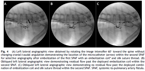
November 2018:
Cavalier's epilepsy is aggravated by tick/flea anti-parasite
drug fipronil and treated with levetiracetam and gabapentin.
 In
an
October 2018 article, a team of Turkish veterinarians (Sinan Kandır
[right], Cengiz Yüce Kayabek, Bilge Kaan Tekelioğlu) report on
the diagnosis and treatment of a 3-year-old neutered female cavalier
King Charles spaniel diagnosed with mitral valve disease which began to
experience epileptic seizures. The epilepsy first was treated with
levetiracetam, which resulted in the epileptic seizures disappearing.
Then, three months later, the cavalier was treated for fleas and ticks
with fipronil (Frontline TopSpot, Fiproguard, Flevox, PetArmor). The
epileptic attacks "dramatically increased and became more severe just
after the fipronil treatment (~7 times per day)." Gabapentin was added
to the levetiracetam treatment protocol, and the epileptic seizures. The
researchers concluded:
In
an
October 2018 article, a team of Turkish veterinarians (Sinan Kandır
[right], Cengiz Yüce Kayabek, Bilge Kaan Tekelioğlu) report on
the diagnosis and treatment of a 3-year-old neutered female cavalier
King Charles spaniel diagnosed with mitral valve disease which began to
experience epileptic seizures. The epilepsy first was treated with
levetiracetam, which resulted in the epileptic seizures disappearing.
Then, three months later, the cavalier was treated for fleas and ticks
with fipronil (Frontline TopSpot, Fiproguard, Flevox, PetArmor). The
epileptic attacks "dramatically increased and became more severe just
after the fipronil treatment (~7 times per day)." Gabapentin was added
to the levetiracetam treatment protocol, and the epileptic seizures. The
researchers concluded:
"Fipronil, phenylpyrazole origin and other antiparasitic drugs should be used very carefully and under veterinary consultant supervision due to their side effects especially in epileptic dogs. Finally, gabapentin could be a good option for the alternative add-on therapy to control epileptic seizures in CKCSs."
June 2018:
Periodontal disease is suspected cause of bacterial
meningoencephalomyelitis in a cavalier.
 In
a
June 2018 article, a team of PennVet clinicians (Alexander E. Tun
[right], Leontine Benedicenti, Evelyn M. Galban) diagnosed
meningoencephalomyelitis caused by the bacteria Pasteurella multocida in
a 5-year-old cavalier King Charles spaniel. (Meningoencephalomyelitis
[MEM] is inflammation of the brain, including its protective membranes,
and of the spinal cord, caused by a bacteria.) The dog had severe
periodontal disease, with contact ulceration and possible necrotizing
stomatitis. Her symptoms included fever, lethargy, inappetence, and
multifocal neurologic signs, mainly dull mentation. Based upon
examination of her cerebrospinal fluid (CSF) and blood culture, and her
response to therapy (anti-emetics and gastroprotectants, an opioid for
analgesia, and dexamethasone sodium phosphate as an antiinflammatory),
P. multocida meningoencephalomyelitis was diagnosed. They opine that the
severe periodontal disease led to a bacteremia causing hematogenous
seeding of a bacterial meningitis originating at the disrupted
blood–spinal cord barrier.They concluded:
In
a
June 2018 article, a team of PennVet clinicians (Alexander E. Tun
[right], Leontine Benedicenti, Evelyn M. Galban) diagnosed
meningoencephalomyelitis caused by the bacteria Pasteurella multocida in
a 5-year-old cavalier King Charles spaniel. (Meningoencephalomyelitis
[MEM] is inflammation of the brain, including its protective membranes,
and of the spinal cord, caused by a bacteria.) The dog had severe
periodontal disease, with contact ulceration and possible necrotizing
stomatitis. Her symptoms included fever, lethargy, inappetence, and
multifocal neurologic signs, mainly dull mentation. Based upon
examination of her cerebrospinal fluid (CSF) and blood culture, and her
response to therapy (anti-emetics and gastroprotectants, an opioid for
analgesia, and dexamethasone sodium phosphate as an antiinflammatory),
P. multocida meningoencephalomyelitis was diagnosed. They opine that the
severe periodontal disease led to a bacteremia causing hematogenous
seeding of a bacterial meningitis originating at the disrupted
blood–spinal cord barrier.They concluded:
"In the future, be aware that a fever with multifocal neurologic signs and severe periodontal disease in a canine patient may suggest a P. multocida infection and both CSF and blood cultures can be submitted for confirmation."
April
2018:
Cavalier with severe periodontal disease develops Pasteurella
Multocida meningoencephalomyelitis.
In an
April 2018 article, a team of University of Pennsylvania
neurologists (Alexander E. Tun [right], Leontine Benedicenti,
Evelyn M. Galban) report on a 5-year-old spayed female cavalier King
Charles spaniel with severe periodontal disease which had developed
Pasteurella Multocida meningo-encephalomyelitis causing signs of a
neurological disorder. Based on cerebrospinal fluid and blood culture,
as well as response to therapy, they opine that the severe periodontal
disease led to a bacteremia causing hematogenous seeding of a bacterial
meningitis originating at the disrupted blood–spinal cord barrier.
April 2018:
Cavaliers rank 11th in diagnosis of arthritis in UK 2013 study
of most frequently affected breeds.
 In
an
April 2018 article, UK researchers (Katharine L. Anderson, Dan G.
O’Neill, David C. Brodbelt, David B. Church, Richard L. Meeson, David
Sargan, Jennifer F. Summers, Helen Zulch, Lisa M. Collins [right])
examining all reported cases of osteoarthritis in a UK dog population
under primary veterinary care during 2013, found that 2.42% were
diagnosed with osteoarthritis. This made the CKCS the 11th most
frequently affected breed in the study. Of 10,143 cavaliers in the
study, 245 were diagnosed with osteoarthritis. The most frequently
affected breeds were: (1) golden retriever, (2) Labrador retriever, (3)
Rottweiler, (4) German shepherd, and (5) border collie. The overall
average of all dogs diagnosed with osteoarthritis was 2.5%.
In
an
April 2018 article, UK researchers (Katharine L. Anderson, Dan G.
O’Neill, David C. Brodbelt, David B. Church, Richard L. Meeson, David
Sargan, Jennifer F. Summers, Helen Zulch, Lisa M. Collins [right])
examining all reported cases of osteoarthritis in a UK dog population
under primary veterinary care during 2013, found that 2.42% were
diagnosed with osteoarthritis. This made the CKCS the 11th most
frequently affected breed in the study. Of 10,143 cavaliers in the
study, 245 were diagnosed with osteoarthritis. The most frequently
affected breeds were: (1) golden retriever, (2) Labrador retriever, (3)
Rottweiler, (4) German shepherd, and (5) border collie. The overall
average of all dogs diagnosed with osteoarthritis was 2.5%.
January 2018:
UK study of 486 dogs shows three years between core vaccine
boosters provide long-lived immunity.
 In
a
January 2018 article by a team of UK researchers (R. Killey, C.
Mynors, R. Pearce, A. Nell, A. Prentis, Michael J. Day [right]), they
used an in-practice test kit to detect protective serum antibody against
canine distemper virus, canine adenovirus and canine parvovirus type 2
in 486 dogs of various breeds. They report finding that 93.6% of the
dogs had protective antibody against all three of the core vaccine
antigens. They surmisded that the small number of other dogs may have
had waning of previous serum antibody or may have been rare genetic
non-responders to that specific antigen. They concluded that:
In
a
January 2018 article by a team of UK researchers (R. Killey, C.
Mynors, R. Pearce, A. Nell, A. Prentis, Michael J. Day [right]), they
used an in-practice test kit to detect protective serum antibody against
canine distemper virus, canine adenovirus and canine parvovirus type 2
in 486 dogs of various breeds. They report finding that 93.6% of the
dogs had protective antibody against all three of the core vaccine
antigens. They surmisded that the small number of other dogs may have
had waning of previous serum antibody or may have been rare genetic
non-responders to that specific antigen. They concluded that:
"UK veterinarians can be reassured that triennial revaccination of adult dogs with core vaccines provides long-lived protective immunity. In-practice serological test kits are a valuable tool for informing decision-making about canine core revaccination."
October
2017:
Cavalier with facial nerve paralysis is successfully treated
with laser treatment at acupuncture points.
 In an
October 2017 abstract, Dr. Dustin Dees (right) of Austin,
Texas reports successfully treating a cavalier King Charles spaniel
suffering from idiopathic facial nerve paralysis by using continuous
wave laser treatments at four acupuncture sites corresponding to a
branch of the facial nerve. The method is called "photobiomodulation".
Treatments were performed twice a week for five weeks. The results were
successful. The dog's blinking ability substantially improved, and
corneal dryness resolved with marked improvement of the dog's Schirmer
testing.
In an
October 2017 abstract, Dr. Dustin Dees (right) of Austin,
Texas reports successfully treating a cavalier King Charles spaniel
suffering from idiopathic facial nerve paralysis by using continuous
wave laser treatments at four acupuncture sites corresponding to a
branch of the facial nerve. The method is called "photobiomodulation".
Treatments were performed twice a week for five weeks. The results were
successful. The dog's blinking ability substantially improved, and
corneal dryness resolved with marked improvement of the dog's Schirmer
testing.
October 2017:
Cavaliers were at highest risk of severe reaction to
incompatible blood transfusions, in Italian study.
 In a
September 2017 article by a team of Italian veterinary blood
specialists (Anyela Andrea Medina Valentin [right], Alessandra Gavazza, George
Lubas), they studied 7,414 dogs, including 103 cavalier King Charles
spaniels, to determine the potential sensitization risk to dogs
receiving blood transfusions consisting of the Dog Erythrocyte Antigen
(DEA) 1 blood group.
In a
September 2017 article by a team of Italian veterinary blood
specialists (Anyela Andrea Medina Valentin [right], Alessandra Gavazza, George
Lubas), they studied 7,414 dogs, including 103 cavalier King Charles
spaniels, to determine the potential sensitization risk to dogs
receiving blood transfusions consisting of the Dog Erythrocyte Antigen
(DEA) 1 blood group.
EDITOR'S NOTE: Dogs have natural occuring antibodies against the DEA 1 antigen in red blood cells (erythrocytes or RBCs). In a dog with DEA 1 negative RBCs and with an immune system which is highly sensitized to DEA 1 positive RBCs, the system will produce alloantibodies following the first transfusion of DEA 1 positive blood. Following a second transfusion of DEA 1 positive blood, a dog which is highly sensitized to DEA 1 positive may develop an acute reaction, including fever, pigmenturia, and lethargy, and its packed cell volume (PCV) will not rise as expected. Death may result from such a mis-matched second transfusion. See this May 1995 article.
In this Italian study, they found that CKCSs were among the few breeds having the highest percentage likelihood to become sensitized (25.0%) following the first mis-matched transfusion, and that cavaliers also were among the breeds having the highest risk (6.2%) of an acute transfusional reaction following a second such transfusion. They therefore recommend:
"Blood typing to identify the presence of DEA 1 and the cross-match to establish full compatibility should be performed before each transfusion in order to reduce the risk of sensitization or immunological reaction between donor and recipient dogs."
March 2017: Two cavaliers are among 14 Texas dogs poisoned by eating sago palm seeds. In a May 2017 article, the researchers (Carolyn Clarke, Derek Burney) report on 14 Texas dogs (including two cavaliers) which suffered toxicosis from eating the seeds or other parts of the cycad (sago) palm tree. Nine of the 14 dogs died as a direct result of cycad palm intoxication. Three of the five survivors had persistently elevated liver enzymes, indicating liver damage. Serum albumin levels and nadir platelet counts were significantly lower in nonsurvivors compared to survivors. The article does not indicate in which category the CKCSs fell.
December 2016:
USA Study of 90,000 dogs show neutering raises risk of several
autoimmune diseases.
 In a
December 2016 article, a team of University of California-Davis
reasearchers (Crystal R. Sundburg, Janelle M. Belanger, Danika L.
Bannasch, Thomas R. Famula, Anita M. Oberbauer [right]) examined the medical
records from 1995 to 2010 of 90,090 dogs of all AKC-recognized breeds
treated at their veterinary hospital, to determine the relation between
neuter status and autoimmune diseases. They report finding that neutered
dogs had a significantly greater risk of:
In a
December 2016 article, a team of University of California-Davis
reasearchers (Crystal R. Sundburg, Janelle M. Belanger, Danika L.
Bannasch, Thomas R. Famula, Anita M. Oberbauer [right]) examined the medical
records from 1995 to 2010 of 90,090 dogs of all AKC-recognized breeds
treated at their veterinary hospital, to determine the relation between
neuter status and autoimmune diseases. They report finding that neutered
dogs had a significantly greater risk of:
• atopic dermatitis (ATOP)
• autoimmune hemolytic anemia (AIHA)
• hypoadrenocorticism (ADD)
• hypothyroidism (HYPO)
• immune-mediated thrombocytopenia (ITP)
• inflammatory bowel disease (IBD)
than intact dogs with neutered females being at greater risk than neutered males for all but AIHA and ADD. Neutered females, but not males, had a significantly greater risk of lupus erythematosus (LUP) than intact females. Pyometra was a greater risk for intact females.
November 2016:
Cavaliers were 15+% of dogs with erosive immune-mediated
polyarthritis (IMPA).
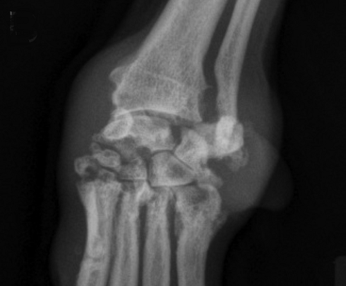 In
a
November 2016 article, a team of Wisconsin and Michigan researchers
(Magen L. Shaughnessy, Susannah J. Sample, Carter Abicht, Caitlin
Heaton, Peter Muir) studied the clinical records of 79 dogs
diagnosed with immune-mediated polyarthritis (IMPA). Of those 79 dogs,
13 had erosive IMPA, of which, two were cavalier King Charles spaniels.
The 13 affected dogs had erosive lesions in their carpal joints. The
estimated median synovial fluid lymphocyte count for dogs with erosive
IMPA was significantly greater than that for dogs with nonerosive IMPA.
Results indicated erosive IMPA most commonly affected the carpal joints
of middle-aged small-breed dogs. (See left carpus of an 8-year-old
spayed female Cavalier King Charles Spaniel with subjectively moderate
subchondral bone lysis accompanied by severe synovial swelling -- in
x-ray at right.)
In
a
November 2016 article, a team of Wisconsin and Michigan researchers
(Magen L. Shaughnessy, Susannah J. Sample, Carter Abicht, Caitlin
Heaton, Peter Muir) studied the clinical records of 79 dogs
diagnosed with immune-mediated polyarthritis (IMPA). Of those 79 dogs,
13 had erosive IMPA, of which, two were cavalier King Charles spaniels.
The 13 affected dogs had erosive lesions in their carpal joints. The
estimated median synovial fluid lymphocyte count for dogs with erosive
IMPA was significantly greater than that for dogs with nonerosive IMPA.
Results indicated erosive IMPA most commonly affected the carpal joints
of middle-aged small-breed dogs. (See left carpus of an 8-year-old
spayed female Cavalier King Charles Spaniel with subjectively moderate
subchondral bone lysis accompanied by severe synovial swelling -- in
x-ray at right.)
November 2016: French study adds cavaliers to list of breeds predisposed to facial nerve paralysis. In a November 2016 abstract, French researchers studied 69 dogs with facial nerve paralysis. The report does not provide a count of cavalier King Charles spaniels included in the study, but the researchers concluded that the CKCS and the French bulldog should be added to the list of predisposed breeds to the disorder. Additionally, they found:
"Idiopathic facial paralysis was diagnosed in 48% of dogs. Vestibular signs were the most common additional clinical signs and were observed in 36% of dogs with idiopathic facial paralysis. Peripheral nervous system disease was diagnosed in 19% of dogs, and central nervous system disease occurred in 30% of dogs. ... Improved diagnostic methods enabled the diagnosis of a higher percentage of inflammatory/infectious diseases, which were absent in the central nervous system aetiologies of a previous similar study, and revealed metabolic (hypothyroidism), inflammatory and neoplastic aetiologies for peripheral nervous system disease."
November 2016:
French study of 1,668 cavalier bitches shows birth, still-born,
and post natal mortality rates.
 In an
October
2016 report by a team of French reproduction specialists (Sylvie
Chastant-Maillard [right], C Guillemot, A Feugier, C Mariani, A Grellet, H
Mila), they studied 2,561 heats of 1,668 cavalier King Charles spaniels
in France. They found:
In an
October
2016 report by a team of French reproduction specialists (Sylvie
Chastant-Maillard [right], C Guillemot, A Feugier, C Mariani, A Grellet, H
Mila), they studied 2,561 heats of 1,668 cavalier King Charles spaniels
in France. They found:
Number of puppies born per litter: 4.4 ± 2.0
Stillbirth rate: 8.8%
Post-natal mortality rate: 7.5%
Number of puppies surviving per whelping: 3.7 ± 2.0
September 2016: Cavaliers show temporomandibular joint dysplasia in a Brazilian study. In an August 2016 study, eleven cavalier King Charles spaniels were included in a review of the temporomandibular joints (TMJs) in 48 dogs, examining the morphologic and morphometric features of the TMJ using computed tomography (CT). (The TMJ is the hinge of the jaw that connects the jaw to the temporal bones of the skull.) The researchers found:
"The TMJs of Cavalier King Charles spaniels showed remarkably lower values associated with shallow mandibular fossae and incongruent articular heads.
"Cavalier King Charles spaniels had the shallowest mandibular fossae of all breeds. The retroarticular process appeared as a bony extension on the medioventral aspect of the mandibular fossa, articulating with the caudomedial aspect of the condylar process. This bony extension was prominent in Labrador retrievers, German shepherds, boxers, and English bulldogs. The retroarticular process was less developed in cocker spaniels, shih tzus, and pug. In Cavalier King Charles spaniels, it was small or absent.
"The wide angles documented in Labrador retrievers, German shepherds, boxers, and English bulldogs were consistent with long and well-defined retroarticular processes, whereas the narrow angles found in cocker spaniels, shih tzus, Cavalier King Charles spaniels, and a pug were due to less developed or absent retroarticular processes. A dramatically reduced ventral extension of the retroarticular process, as observed in cocker spaniels and Cavaliers King Charles spaniels in this study, may permit slight caudal dislocation of the head of the condylar process and predispose to TMJ instability.
"German shepherds, boxers, and English bulldogswere consistent with deeper (more concave) mandibular fossae and more prominent retroarticular processes, whereas the narrow angles found in cocker spaniels, shih tzus, CavalierKing Charles spaniels, and the pug were due to more shallower (less concave) mandibular fossae and less prominent or absent retroarticular processes.
"The present study agrees with the asymptomatic dysplastic condition of the TMJ previously reported in the Cavalier King Charles spaniel and suggests that this morphological features may exist in certain dog breeds such as cocker spaniels and small breeds such as shih tzus and pugs."
Diagram C of Figure 11 below shows the severe shallowness of the CKCS's mandibular fossa, compared to those of the Labrador retriever, German shepherd boxer, and English bulldog (diagram A) and those of the Cocker spaniel, pug, and Shih Tzus (diagram C).
July 2016: Gestation period of the CKCS is found to be shorter than that of the average canine bitch. In a July 2016 poster abstract, a team of N.C. State veterinary theriogenology researchers (K. S. Baltutis, T. M. Beachler, S. K. Lyle, C. S. Bailey) compared the gestation periods of 17 cavaliers and 17 bitches of other breeds. They found that the CKCSs' average gestation period of 62.8 days was 3.2% shorter than the reported canine gestation length of 65 days and roughly two days shorter than that of the control group. They concluded that the implications of this study include recommendations for scheduling a timed Caesarean section and approaches to managing late-term complications. See, also this June 2016 post.
July 2016:
Dr. Simon Swift inserts wire stent to relieve pulmonic stenosis
in a Havanese. In a
July
2016 newspaper
article, veterinary cardiologist Simon Swift of the University of
Florida's veterinary college reported a case of a Havanese spaniel with
pulmonic stenosis which for which a valvuloplasty procedure was
unsuccessful because the dog had an "unusually thick right ventricle and
abnormal valve". Dr. Swift and a surgical team, which included the
university's pediatric cardiology specialists, put a stent, a metal tube
made of wire mesh (see photo at left), on a balloon and entered
the chest straight into the heart. Then, they expanded the balloon to
open the stent and relieve the blockage in his artery, Swift said.
July 2016:
Thai veterinarians successfully relieve pulmonic stenosis in a
cavalier, using valvulopasty. In a
July 2016 article, a team of Thailand veterinary surgeons and other
specialists (Chollada Buranakarl, Anusak Kijtawornrat, Wasan
Udayachalerm, Sirilak Disatian Surachetpong, Saikaew Sutayatram,
Pasakorn Briksawan, Sumit Durongphongtorn, Rampaipat Tungjitpeanpong,
Nardtiwa Chaivoravitsakul) report a successful insertion of a balloon in
the pulmonary valves of three dogs (valvuloplasty), including a cavalier King
Charles
spaniel (#2 in the report), affected by pulmonic stenosis. Pulmonic
stenosis is a common congenital cardiac defect, characterized by the
narrowing and obstruction of blood through the heart's pulmonary valve,
which connects the pulmonary artery to the right ventricle chamber. In
this study, the balloon was inserted by a catheter and then inflated at
the location of the narrowing of the pulmonary valve. (See examples
of balloon catheters at right.) Once the balloon was inflated, the
narrowing of the valve disappeared and the pressure through the valve
greatly decreased in the CKCS.
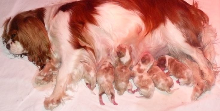 June 2016:
Study of six cavalier pregnancies shows average ovulation periods and
progesterone concentrations. In a June 2016 article
comparing the progesterone (P4) concentrations of cavalier King Charles
spaniels (CKCS) and Bernese mountain dogs, the Danish researchers (K. Thejll
Kirchhoff, S. Goericke-Pesch) found that six CKCSs delivered from one
and five puppies, between 62 and 65 days after ovulation. The bitches' P4
concentrations continuously decreased from the first to the last
sampling during pregnancy.
June 2016:
Study of six cavalier pregnancies shows average ovulation periods and
progesterone concentrations. In a June 2016 article
comparing the progesterone (P4) concentrations of cavalier King Charles
spaniels (CKCS) and Bernese mountain dogs, the Danish researchers (K. Thejll
Kirchhoff, S. Goericke-Pesch) found that six CKCSs delivered from one
and five puppies, between 62 and 65 days after ovulation. The bitches' P4
concentrations continuously decreased from the first to the last
sampling during pregnancy.
November 2015: Study shows cavalier puppies' emotional and behavioral development are later than two other breeds. In a July 2015 study of the level of fear avoidance among puppies of different breeds, it has been shown that cavalier puppies from 4 to 10 weeks of age exhibited a later onset of fear-related avoidance behavior when compared with German shepherd dog puppies and Yorkshire terrier puppies. In the same study, CKCSs demonstrated the highest incidence of crouching in response to a loud noise, followed by the Yorkshire terriers. Breed differences in puppy mobility were observed beginning at 6 weeks of age, with German shepherd dogs demonstrating the most mobility and cavaliers the least. The authors concluded that the results of their study support the hypothesis that emotional and behavioral development, as well as the onset of fear-related avoidance behavior, varies among breeds of domestic dogs.
September 2015:
International Veterinary Epilepsy Task Force reports on
myoclonus in
cavaliers.
 In a
September 2014 article, the International
Veterinary Epilepsy Task Force, consisting of neurologists and other
veterinary specialists from the US, UK, and throughout Europe
(Velia-Isabel Hülsmeyer, Andrea Fischer, Paul J.J. Mandigers [right], Luisa
DeRisio, Mette Berendt, Clare Rusbridge, Sofie F.M. Bhatti, Akos
Pakozdy, Edward E. Patterson, Simon Platt, Rowena M.A. Packer, Holger A.
Volk) issues their massive "current understanding of idiopathic epilepsy
of genetic or suspected genetic origin in purebred dogs", including, of
course, the cavalier King Charles spaniel. Their primary findings
regarding the CKCS included "myoclonus" (spasmodic jerky
contraction of groups of muscles) as a potential breed-specific disease
which may mimic idiopathic epilepsy. They stated:
In a
September 2014 article, the International
Veterinary Epilepsy Task Force, consisting of neurologists and other
veterinary specialists from the US, UK, and throughout Europe
(Velia-Isabel Hülsmeyer, Andrea Fischer, Paul J.J. Mandigers [right], Luisa
DeRisio, Mette Berendt, Clare Rusbridge, Sofie F.M. Bhatti, Akos
Pakozdy, Edward E. Patterson, Simon Platt, Rowena M.A. Packer, Holger A.
Volk) issues their massive "current understanding of idiopathic epilepsy
of genetic or suspected genetic origin in purebred dogs", including, of
course, the cavalier King Charles spaniel. Their primary findings
regarding the CKCS included "myoclonus" (spasmodic jerky
contraction of groups of muscles) as a potential breed-specific disease
which may mimic idiopathic epilepsy. They stated:
"Older Cavalier King Charles spaniels (>5 years old) have a high prevalence of myoclonus, which manifests most commonly as a brief jerking of the head and forelimbs when the dog is standing or sitting. Initially the syndrome is relatively benign but can be progressive with affected dogs suffering frequent jerks which may interfere with function, for example cause the dog to fall or stumble. The syndrome can be confused with focal epileptic seizures but generally does not respond to AEDs [antiepileptic drugs] licenced for dogs although may respond to levetiracetam."
August 2015:
Japanese researchers find porencephaly in a fly-biting cavalier King
Charles spaniel.
![]() In a
July 2015 article, a team of Japanese veterinary researchers (Ai
Hori, Kiwamu Hanazono, Kenjirou Miyoshi, Tetsuya Nakade) report
discovering porencephaly -- a congenital cerebral cavity, filled with
cerebrospinal fluid (CSF) -- in a 9 month old female cavalier King
Charles spaniel. The dog exhibited symptoms of chewing and excitement
before secondary generalized seizures and fly-biting after the seizures
for 5–6 min. (At right, see MRI scan of CKCS' lesion at arrow.)
They examined a total of two affected dogs and one affected
cat. Their aim of the study was to find if there was any hippocampal
atrophy in cases of porencephaly, and they found in all three cases,
less hippocampal volume or hippocampal loss at the lesion side or the
larger defect side. They also noted that the severity of seizure
symptoms was attributed to cyst ratio and asymmetric ratio. Both the
cyst ratio and asymmetric ratio had correlation with the seizure
symptoms. They concluded that porencephaly may coexist with hippocampal
atrophy, and that clinicians should evaluate carefully the hippocampal
volume and asymmetry in MRI, because the atrophy may have relationships
with porencephaly-related seizures.
In a
July 2015 article, a team of Japanese veterinary researchers (Ai
Hori, Kiwamu Hanazono, Kenjirou Miyoshi, Tetsuya Nakade) report
discovering porencephaly -- a congenital cerebral cavity, filled with
cerebrospinal fluid (CSF) -- in a 9 month old female cavalier King
Charles spaniel. The dog exhibited symptoms of chewing and excitement
before secondary generalized seizures and fly-biting after the seizures
for 5–6 min. (At right, see MRI scan of CKCS' lesion at arrow.)
They examined a total of two affected dogs and one affected
cat. Their aim of the study was to find if there was any hippocampal
atrophy in cases of porencephaly, and they found in all three cases,
less hippocampal volume or hippocampal loss at the lesion side or the
larger defect side. They also noted that the severity of seizure
symptoms was attributed to cyst ratio and asymmetric ratio. Both the
cyst ratio and asymmetric ratio had correlation with the seizure
symptoms. They concluded that porencephaly may coexist with hippocampal
atrophy, and that clinicians should evaluate carefully the hippocampal
volume and asymmetry in MRI, because the atrophy may have relationships
with porencephaly-related seizures.
June 2015:
Cavalier displays tremors, facial twitches, and salivation syndromes due to pyrethrin
poisoning from insecticides.
In a
September 2014 article, a
team of Israeli veterinarians and researchers (Sigal Klainbart, Yael
Merbl, Efrat Kelmer, Olga Cuneah, Nir Edery, Jakob A. Shimshon) reported on the diagnosis
and treatment of a cavalier King Charles spaniel suffering from
generalized body tremors, facial twitching, and salivation. Biospotix
spray (geraniol essential oils) and Advantage spot on had been applied
on the dog’s skin, and the owner’s household was sprayed against insects
using a commercial pyrethroid preparation (Admiral) 24 hours before
clinical signs appeared. The dog was otherwise healthy, fully
vaccinated, lived in an apartment and leashwalked. They diagnosed
pyrethroid toxicosis. They treated with diazepam, methocarbamol and IV
fluids, followed by general anesthesia with isofluran and diazepam CRI.
After twenty-four hours, the dog was no longer under general anesthesia.
Seventy two hours after admission the dog was discharged, was alert and
responsive when stimulated, and walked and ate normally.
April 2014: Epileptic CKCS has leg twitching and loss of muscle control reactions to potassium bromide treatment. In an April 2014 report, a spayed female cavalier diagnosed with idiopathic epilepsy was treated with potassium bromide and phenobarbital. Eight days later, the veterinarians found her suffering from a lack of muscle control and repetitive twitching of the limbs. They concluded she was overdosed with the bromide solution. She had no similar episodes following a reduction in the dosing.
RETURN TO TOP
Veterinary Resources
Prevalence of disorders recorded in Cavalier King Charles Spaniels attending primary-care veterinary practices in England. Jennifer F Summers, Dan G O’Neill, David B Church, Peter C Thomson, Paul D McGreevy, David C Brodbelt. Canine Genetics & Epidemiology. April 2015;2:4. Quote: Background: Concerns have been raised over breed-related health issues in purebred dogs, but reliable prevalence estimates for disorders within specific breeds are sparse. Electronically stored patient health records from primary-care practice are emerging as a useful source of epidemiological data in companion animals. This study used large volumes of health data from UK primary-care practices participating in the VetCompass animal health surveillance project to evaluate in detail the disorders diagnosed in a random selection of over 50% of dogs recorded as Cavalier King Charles Spaniels (CKCSs). Confirmation of breed using available microchip and Kennel Club (KC) registration data was attempted. Results: In total, 3624 dogs were recorded as CKCSs within the VetCompass database of which 143 (3.9%) were confirmed as KC-registered via microchip identification linkage of VetCompass to the KC database. 1875 dogs (75 KC registered and 1800 of unknown KC status, 52% of both groups) were randomly sampled for detailed clinical review. Clinical data associated with veterinary care were recorded in 1749 (93.3%) of these dogs. The most common specific disorders recorded during the study period were heart murmur (541 dogs, representing 30.9% of study group), diarrhoea of unspecified cause (193 dogs, 11.0%), dental disease (166 dogs, 9.5%), otitis externa (161, 9.2%), conjunctivitis (131, 7.4%) and anal sac infection (129, 7.4%). The five most common disorder categories were cardiac (affecting 31.7% of dogs), dermatological (22.2%), ocular (20.6%), gastrointestinal (19.3%) and dental/periodontal disorders (15.2%). Discussion and conclusions: Study findings suggest that many of the disorders commonly affecting CKCSs are largely similar to those affecting the general dog population presented for primary veterinary care in the UK. However, cardiac disease (and MVD in particular) continues to be of particular concern in this breed. Further work: This work highlights the value of veterinary practice based breed-specific epidemiological studies to provide targeted and evidence-based health policies. Further studies using electronic patient records in other breeds could highlight their potential disease predispositions.
Addison's Disease --hypoadrenocorticism:
Naturally Occurring Adrenocortical Insufficiency – An Epidemiological Study Based on a Swedish-Insured Dog Population of 525,028 Dogs. J.M. Hanson, K. Tengvall, B.N. Bonnett, Å. Hedhammar. J. Vet. Int. Med. January 2016;30(1):76-84. Quote: "Background: Naturally occurring adrenocortical insufficiency (NOAI) in dogs is considered an uncommon disease with good prognosis with hormonal replacement treatment. However, there are no epidemiological studies with estimates for the general dog population. Objectives: To investigate the epidemiological characteristics of NOAI in a large population of insured dogs. Animals: Data were derived from 525,028 client-owned dogs insured by a Swedish insurance company representing 2,364,652 dog-years at risk (DYAR) during the period between 1995–2006. Methods: Retrospective cohort study. Incidence rates, prevalences, and relative risks for dogs with NOAI (AI with no previous claim for hypercortisolism), were calculated for the whole dog population, and for subgroups divided by breed and sex. ... The majority of dogs with NOAI have primary AI (ie, Addison′s disease), which is most commonly considered to be associated with adrenalitis and adrenocortical atrophy, supported by evidence from pathology-based reports of dogs with AI. ... Mortality rates were calculated and compared in dogs with NOAI and the remaining dogs overall. Results: In total 534 dogs were identified with NOAI. The overall incidence was 2.3 cases per 10,000 DYAR. [The cavalier King Charles spaniel (CKCS) incidence rate was 1.39 cases per 10,000 DYAR.] The relative risk of disease was significantly higher in the Portuguese Water Dog, Standard Poodle, Bearded Collie, Cairn Terrier, and Cocker Spaniel compared with other breeds combined. Female dogs overall were at higher risk of developing AI than male dogs (RR 1.85; 95% CI, 1.55–2.22; P < .001). [For female CKCSs, the RR was 1.30(0.220 - 8.87).] The relative risk of death was 1.9 times higher in dogs with NOAI than in dogs overall. [For CKCSs, the relative risk of death was 1.39 times higher for dogs with NOAI.] Conclusion and Clinical Importance: The data supports the existence of breed-specific differences in incidence rates of NOAI in dogs."
Hydrocortisone in the management of acute hypoadrenocorticism in dogs: a retrospective series of 30 cases. E. Gunn, R. E. Shiel, C. T. Mooney. J. Small Anim. Pract. April 2016;57(5):227-233. Quote: "Objectives: The objectives of this study were to describe the efficacy, outcome and adverse effects of intravenous hydrocortisone and fluid therapy for the management of acute hypoadrenocorticism in dogs. Methods: A retrospective review of dogs with primary hypoadrenocorticism receiving intravenous hydrocortisone and fluid therapy was performed. Results: Thirty newly-diagnosed dogs were included. There was an excellent clinical response, with all dogs surviving to discharge within a median of 2 days. In 23 cases with complete data, the mean rate of change of sodium over 24 hours was 0·48 (±0·28) mmol/L/hour, while the mean rate of change of potassium was −0·12 (±0·06) mmol/L/hour. Circulating potassium concentration normalised in 68·4% and 100% of cases of by 12 and 24 hours, respectively. Additional treatment for hyperkalaemia was not found necessary. Plasma sodium concentration increased by >12 mmol/L/24 hours on 7 of 23 (30·4%) occasions. One dog exhibited associated temporary neurological signs. Clinical Significance: Intravenous hydrocortisone infusion and fluid therapy for the management of acute hypoadrenocorticism is associated with a rapid resolution of hyperkalaemia and is well tolerated with few adverse effects. Regular electrolyte monitoring is required to ensure that rapid increases in sodium concentration are avoided."
Juvenile Addison´s Disease in the Cavalier King Charles Spaniel. Veronika Šimerdová, Ivana Fazekašová, Ivo Hájek. Sibra Center, Bratislava. April 2016. Quote: An eight-week-old Cavalier King Charles spaniel (unneutered female, 1.85 kg) was presented to the Sibra Center due to anorexia, lethargy and intermittent diarrhea lasting one week. The patient's vaccination was incomplete. The administration of antibiotics and infusion therapy at the previous workplace did not lead to a permanent improvement of the clinical condition. Clinical examination revealed apathy, general weakness, hypothermia and prolonged CRT. Hematemesis occurred during the clinical examination. Hematological examination revealed leukocytosis, lymphocytosis and eosinophilia, and biochemical examination of peripheral blood showed urea elevation, hypoglycemia, hyponatremia and hyperkalemia. ... Symptomatic therapy was started for the patient, consisting of placement on a heated pad, administration of glucose, infusion therapy and protective treatment of the mucous membranes. This treatment resulted in modest clinical improvement. Addison's disease was suspected due to persistent changes in sodium and potassium concentration (sodium:potassium ratio 19:1, normal value 27:1 to 40:1). Performing an ACTH-stimulation test confirmed the suspected diagnosis. Synthetic ACTH (tetracosactide; Synacthen, Novartis) was administered at a dose of 5 μg/kg intravenously, and blood for cortisol determination was collected before and one hour after administration. ... Although the patient improved clinically significantly after the administration of corticoids, the owner rejected the need for life-long medication and chose euthanasia. Discussion: The median age of diagnosis of Addison's disease in dogs is 4 years.4 The juvenile form of this disease has been described in Nova Scotia duck tolling retrievers with onset starting at 7.5 weeks of age. Half of Nova Scotia duck tolling retrievers are younger than 2.5 years at the time of diagnosis. Addison's disease is hereditary in this breed. A predisposition to Addison's disease or juvenile Addison's disease has not yet been described in Cavalier King Charles spaniels. Conclusion: This communication is the first description of juvenile Addison's disease in a Cavalier King Charles spaniel. Addison's disease should be part of the differential diagnosis of lethargy and intermittent gastrointestinal symptoms even in very young dogs.
A Case of Canine Polyglandular Deficiency Syndrome with Diabetes Mellitus and Hypoadrenocorticism. Sho Furukawa, Natsuko Meguri, Kazue Koura, Hiroyuki Koura, Akira Matsuda. Vet. Sci. March 2021; doI: 10.3390/vetsci8030043. Quote: This report describes the first clinical case, to our knowledge, of a dog with polyglandular deficiency syndrome with diabetes mellitus and hypoadrenocorticism. A six-year-old female Cavalier King Charles Spaniel presented with a history of lethargy and appetite loss. ... Polyglandular deficiency syndrome (PDS), also described as autoimmune polyglandular syndrome (APS), is characterized by multiple endocrine end-organ failure. ... To date, although the combination of PDS and APS is clinically recognized frequently today, only a few cases of PDS with hypoadrenocorticism and hypothyroidism in canines have been reported in the literature. ... Primary hypoadrenocorticism is an uncommon disease in dogs. Commonly, the zonae glomerulosa, fasciculata, and reticularis in the adrenal cortex are destroyed, resulting in a lack of both cortisol and aldosterone. Its etiology is not fully understood, but in some animals, an immune-mediated adrenal cortical destruction has been observed. Most of the clinical signs, including lethargy, anorexia, decreased appetite, vomiting, and weight loss are intermittent in dogs with primary hypoadrenocorticism; however, some dogs present with hypovolemic shock with severe hyperkalemia, needing acute management with aggressive intravenous fluid resuscitation. Lifelong glucocorticoid and mineralocorticoid supplementation is warranted in chronic therapy for canine primary hypoadrenocorticism. Either daily fludrocortisone oral administration or monthly desoxycorticosterone pivalate (DOCP) injection is used to supplement the mineralocorticoids. Fludrocortisone is a shorter-acting oral mineralocorticoid with some glucocorticoid activity, whereas DOCP has no glucocorticoid activity; thus, all dogs receiving DOCP require additional glucocorticoid administration. Prednisolone is most frequently used for supplementing glucocorticoid at the physiological dose. ... The dog was diagnosed with diabetic ketoacidosis based on hyperglycemia and renal glucose and ketone body loss. The dog’s condition improved on intensive treatment of diabetes mellitus; daily subcutaneous insulin detemir injection maintained an appropriate blood glucose level over half a year. However, the dog’s body weight gradually decreased from day 207, and on day 501, it presented with a decreased appetite; the precise cause could not be determined. Based on mild hyponatremia and hyperkalemia, hypoadrenocorticism was suggested; the diagnosis was made using an adrenocorticotropic hormone stimulation test. Daily fludrocortisone with low-dose prednisolone oral administration resulted in poor recovery of the blood chemistry abnormalities; however, monthly desoxycorticosterone pivalate (DOCP) subcutaneous injection with daily low-dose prednisolone oral administration helped in the significant recovery of the abnormalities. Therefore, clinicians should consider the possibility of coexistence of hypoadrenocorticism in dogs with diabetes mellitus presenting with undifferentiated weight loss. Additionally, DOCP (not fludrocortisone) may be useful in treating dogs with diabetes mellitus complicated with hypoadrenocorticism.
anal sac disorders:
Breed, gender and neutering status of British dogs with anal sac gland carcinoma. G. A. Polton, V. Mowat, H. C. Lee, K. A. Mckee, T. J. Scase. Vet. & Comparative Onocology. September 2006; doi: 10.1111/j.1476-5829.2006.00100.x. Quote: This study details the breed, gender and neutering status of a large cohort of British canine patients suffering from histologically confirmed anal sac gland carcinoma [ASGC]. Estimates of the relative risk for the development of this disease attributable to these factors are calculated. To reduce the impact of sampling errors, cases were selected from veterinary histopathology laboratories rather than referral hospital databases, and multiple estimates of the general British canine population were used. The weaknesses of the statistical assumptions made are discussed. There was no evidence to support a gender predisposition for the development of this condition. English cocker spaniels are significantly over-represented, with a mean relative risk estimate of 7.3. ... The data indicated that English cocker spaniels, springer spaniels and cavalier King Charles spaniels exhibited statistically significant probabilities of an increased incidence of ASGC; the cocker spaniels demonstrated absolute consistency across the reference populations, P < 0.001. These three breeds were distinguished and the Fisher’s exact tests repeated so that ‘at-risk’ breeds were excluded from the ‘not-at-risk’ groups. Odds ratios and their 95% confidence intervals were then calculated for the at-risk breeds using the modified groupings (Table 3). The data indicate a marked predisposition for the development of ASGC in English cocker spaniels, with an average odds ratio of 7.3; average odds ratios for springer spaniels and cavalier King Charles spaniels were 2.2 and 2.6, respectively. ... The mean relative risk estimate associated with being neutered was 1.4; the effect of neutering appeared to be more significant in male dogs compared with that in female dogs.
Anal sac tumours of the dog and their response to cytoreductive surgery and chemotherapy. S.G. Emms. Austr Vet J; June 2005;83(6):340-343. Quote: "A retrospective study of anal sac tumours without pulmonary metastases, from the author's clinical records for the period July 1989 to July 2002, was conducted to establish the response to treatment with surgery and melphalan chemotherapy. Of 21 dogs [including cavalier King Charles spaniels] with tumours of the anal sacs 19 had apocrine gland adenocarcinomas of anal sac origin, one had a benign papillary cystadenoma and another had a malignant melanoma. Two of the 19 dogs had bilateral anal sac adenocarcinomas. Ten of the 19 dogs with apocrine gland adenocarcinomas of anal sac origin had sublumbar lymphadenopathy. Five dogs were excluded by their owners from recommended treatment. Fourteen dogs with apocrine gland adenocarcinomas of anal sac origin were treated by surgical cytoreduction and chemotherapy with melphalan. Seven of the 14 dogs had regional lymph node metastases. Cytoreduction was by local excision of the anal sac in all 14 dogs and concurrent removal of the sublumbar retroperitoneal lymph nodes in the seven dogs with regional lymph node metastases. The median survival time of dogs with sublumbar nodal metastasis was 20 months and for dogs with tumour localised to the anal sac the median survival time was 29.3 months. There was no difference in median survival of those dogs with sublumbar metastases compared to those without. This study suggests there is a role for melphalan in the treatment of dogs with anal sac adenocarcinoma when combined with cytoreductive surgery, with treatment survival times and the local recurrence rate of the primary tumour comparing favourably with previously published treatment regimes."
Examining the heritability of anal sac gland carcinoma in cocker spaniels. Gerry Polton. J Sm Animal Prac; Jan 2009;50(1):57. Quote: "English cocker spaniels, and to a lesser extent springer and cavalier King Charles spaniels, are at higher risk of developing anal sac gland tumours than other dogs. ... If colleagues have encountered this tumour in an English cocker spaniel, a springer spaniel or a cavalier King Charles spaniel, it would be of great value to this project if a blood sample and/or the affected patient’s pedigree certificate could be submitted for inclusion in the analyses. ... For further information or to submit blood or pedigrees please contact Gerry Polton at North Downs Specialist Referrals, Friesian Building 3&4, Brewerstreet Dairy Business Park, Brewer Street, Bletchingley, Surrey RH1 4QP, UK. Tel: +44 (0) 1883 741440, Fax: +44 (0) 1883 347030. E-mail: gerrypolton@gmail.com website: www.ndsr.co.uk"
Evaluation of an anal sac adenocarcinoma tumor in a Spitz dog. Javad Javanbakht, Abbas Tavassoli, Atefeh Sabbagh, Mehdy Aghamohammmad Hassan, Shohreh Alian Samakkhah, Radmehr Shafiee, Ali Lakzian, Vahideh Rahmani Ghalee, and Sonia Shoja Gharebagh. Asian Pac J Tropical Biomed. Jan. 2013; 3(1):74-78. Quote: "Anal Gland Adenocarcinoma is a cancer of the apocrine gland, located inside of the anal sac. Canine anal glands are scent glands that produce an oily secretion. They are found on either side of the dog's anus between the internal and external sphincter. These tumors tend to be locally invasive and can metastasize early. However, this cancer is associated with a prolonged clinical course. Breeds more commonly affected include English Cocker Spaniels, Cavalier King Charles Spaniels, German Shepherds and English Springer Spaniels. Both genders of dogs are equally affected. The cause of this tumor is unknown."
Breed-Predispositions to Cancer in Pedigree Dogs. Jane M. Dobson. ISRN Vet.Sci. 2013. Quote: "Cancer is a common problem in dogs and although all breeds of dog and crossbred dogs may be affected, it is notable that some breeds of pedigree dogs appear to be at increased risk of certain types of cancer suggesting underlying genetic predisposition to cancer susceptibility. Although the aetiology of most cancers is likely to be multifactorial, the limited genetic diversity seen in purebred dogs facilitates genetic linkage or association studies on relatively small populations as compared to humans, and by using newly developed resources, genome-wide association studies in dog breeds are proving to be a powerful tool for unravelling complex disorders. This paper will review the literature on canine breed susceptibility to histiocytic sarcoma, osteosarcoma, haemangiosarcoma, mast cell tumours, lymphoma, melanoma, and mammary tumours including the recent advances in knowledge through molecular genetic, cytogenetic, and genome wide association studies. ... In our clinic (Cambridge, UK) ... cavalier King Charles spaniels ... are under-represented [for mast cell tumors]. ... Canine anal sac gland carcinoma (ASGC) is a relatively uncommon malignancy arising from the apocrine glands in the walls of the anal sacs. This tumour is invasive and metastatic in nature and is often associated with a paraneoplastic hypercalcaemia. Although ASGC [anal sac gland carcinoma] may arise in any breed of dog the English cocker spaniel and to a lesser degree other spaniel types (English springer and cavalier King Charles) have been reported to be predisposed to development of this tumour."
Risk factors for postoperative complications following bilateral closed anal sacculectomy in the dog. T. M. Charlesworth. J. Sm. Anim. Pract. July 2014; doi: 10.1111/jsap.12217. Quote: Objectives: To report the complication rate for bilateral closed anal sacculectomy in the dog and to evaluate potential risk factors for the development of postoperative complications. To identify breed groups at risk of requiring anal sacculectomy. Methods: A retrospective review of medical records of dogs undergoing bilateral closed anal sacculectomy between 2003 and 2013. Results: Sixty-two dogs were included in the study of which 32·3% developed mild and self-limiting complications including 14·5% dogs that experienced postoperative defaecatory complications. No dog developed permanent faecal incontinence. Dogs less than 15 kg bodyweight were more likely to develop postoperative complications. Dogs that used gel to distend the anal sac were more likely to have postoperative complications than those that did not. Previous abscess formation, recurrent disease and pretreatment with antibiotics had no significant effect on postoperative complication rates. Cavalier King Charles spaniels and Labrador-type dogs were over-represented within this study population. ... the CKCS and Labrador/Labrador crosses were likely to be truly over-represented and may be at increased risk of requiring anal sacculectomy. ... Clinical Significance: Anal sacculectomy is a safe procedure with a relatively high rate of short-term but self-limiting, minor, postoperative complications. Smaller (<15 kg) dogs are more likely to experience postoperative complications but the risk of permanent faecal incontinence is low.
Surgical management of primary, metastatic and recurrent anal sac adenocarcinoma in the dog: 52 cases. D. C. Barnes, J. L. Demetriou. J. Sm. Anim. Pract. May 2017;58(5):263-268. Quote: Objectives: To report the outcomes and complications of a cohort of dogs with primary and recurrent anal sac adenocarcinoma managed with surgery as the first-line treatment. To report the use of lymph node cytology for identification of metastatic disease. Methods: Retrospective review of case records of a single referral centre population of dogs diagnosed with anal sac adenocarcinoma. Results: Fifty-two clinical cases were identified [including one cavalier King Charles spaniel]. Altered ultrasonographic appearance of lymph nodes was highly consistent with metastatic disease as assessed by cytology and histopathology. Seven of 58 (12%) perineal surgeries had reported minor complications and seven (12%) others required further surgical intervention. Minor controllable intra-operative bleeding was the only complication noted associated with lymph node extirpation in two of 39 (5%) metastectomy procedures. Six dogs (12%) suffered local recurrence and 22 (42%) developed subsequent or recurrent nodal metastatic disease. From the time of detection of disease recurrence, median additional survival associated with a second surgical intervention was 283 days. Clinical Significance: Coeliotomy for lymph node metastatectomy in dogs with adenocarcinoma of the anal sac has low morbidity and should be considered in patients presenting with evidence of regional metastatic disease both at initial presentation and with recurrent disease.
Non-neoplastic anal sac disorders in UK dogs: Epidemiology and management aspects of a research-neglected syndrome. Dan G. O'Neill, Anke Hendricks, Jennifer A. Phillips, Dave C. Brodbelt, David B. Church, Anette Loeffler. Vet. Rec. July 2021: doi: 10.1002/vetr.203. Quote: Background: Non-neoplastic anal sac disorders (ASD) are frequent presentations for dogs in primary-care practice but evidence-based information on disease occurrence and risk is sparse. This study estimates prevalence, breed associations and other risk factors as well as reporting on clinical management. Methods: A cohort study of dogs attending VetCompass practices between 1 January 2013 and 31 December 2013. Risk factor analysis used multivariable logistic regression methods. Results: Of 104,212 dogs attending 110 UK practices, the 1-year period prevalence of ASD was 4.40% (95% CI: 4.22–4.57). Compared to crossbreds, six breeds showed increased odds of ASD (Cavalier King Charles spaniel, King Charles spaniel, Cockapoo, Shih-tzu, Bichon Frise and Cocker spaniel), and six breeds showed reduced odds (Labrador Retriever, Border collie, Staffordshire Bull Terrier, Lurcher, German Shepherd Dog and Boxer). Brachycephalic types had 2.6 times the odds for ASD compared to dolichocephalic types. Medication prescribed for ASD included antimicrobials (n = 480, 20.24%) and analgesics (n = 284, 11.97%). Anal sacculectomy was performed in under 1% of cases. ... There is also some limited evidence on breed as a risk factor for ASD. Cavalier King Charles Spaniels were reported with prevalence estimates up to 19% for unspecified anal sac disease. ... The prevalence was well over [three times] the 4.4% level for the Cavalier (14.83%) and King Charles Spaniels (13.78%), while the prevalence was less than half of 4.4% for Boxer (1.53%) and German Shepherd Dog (1.72%). ... Conclusions: High prevalence, strong breed predispositions and evidence of severity suggested from the antimicrobial and analgesic therapies combined with current substantial knowledge gaps identify ASD as a key research-neglected syndrome in dogs.
artery fistulas:
Clinical, echocardiographic and advanced imaging characteristics of 13 dogs with systemic-to-pulmonary arteriovenous fistulas. M. Claretti, D. Pradelli, S. Borgonovo, E. Boz, C.M. Bussadori. J. Vet. Cardio. December 2018(6);20:415-424. Quote: Objectives: The objective is to describe the clinical, radiographic, echocardiographic and angiographic findings in dogs with systemic-to-pulmonary arteriovenous fistula (SPAVF). Animals: Thirteen medical records of client-owned dogs with a diagnosis of SPAVF were reviewed/analysed. ... The breeds included Cavalier King Charles spaniels (N = 4), Labrador retrievers (N = 2) and one each of Newfoundland, schnauzer, Breton, Italian greyhound, Border collie, French bulldog and German shepherd dog. ... Methods: This is a retrospective study of case records. Thoracic radiography, transthoracic echocardiography (TTE), transesophageal echocardiography (TEE), three-dimensional TEE, intracardiac echocardiography, fluoroscopy-guided or computed tomography (CT) angiography were carried out. Results: Based on the TTE, SPAVF was identified in seven of the included dogs. ... In addition, one Cavalier King Charles spaniel (case 3) showed SPAVFs with concurrent mitral insufficiency. ... In eight cases, TEE and angiography were both performed and confirmed the diagnosis. Computed tomography angiography was performed in three dogs [including a Cavalier King Charles Spaniel]. A case was diagnosed by TEE alone, another one by three-dimensional TEE and the latter by intracardiac echocardiography. Conclusions:Transthoracic echocardiography identified seven cases of SPAVF, while definitive diagnosis in the remaining dogs required selective angiography or computed tomography angiography.
Transcatheter embolization of systemic-to-pulmonary artery fistulas in a dog using embolization coils and silk suture. R.L. Winter, J.A. Horton, D.K. Newhard, M. Holland. J. Vet. Cardiol. March 2019;23(2):104-111. Quote: A 4-month-old intact female Cavalier King Charles spaniel presented for evaluation of a [grade II/VI] left, basilar continuous murmur. Thoracic radiographs disclosed mild left atrial enlargement with a vertebral heart size of 10.0 (normal ≤ 10.5). Transthoracic echocardiographic findings included subjectively normal right atrial and right ventricular size. Left ventricular internal dimensions in systole and in diastole were normal. There was a mild increase in the left atrial size, with a left atrial to aortic root ratio of 1.7. ... The left atrium was enlarged when compared with an adult-derived reference range, and the appearance of the left atrium was subjectively enlarged. The presence of left atrial enlargement, hyperdynamic femoral arterial pulse, and the size of the fistulas suggested these to be hemodynamically important shunts. The overall heart size was normal, and there was no obvious pulmonary overcirculation, which would suggest it was not necessary to embolize these fistulas. Weighing these factors plus considering the breed of this dog and the likelihood of future left heart volume overload due to myxomatous mitral valve disease, the owner elected to pursue embolization before any further cardiovascular remodeling might occur. The decrease in the left atrial size after embolization suggests the shunts were hemodynamically significant. ... The dog in this report had two SPAFs [systemic-to-pulmonary artery fistulas], which were likely congenital in origin based on their morphology. Both AVF [arteriovenous fistulas] and SPAF may induce volume overload to the pulmonary arteries and veins. Systemic-topulmonary artery fistulas may have similar presenting clinical signs as the more commonly diagnosed disease patent ductus arteriosus, namely a continuous heart murmur and evidence of left heart enlargement or pulmonary overcirculation. ... Transthoracic echocardiography suggested anomalous vessels around the main pulmonary artery, and computed tomography angiography revealed two systemic-to-pulmonary artery fistulas. Transcatheter embolization of these fistulas was achieved with a combination of embolization coils and silk suture threads delivered through a microcatheter. ... In the dog in this report, a combination of embolization coils and silk suture threads was used to embolize the fistulas. Immediate, complete embolization was observed, but a trivial amount of flow had returned at the recheck. Further examinations will determine if additional action is required. ... The vertebral heart size had decreased and was 9.2, and the left atrium was decreased in size compared with the previous examination with a left atrial to aortic root ratio of 1.1. All other findings were unchanged compared with the previous echocardiographic examination. Thoracic radiography revealed that the embolization coils had not migrated. Cardiac auscultation still revealed no heart murmur. ... The decrease in the left atrial size after embolization suggests the shunts were hemodynamically significant. ... In conclusion, we describe a case of transcatheter embolization of SPAF in a dog using a combination of embolization coils and silk suture. Excellent visualization of the fistulas was achieved using CTA [computed tomography angiography], optimally obliqued fluoroscopic angles and road mapping angiography. Embolization of the fistulas was achieved with a combination of embolization coils and silk suture, which to the authors’ knowledge has not been previously reported in the dog. The injection of silk suture threads into an anomalous vessel partially occluded with embolization coils may be a safe, expedient, and effective way to approach embolization in similar cases.
arthritis:
Idiopathic, non-infectious, non-erosive arthritis in a bitch. C.R. Hutchings, G.A. Verkerk, K.J. Kissling. New Zealand Vet. J. 1980;28(5). Quote: A 2 1/2-year-old Cavalier King Charles Spaniel bitch was examined because of recurring episodes of lameness and joint swelling over a period of 2 years. Clinical examination, radiography, serology, joint-fluid analysis and histopathology of the joint capsule led to a diagnosis of idiopathic, non-infectious, non-erosive arthritis. Combination therapy with prednisolone, cyclophosphamide and azothioprine produced a rapid improvement in the dog's condition. The features of this disease and its relationship to rheumatoid arthritis and systemic lupus erythematosis are discussed.
Progressive lameness in a Cavalier King Charles Spaniel. Mark Longley, Kinley Smith. BSAVA Companion. April 2011; doi: 10.22233/20412495.0411.10. Quote: An 8-year-old Cavalier King Charles Spaniel presented with a 6-month history of progressive forelimb lameness. The diagnosis and treatment of rheumatoid arthritis is described.
Micronized/ultramicronized palmitoylethanolamide displays superior oral efficacy compared to nonmicronized palmitoylethanolamide in a rat model of inflammatory pain. Daniela Impellizzeri, Giuseppe Bruschetta, Marika Cordaro, Rosalia Crupi, Rosalba Siracusa, Emanuela Esposito, Salvatore Cuzzocrea. Neuroinflammation. August 2014; doi: 10.1186/s12974-014-0136-0. Quote: Background: The fatty acid amide palmitoylethanolamide (PEA) has been studied extensively for its anti-inflammatory and neuroprotective actions. The lipidic nature and large particle size of PEA in the native state may limit its solubility and bioavailability when given orally, however. Micronized formulations of a drug enhance its rate of dissolution and reduce variability of absorption when orally administered. The present study was thus designed to evaluate the oral anti-inflammatory efficacy of micronized/ultramicronized versus nonmicronized PEA formulations. Methods: Micronized/ultramicronized PEA was produced by the air-jet milling technique, and the various PEA preparations were subjected to physicochemical characterization to determine particle size distribution and purity. Each PEA formulation was then assessed for its anti-inflammatory effects when given orally in the carrageenan-induced rat paw model of inflammation, a well-established paradigm of edema formation and thermal hyperalgesia. Results: Intraplantar injection of carrageenan into the right hind paw led to a marked accumulation of infiltrating inflammatory cells and increased myeloperoxidase activity. Both parameters were significantly decreased by orally given micronized PEA (PEA-m; 10 mg/kg) or ultramicronized PEA (PEA-um; 10 mg/kg), but not nonmicronized PeaPure (10 mg/kg). Further, carrageenan-induced paw edema and thermal hyperalgesia were markedly and significantly reduced by oral treatment with micronized PEA-m and ultramicronized PEA-um at each time point compared to nonmicronized PeaPure. However, when given by the intraperitoneal route, all PEA formulations proved effective. Conclusions: These findings illustrate the superior anti-inflammatory action exerted by orally administered, micronized PEA-m and ultramicronized PEA-um, versus that of nonmicronized PeaPure, in the rat paw carrageenan model of inflammatory pain.
Nerve Growth Factor: A Focus on Neuroscience and Therapy. Luigi Aloe, Maria Luisa Rocco, Bijorn Omar Balzamino, Alessandra Micera. Curr. Neuropharmacol. May 2015; doi: 10.2174/2F1570159X13666150403231920 Quote: Nerve growth factor (NGF) is the firstly discovered and best characterized neurotrophic factor, known to play a critical protective role in the development and survival of sympathetic, sensory and forebrain cholinergic neurons. NGF promotes neuritis outgrowth both in vivo and in vitro and nerve cell recovery after ischemic, surgical or chemical injuries. Recently, the therapeutic property of NGF has been demonstrated on human cutaneous and corneal ulcers, pressure ulcer, glaucoma, maculopathy and retinitis pigmentosa. NGF eye drops administration is well tolerated, with no detectable clinical evidence of systemic or local adverse effects. The aim of this review is to summarize these biological properties and the potential clinical development of NGF.
Clinical features and pathological joint changes in dogs with
erosive immune-mediated polyarthritis: 13 cases (2004–2012).
Magen L. Shaughnessy, Susannah J. Sample, Carter Abicht, Caitlin
Heaton, Peter Muir. JAVMA. November 2016;249(10):1156-1164. Quote:
Objective: To evaluate the clinical features and pathological joint
changes in dogs with erosive immune-mediated polyarthritis (IMPA).
Design: Retrospective case series. Animals: 13 dogs with erosive
IMPA [including two cavalier King Charles spaniels
(15.4%)] and 66 dogs with nonerosive IMPA. Procedures: The medical
record database of a veterinary teaching hospital was reviewed to
identify dogs with IMPA that were examined between October 2004 and
December 2012.
For
each IMPA-affected dog, information extracted from the medical
record included signalment, diagnostic test results, radiographic
findings, and treatments administered. Dogs were classified as
having erosive IMPA if review of radiographs revealed the presence
of bone lysis in multiple joints, and descriptive data were
generated for those dogs. All available direct smears of synovial
fluid samples underwent cytologic evaluation. The synovial fluid
total nucleated cell count and WBC differential count were estimated
and compared between dogs with erosive IMPA and dogs with nonerosive
IMPA. Results: 13 of 79 (16%) dogs had erosive IMPA. Dogs with
erosive IMPA had a mean ± SD age of 7.1 ± 2.4 years and body weight
of 8.3 ± 3.4 kg (18.3 ± 7.5 lb). All 13 dogs had erosive lesions in
their carpal joints. (See left carpus of an 8-year-old spayed
female Cavalier King Charles Spaniel with subjectively moderate
subchondral bone lysis accompanied by severe synovial swelling -- in
x-ray at right).The estimated median synovial fluid lymphocyte
count for dogs with erosive IMPA was significantly greater than that
for dogs with nonerosive IMPA. All dogs received immunosuppressive
therapy with leflunomide (n = 9), prednisone (3), or
prednisone-azathioprine (1). Conclusions & Clinical Relevance:
Results indicated erosive IMPA most commonly affected the carpal
joints of middle-aged small-breed dogs. Further genetic analyses and
analysis of lymphocyte-subsets are warranted for dogs with erosive
IMPA.
Prevalence, duration and risk factors for appendicular osteoarthritis in a UK dog population under primary veterinary care. Katharine L. Anderson, Dan G. O’Neill, David C. Brodbelt, David B. Church, Richard L. Meeson, David Sargan, Jennifer F. Summers, Helen Zulch, Lisa M. Collins. Sci. Repts. April 2018;8:5641. Quote: Osteoarthritis is the most common joint disease diagnosed in veterinary medicine and poses considerable challenges to canine welfare. This study aimed to investigate prevalence, duration and risk factors of appendicular osteoarthritis in dogs under primary veterinary care in the UK. The VetCompassTM programme collects clinical data on dogs attending UK primary-care veterinary practices. The study included all VetCompassTM dogs under veterinary care during 2013. Candidate osteoarthritis cases were identified using multiple search strategies. A random subset was manually evaluated against a case definition. Of 455,557 study dogs, 16,437 candidate osteoarthritis cases were identified; 6104 (37%) were manually checked and 4196 (69% of sample) were confirmed as cases. Additional data on demography, clinical signs, duration and management were extracted for confirmed cases. Estimated annual period prevalence (accounting for subsampling) of appendicular osteoarthritis was 2.5% (CI95: 2.4–2.5%) equating to around 200,000 UK affected dogs annually. Risk factors associated with osteoarthritis diagnosis included breed (e.g. Labrador, Golden Retriever), being insured, being neutered, of higher bodyweight and being older than eight years. Duration calculation trials suggest osteoarthritis affects 11.4% of affected individuals’ lifespan, providing further evidence for substantial impact of osteoarthritis on canine welfare at the individual and population level. ... Prevalence estimate. The estimated annual period prevalence of osteoarthritis diagnosis in dogs under primary veterinary care in the UK during 2013 was 2.5% (CI95: 2.4–2.5%). Prevalence of the most frequently affected breeds was also calculated with the most prevalent breeds being large breeds, specifically: Golden Retriever (7.7% of all Golden retrievers), Labrador Retriever (6.1% of all Labradors), Rottweiler (5.4% of all Rottweilers) and German Shepherd Dog (4.9% of all German Shepherds). ... Cavalier King Charles (2.42%).
Spontaneous Septic Arthritis of Canine Elbows: Twenty-One Cases. Ben Mielke, Eithne Comerford, Kate English, Richard Meeson. Vet Comp Orthop Traumatol. November 2018;31(6):488-493. Quote: Objective: This study provides information on clinical features, diagnosis, treatment and associated risk factors of spontaneous septic elbow arthritis in the dog. Methods: Medical records between March 2007 and June 2015 were searched for cases of spontaneous septic elbow arthritis with a diagnosis based on clinical signs, arthrocentesis, cytological and microbiological analysis of elbow joint synovial fluid, radiography and outcome following treatment. Results: Twenty-one cases of septic arthritis were identified [including one cavalier King Charles spaniel]. Pre-existing osteoarthritis was present in 14/15 elbows for which diagnostic imaging was available. Although all cases had increased neutrophil count on synovial fluid cytology, culture was only positive in 12/21. Despite initial improvement in lameness scores (pre-treatment 9/10 [range: 1-10] versus post-treatment 3/10 [range: 1-5]), 11/12 had residual long-term lameness. Recurrence of infection was noted in 3/12 elbows for which long-term (>8 weeks) follow-up was available. There was an acute mortality rate of 2/21 associated with severe systemic sepsis. Clinical significance: Septic arthritis, even in the absence of pyrexia, should be considered as a major differential diagnosis in middle aged, large breed dogs, with pre-existing elbow arthritis, that suffer an acute onset lameness, with elbow joint effusion and discomfort. Antibiotic therapy alone was effective for treatment with high initial response rates. Chronic lameness post-treatment was common, and a high rate of recurrence was seen with 3/12 dogs suffering more than one episode.
The Use of Cannabidiol-Rich Hemp Oil Extract to Treat Canine Osteoarthritis-Related Pain: A Pilot Study. Lori Kogan, Peter Hellyer, Robin Downing. AHVMA J. March 2020;58(Spring):42-45. Quote: The objective of this 90-day pilot clinical trial was to assess the impact of a full-spectrum product containing hemp extract and hemp seed oil on dogs with chronic mal adaptive pain. A total of 37 dogs diagnosed with chronic maladaptive pain primarily as a result of osteoarthritis were enrolled in the study. The dogs were given an initial physical examination that included systematic pain palpation, mapping of pain patterns, informal gait analysis, metabolic profile, and owner interview. The same palpa tions and mappings were performed during each biweekly assessment to identify trends, chart progress, and inform dose adjustments. ... Among these 30 dogs, the dose of CBD needed to achieve a positive effect ranged from 0.3 up to 4.12 mg/kg BID. The 2 dogs in the study requiring the highest dose of the CBD product were both Cavalier King Charles spaniels (not related to one another), and neither of these dogs experienced any changes/elevations in liver enzymes. It is unclear why some patients responded to a very small dose of the CBD product (0.3 mg/kg per dose), whereas the majority required dosing in the range of 1 to 2 mg/kg per dose. ... The metabolic parameters were repeated at the end of the study. Of the 32 dogs that completed the study, 30 dogs demonstrated improved pain support. Of the 23 dogs in the study that were taking gabapentin at the time of enrollment, 10 dogs were able to discontinue the gabapentin, and an additional 11 dogs were able to have their daily dose reduced with the addition of the cannabidiol (CBD) oil. Conclusion: The addition of a hemp- derived CBD oil appears to positively affect dogs with chronic maladaptive pain by decreasing their pain, thereby improving their mobility and quality of life. The reduction in gabapentin dose may be the result of changes in analgesia and/or sedation with the addition of the hemp oil extract.
Oral Transmucosal Cannabidiol Oil Formulation as Part of a Multimodal Analgesic Regimen: Effects on Pain Relief and Quality of Life Improvement in Dogs Affected by Spontaneous Osteoarthritis. Federica Alessandra Brioschi, Federica Di Cesare, Daniela Gioeni, Vanessa Rabbogliatti, Francesco Ferrari, Elisa Silvia D’Urso, Martina Amari, Giuliano Ravasio. Animals. August 2020; doi: 10.3390/ani10091505. Quote: The aim of this study was to evaluate the efficacy of oral transmucosal (OTM) cannabidiol (CBD), in addition to a multimodal pharmacological treatment for chronic osteoarthritis-related pain in dogs. Twenty-one dogs were randomly divided into two groups: in group CBD (n = 9), OTM CBD (2 mg kg−1 every 12 h) was included in the therapeutic protocol (anti-inflammatory drug, gabapentin, amitriptyline), while in group C (n = 12), CBD was not administered. Dogs were evaluated by owners based on the Canine Brief Pain Inventory scoring system before treatment initiation (T0), and one (T1), two (T2), four (T3) and twelve (T4) weeks thereafter. Pain Severity Score was significantly lower in CBD than in C group at T1 (p = 0.0002), T2 (p = 0.0043) and T3 (p = 0.016). Pain Interference Score was significantly lower in CBD than in C group at T1 (p = 0.0002), T2 (p = 0.0007) and T4 (p = 0.004). Quality of Life Index was significantly higher in CBD group at T1 (p = 0.003). The addition of OTM CBD showed promising results. Further pharmacokinetics and long-term studies in larger populations are needed to encourage its inclusion into a multimodal pharmacological approach for canine osteoarthritis-related pain.
A randomized, double-blind, placebo-controlled study of daily cannabidiol for the treatment of canine osteoarthritis pain. Chris D. Verrico, Shonda Wesson, Vanaja Konduri, Colby J. Hofferek, Jonathan Vazquez-Perez, Emek Blair, Kenneth Dunner Jr, Pedram Salimpour, William K. Decker, Matthew M. Halpert. Pain. September 2020; doi: 10.1097/j.pain.0000000000001896. Quote: Over the last 2 decades, affirmative diagnoses of osteoarthritis (OA) in the United States have tripled due to increasing rates of obesity and an aging population. Hemp-derived cannabidiol (CBD) is the major nontetrahydrocannabinol component of cannabis and has been promoted as a potential treatment for a wide variety of disparate inflammatory conditions. Here, we evaluated CBD for its ability to modulate the production of proinflammatory cytokines in vitro and in murine models of induced inflammation and further validated the ability of a liposomal formulation to increase bioavailability in mice and in humans. Subsequently, the therapeutic potential of both naked and liposomally encapsulated CBD was explored in a 4-week, randomized placebo-controlled, double-blinded study in a spontaneous canine model of OA. In vitro and in mouse models, CBD significantly attenuated the production of proinflammatory cytokines IL-6 and TNF-α while elevating levels of anti-inflammatory IL-10. In the veterinary study, CBD significantly decreased pain and increased mobility in a dose-dependent fashion among animals with an affirmative diagnosis of OA. Liposomal CBD (20 mg/day) was as effective as the highest dose of nonliposomal CBD (50 mg/day) in improving clinical outcomes. Hematocrit, comprehensive metabolic profile, and clinical chemistry indicated no significant detrimental impact of CBD administration over the 4-week analysis period. This study supports the safety and therapeutic potential of hemp-derived CBD for relieving arthritic pain and suggests follow-up investigations in humans are warranted.
Efficacy and safety of cannabidiol for the treatment of canine osteoarthritis: a systematic review and meta-analysis of animal intervention studies. Chanthawat Patikorn, Osot Nerapusee, Kumpanart Soontornvipart, Kanta Lawonyawut, Kachapong Musikpodok, Kanisorn Waleethanaphan, Puree Anantachoti. Front. Vet. Sci. September 2023; doi: 10.3389/fvets.2023.1248417. Quote: Introduction: Canine osteoarthritis (OA) is a degenerative disease with chronic inflammation of internal and external joint structures in dogs. Cannabis spp. contains cannabidiol (CBD), a substance known for various potential indications, such as pain relief and anti-inflammatory in various types of animals, including dogs with OA. As CBD is increasingly in the spotlight for medical use, we aimed to perform a systematic review and meta-analysis to evaluate the efficacy and safety of CBD in treating canine OA. Methods: We searched PubMed, Embase, Scopus, and CAB Direct for animal intervention studies investigating the effects of CBD for canine OA from database inception until February 28, 2023. Study characteristics and findings were summarized. A risk of bias in the included studies was assessed. Meta-analyses were performed using a random-effects model to estimate the effects of CBD on pain scores (0–10), expressed as mean difference (MD) and 95% confidence interval (95% CI). Certainty of evidence was assessed using GRADE. Results: Five articles were included, which investigated the effects of CBD in 117 dogs with OA. All studies were rated as having a high risk of bias. CBD products varied substantially, i.e., oral full-spectrum CBD oil in four studies, and isolated CBD oil and liposomal CBD oil in another study. Treatment duration varied from 4–12 weeks. Meta-analyses of three studies found that, in dogs with OA, treatment with oral full-spectrum CBD oil may reduce pain severity scores (MD; −0.60, 95% CI; −1.51 to 0.31, I2 = 45.64%, p = 0.19) and pain interference scores (MD; −1.52, 95% CI; −3.84 to 0.80, I2 = 89.59%, p = 0.20) but the certainty of evidence was very low. CBD is generally considered safe and well-tolerated in the short-run, with few mild adverse events observed, such as vomiting and asymptomatic increase in alkaline phosphatase level. Conclusion: CBD is considered safe for treating canine OA. CBD may reduce pain scores, but the evidence is very uncertain to conclude its clinical efficacy. High-quality clinical trials are needed to further evaluate the roles of CBD in canine OA.
Librela (Beransa) – Wonder Drug Or Disaster In The Making? Edward Bassingthwaighte. Dogs Naturally. October 2023. Quote: In my professional opinion, there is no way that Librela should ever be considered as a first treatment or intervention for arthritis in dogs.
RETURN TO TOP
blood pressure:
Epidemiological study of blood pressure in domestic dogs. A. R. Bodey, A. R. Michell. J. Sm. Anim. Pract. March 1996; doi: 10.1111/j.1748-5827.1996.tb02358.x Quote: Previous experience has shown that a noninvasive (indirect) technique using an oscillomet-ric monitor in conjunction with a tail cuff makes routine clinical blood pressure measurement practicable in dogs. The relationship between indirect and direct readings has been evaluated in both anaesthetised and conscious dogs (Bodey and others 1994, 1996). In this study, more than 2000 pressure measurements were taken from 1903 dogs. It was found that systolic is the most variable pressure parameter and that it depends on age, breed, sex, temperament, disease state, exercise regime and, to a minor extent, diet. Diet was not a significant determinant of diastolic and mean arterial pressure. Age and breed were the major predictors for all parameters. Heart rate was primarily affected by the temperament of the animal, though other factors also play a part in prediction. The distribution of systolic, diastolic, mean arterial pressure and heart rate across the dog population approximates to a log normal distribution. On the basis of these results it is possible to describe normal ranges for canine blood pressure; definition of hypertension, though, demands attention to age and breed normal values. The existence of statistically defined hypertension in an individual or breed does not imply adverse effects justifying therapy. Among the secondary causes of hypertension, such as diabetes, obesity and hyperadrenocorticism, hepatic disease was a new addition also undocumented in humans. The hypothesis that dogs, though classic model animals for hypertension, are resistant to its development found support from the modest increase in mean pressure values observed among dogs with renal disease, notably those with substantial reduction of glomerular filtration rate. The existence of breeds such as deerhounds with average pressures in the borderline range for hypertension in humans (and many individuals, therefore, well above) suggests that dogs may also be resistant to some of the adverse effects of high blood pressure.
Blood pressure in the Cavalier King Charles Spaniel. Anna Franzén. Uppsala Univ. March 2007. Quote: Maintenance of blood pressure is one of the body’s main priorities as it controls blood supply to vital organs. Blood pressure is a result of cardiac output and total peripheral resistance (Egner et al., 2003). Cardiac output in particular is affected by systolic dysfunction such as myxomatous mitral valve disease and the resulting compensatory mechanisms (Miller et al., 1995). The Veterinary Blood Pressure Society recommends regular monitoring of blood pressure in patients with heart disease both with and without therapy (Egner et al., 2003). Despite these 2 recommendations this is rarely performed and very limited literature exists on effects of heart disease on blood pressure. The Cavalier King Charles spaniel breed has a high prevalence of myxomatous mitral valve disease (Häggström, 1996) and this study was designed partly to investigate the effect of increasing mitral regurgitation on blood pressure. Blood pressure reference values are breed specific (Bodey et al., 1996a) yet few reference values for individual breeds exist. This study was also aimed at obtaining such values for the Cavalier King Charles spaniel.
RETURN TO TOP
blood transfusons:
An acute hemolytic transfusion reaction caused by dog erythrocyte antigen 1.1 incompatibility in a previously sensitized dog. Giger U., Gelens C. J., Callan M.B., Oakley D.A., JAVMA. May 1995;206(9):1358-1362. Quote: An acute hemolytic transfusion reaction resulting from dog erythrocyte antigen (DEA) 1.1 incompatibility developed in a dog previously sensitized to DEA 1.1 by a transfusion 3 years earlier. The dog developed fever, pigmenturia, and lethargy, and its PCV [packed cell volume] did not rise as expected. The donor blood was type DEA 1.1 positive, whereas the recipient's blood was type DEA 1.1, DEA 1.2, and DEA 7 negative. A major crossmatch was later found to be strongly incompatible. Studies of the recipient's plasma revealed a specific anti-DEA 1.1 alloantibody of the IgG class with high hemolysin and agglutinin activity. Such acute hemolytic transfusion reactions can be avoided by crossmatching previously transfused dogs and by using dogs that are type DEA 1.1 negative (and preferably also type DEA 1.2 and DEA 7 negative) as blood donors.
Prevalence of Dog Erythrocyte Antigen 1 in 7,414 Dogs in Italy. Anyela Andrea Medina Valentin, Alessandra Gavazza, George Lubas. Vet. Med. Int'l. September 2017. DOI:10.1155/2017/5914629. Quote: The study aim was to establish the prevalence of DEA 1, the most immunogenic and clinically important blood group in canine blood transfusion, in 7,414 dogs from Italy. The potential sensitization risk following a first transfusion and the acute reaction risk following a second transfusion given without a cross-matching and blood typing test were also calculated. Dogs tested were purebred (4,798) and mongrel (2,616); 38.8% were DEA 1 negative and 61.2% were DEA 1 positive. [Cavalier King Charles spaniels: total CKCSs tested: 103; 51.5% were DEA 1 negative; 48.5% were DEA 1 positive.] ... Just 48.8% of purebred and 13.9% of mongrel dogs were considered as prospective blood donors based upon their blood type. Most of the breeds had a sensitization risk of 20.0–25.0%. Rottweiler and Ariegeois had less risk of sensitzation (9.4 and 4.2%) and the minor risk of an acute transfusional reaction (0.9–0.2%). ... On the other hand, Beagle, Cane Corso, and Cavalier King Charles Spaniel had the highest percentage likelihood to become sensitized (25.0%). ... Regarding the risk of an acute transfusional reaction documented as hemolysis and/or agglutination following the second transfusion, Ariegeois and Rottweiler breed showed the minor risk (0.2–0.9%). On the contrary Cane Corso, Beagle, Cavalier King Charles Spaniel, American Staffordshire Terrier, Border Collie, Jack Russell Terrier, and Maltese showed the highest risk (6.3 to 6.1%). ... The prevalence of DEA 1 positive and negative dogs in Italy agrees with most of the data already reported in the literature. ... Sensitization of the recipient and production of alloantibodies can result in a severe acute hemolytic transfusion reaction and even death if a second DEA 1 positive RBC transfusion is administered to the same patient. The risk of alloantibody production and transfusion reactions against antigens other than DEA 1 is not yet well defined and there is no documented clinical evidence of a hemolytic reaction caused by DEA 1.2, 3, 5, and 7 in mismatched transfusions. Blood typing to identify the presence of DEA 1 and the cross-match to establish full compatibility should be performed before each transfusion in order to reduce the risk of sensitization or immunological reaction between donor and recipient dogs.
RETURN TO TOP
bromide toxicity:
What is your diagnosis? Marked hyperchloremia in a dog. Ida Piperisova, Jennifer A. Neel, Mark G. Papich. Vet. Clin. Pathol. September 2009; doi: 10.1111/j.1939-165X.2009.00124.x. Quote: A 5-year-old neutered male Cavalier King Charles Spaniel was evaluated for a 3-week history of progressive paresis. The dog had been receiving potassium citrate capsules to acidify urine for the past 2 years because of an earlier history of urolithiasis. Results of neurologic examination, spinal cord radiography, and magnetic resonance imaging of the skull and spinal cord revealed no lesions that could have accounted for the neurologic signs. The main abnormalities on a clinical chemistry profile were marked hyperchloremia (179 mmol/L, reference interval 108–122 mmol/L) and an anion gap of −50.4 mmol/L (reference interval 16.3–28.6 mmol/L). Because of the severe hyperchloremia, serum bromide concentration was measured (400 mg/dL; toxic concentration >150 mg/dL; some dogs may tolerate up to 300 mg/dL). Analysis of the potassium citrate capsules, which had been compounded at a local pharmacy, yielded a mean bromide concentration of 239 mg/capsule. Administration of the capsules was discontinued and there was rapid resolution of the dog's neurologic signs. This case of extreme bromide toxicity, which apparently resulted from inadvertent use of bromide instead of citrate at the pharmacy, illustrates the importance of knowing common interferents with analyte methodologies and of pursing logical additional diagnostic tests based on clinical and laboratory evidence, even when a patient's history appears to rule out a potential etiology.
Unusual manifestation of bromide toxicity (bromism) in an idiopathic epileptic dog already treated with phenobarbital. Fabio Stabile, Alberta de Stefani, and Luisa De Risio. Veterinary Record Case Report. April 2014;2(1). Quote: A three-year seven-month-old female spayed Cavalier King Charles spaniel was diagnosed with idiopathic epilepsy. Because of poor seizure control, the dog was started on oral treatment with potassium bromide as adjunctive treatment to phenobarbital. The dog presented eight days following bromide loading, having developed sedation, general proprioceptive ataxia and generalised appendicular repetitive myoclonus [twitching of the limbs]. The serum bromide concentration was 15.9 mg/ml (target range 1 mg/ml to 2.5 mg/ml), which was suggestive of a bromide overdose. The dog improved after reduction of bromide dosing and no similar episodes were reported by the owners at a follow up of 26 months. To the authors’ knowledge this is the first report describing generalised repetitive myoclonus related to bromide toxicity.
RETURN TO TOP
cervical spondylomyelopathy:
Dorsolateral spinal cord compression at the C2-C3 junction in two Cavalier King Charles spaniels. K. P. Harris, T. C. Saveraid, and S. Rodenas. Vet. Rec. October 2011;169(16):416; doi: 10.1136/vr.d4650. Quote: Due to the high incidence of Chiari-like malformation (CM) and syringomyelia (SM) in Cavalier King Charles spaniels (CKCS), the presentation of this breed with a history of apparent neck pain often prompts an early suspicion for CM-SM. Conversely, CM-SM findings upon MRI have been reported in many asymptomatic CKCS and thus other differentials for neck pain such as vertebral trauma, meningioencephalomyelitis or myelocompression must be exhaustively excluded. Cervical spondylomyelopathy (CSM), also referred to as cervical vertebral malformation-malarticulation syndrome, cervical vertebral stenotic myelopathy and wobbler syndrome among other terms, is a common multifactorial neurological disorder affecting mainly large/giant breed dogs. Cases of CSM have been broadly classified by their mechanism of spinal cord and/or nerve root compression as either disc-associated (DA-CSM) or osseous-associated (OA-CSM). This short communication describes dorsolateral compressive lesions of the spinal cord at the C2-C3 junction, similar to those described in giant breed dogs with OA-CSM, in two CKCS with concurrent CM, being evaluated with MRI for neck pain. To the author's knowledge, this is the first report of such MRI findings in this breed. In case 1, a 13.9 kg two-year-old male neutered CKCS presented with a three-month history of intermittent periods of apparent discomfort manifested as very abrupt rising from rest. Two weeks earlier, the owner had observed an isolated episode of ataxia during which the dog fell to the left, had a left-sided head tilt and facial twitching. The dog remained responsive to the owner throughout and returned to normal within 30 minutes. The owner also reported that the dog frequently scratched at one ear (the side unknown). Neurological and otoscopic examination revealed no abnormalities. Moderate to severe hyperaesthesia was evident upon manipulation of the cervical spine. In case 2, a 13.3 kg five-year-old male neutered CKCS presented with a two-week history of suspected neck pain and excessive headshaking. The owner reported several episodes of apparent discomfort after strenuous exercise prior to these two weeks. Neurological examination revealed no abnormalities. Manipulation and deep palpation of the cervical spine was resented. Otoscopic examination revealed an increased volume of ceruminous discharge in the vertical canals. ... In case 1, the MRI revealed bilateral dorsolateral compression of the spinal cord at the level of the C2-C3 junction (Fig 1). Malformation/hypertrophy of the C2-C3 articular processes was considered to be causing spinal canal stenosis with a more minor component of ligamentous hypertrophy. Two cysts, both approximately 3 mm in diameter, were evident, one adjacent to the right C2-C3 articular facet and immediately cranial to the site of cord compression and one adjacent to the left C2-C3 articular facet and immediately caudal to the site of cord compression. Mild CM was evident and a region of T2-hyperintensity 3.1 mm high, 1.6 mm wide and 9.3 mm long was identified within the spinal cord parenchyma, level with the cranial aspect of the C2 vertebral body, consistent with SM. The cervical intervertebral discs and the rest of the brain were normal. T2-hyperintense material was observed in the right tympanic bulla consistent with an effusion or soft tissue structure such as a polyp. In case 2, the MRI revealed spinal canal stenosis and bilateral dorsolateral compression of the spinal cord at the level of C2-C3, thought to be caused by either enlargement of the C2-C3 articular processes or hypertrophy of the ligamentum flavum (Fig 2). All the cervical intervertebral discs were degenerate although none were protruding. Mild CM was noted without SM. ... Cervical hyperaesthesia and scratching at one ear as reported in case 1 are both commonly reported clinical signs of CM-SM. For case 1, the maximal syrinx height expressed as a percentage of cord height at that level (60 per cent) exceeds the mean reported for a group of CKCS clinically affected by CM-SM (52.33 per cent). The clinical signs in case 2 could be attributed to the CM, even in the absence of SM (Loderstedt and others 2011). Whether the myelocompression in these two dogs is a comorbidity or simply an incidental finding is very difficult to establish. Without surgical or histopathological evaluation, the aetiology of the C2-C3 compression is also uncertain. Dorsal compressive lesions at the C1-C2 junction due to hypertrophy or fibrosis of the dura or ligamentum flavum have been described as an incidental finding in a subset of CKCS with CM. Alternatively, it is possible that the compression in these two CKCS arises from articular facet malformation/hypertrophy such as is reported in young adult giant breed dogs. OA-CSM has not previously been reported in the CKCS, although a single report of thoracolumbar articular facet dysplasia in a young CKCS describes similar bony changes. Although the MR images for the two CKCS share similarities with the published images of giant breed dogs with OA-CSM, in particular the change to the transverse shape of the cord, the cranial location of the myelocompression and the absence of neurological deficits in the two CKCS are at odds with previous reports of this disease complex.
Cystic Abnormalities of the Spinal Cord and Vertebral Column. Ronaldo C. da Costa, Laurie B. Cook. Vet. Clinics: Sm. Anim. Pract. December 2015; doi: 10.1016/j.cvsm.2015.10.010. Quote: Cystic lesions of the vertebral column and spinal cord are an important differential diagnosis in dogs with signs of spinal cord disease. Synovial cysts are commonly associated with degenerative joint disease and commonly affect the cervical and lumbosacral regions. Arachnoid diverticulum (previously known as cysts) is common in the cervical region of large breed dogs and thoracolumbar region of small breed dogs. This article reviews the causes, diagnosis, and treatment of these and other, less common, cystic lesions. ... There are 2 isolated reports of synovial cysts occurring with cranial cervical malformations. The first was a case in a Chihuahua with a synovial cyst arising from the atlantoaxial articulation. The other was a case in a Cavalier King Charles spaniel with bilateral synovial cysts associated with degenerative arthropathy of the cervical vertebra (C) 2 to C3 articular facets. The mechanism of formation in thoracolumbar and lumbosacral extradural synovial cysts is also not clear; however, increased mechanical stresses and instability have been proposed. Some of the affected dogs with lumbosacral cysts had transitional vertebrae and this may be a risk factor.
Osseous associated cervical spondylomyelopathy at the C2-C3 articular facet joint in 11 dogs. C. Cooper, R. Gutierrez-Quintana, J. Penderis, R. Gonçalves. Vet. Rec. November 2015; doi: 10.1136/vr.103104. Quote: In dogs, vertebral canal stenosis at C2-C3 due to articular facet joint degeneration is only sporadically identified. The authors’ aims were to review the clinical presentation, MRI characteristics, treatment and outcome of dogs presenting with this condition. Eleven cases were eligible for inclusion [including two cavalier King Charles spaniels]. Neurological examination revealed tetraparesis and proprioceptive ataxia in all 4 limbs in 3/11, proprioceptive tetra-ataxia only in 4/11, pelvic limb proprioceptive ataxia in 2/11 and no gait abnormalities in 2/11 dogs. Cervical hyperaesthesia was present in 7/11 dogs. MRI revealed bilateral articular facet joint degeneration in 10/11 cases and unilateral degeneration in one. Surgery was performed in six cases and medical management elected in five. Long-term follow-up information was available for 11 animals. Four of the surgical cases are alive and have no neurological deficits, one was euthanased for an unrelated condition and one lost to follow-up. Of the cases managed medically, three are alive showing no neurological deficits, one is alive still displaying neurological deficits and one euthanased for an unrelated condition whilst still ataxic. This study shows that both medical and surgical management can result in good outcomes in dogs with vertebral canal stenosis resulting from articular facet joint degeneration at the level of C2-C3. ... In all CKCS, CM was identified on MRI but this was not associated with SM. ... In the two CKCS reported in the present study, CM was identified on MRI; this was not thought likely to cause the degree of neurological deficits present (all cases were tetraparetic) so these cases were considered appropriate for inclusion. ... The only small breed identified with this abnormality was the CKCS, reported both in this paper and in one previous report (Harris and others 2011).
Instrumented cervical fusion using patient specific end-plate conforming interbody devices with a micro-porous structure in nine dogs with disk-associated cervical spondylomyelopathy. Colin J. Driver, Victor Lopez, Ben Walton, Dan Jones, Rory Fentem, Andrew Tomlinson, Jeremy Rose. Front. Vet. Sci. June 2023; doi: 10.3389/fvets.2023.1208593. Quote: Objective: To report the medium and long-term outcome of nine dogs with disk-associated cervical spondylomyelopathy (DA-CSM), treated by instrumented interbody fusion using patient specific end-plate conforming device that features a micro-porous structure to facilitate bone in-growth. Study design: A retrospective clinical study. Animals: Nine medium and large breed dogs. Methods: Medical records at two institutions were reviewed between January 2020 and 2023. Following magnetic resonance imaging (MRI) diagnosis of DA-CSM, pre-operative computed tomography (CT) scans were exported to computer software for in-silico surgical planning. Interbody devices were 3D-manufactured by selecting laser melting in titanium alloy. These were surgically implanted at 13 segments alongside mono-or bi-cortical vertebral stabilization systems. Follow-up included neurologic scoring and CT scans post-operative, at medium-term follow up and at long-term follow-up where possible. Interbody fusion and implant subsidence were evaluated from follow-up CT scans. Results: Nine dogs were diagnosed with DA-CSM between C5-C7 at a total of 13 operated segments. Medium-term follow up was obtained between 2 and 8 months post-operative (3.00 ± 1.82 months). Neurologic scoring improved (p = 0.009) in eight of nine dogs. Distraction was significant (p < 0.001) at all segments. Fusion was evident at 12/13 segments. Subsidence was evident at 3/13 operated segments but was only considered clinically relevant in one dog that did not improve; as clinical signs were mild, revision surgery was not recommended. Long-term follow up was obtained between 9 and 33 months (14.23 ± 8.24 months); improvement was sustained in 8 dogs. The dog that suffered worsened thoracic limb paresis at medium-term follow up was also diagnosed with immune-mediated polyarthropathy (IMPA) and was euthanased 9 months post-operative due to unacceptable side-effects of corticosteroid therapy. Conclusion: End-plate conforming interbody devices with a micro-porous structure were designed, manufactured, and successfully implanted in dog with DA-CSM. This resulted in CT-determined fusion with minimal subsidence in the majority of operated segments. Clinical significance: The technique described can be used to distract and fuse cervical vertebrae in dogs with DA-CSM, with favorable medium-and long-term outcomes.
RETURN TO TOP
coronavirus:
Myocardial Injury Complicated by Systolic Dysfunction in a COVID-19-Positive Dog. Giovanni Romito, Teresa Bertaglia, Luigi Bertaglia, Nicola Decaro, Annamaria Uva, Gianluca Rugna, Ana Moreno, Giacomo Vincifori, Francesco Dondi, Alessia Diana, Mario Cipone. Animals. December 2021; doi: 10.3390/ani11123506. Quote: A six-year-old Cavalier King Charles spaniel was referred with a two-month history of severe exercise intolerance and syncope. ... Cardiac auscultation revealed a grade II/VI left apical systolic murmur. ... Thoracic radiographs revealed mild generalised enlargement of the cardiac silhouette (vertebral heart scale 11.5, breed-specific reference interval 10.60 ± 0.50. ... A transthoracic echocardiography was also performed by a board-certified cardiologist (GR) ...This showed LV volume overload and global systolic dysfunction without concomitant left atrial dilation (Table 1). Although the mitral valve leaflets were structurally and functionally normal, a mild mitral regurgitation with central jet was present. ... No other echocardiographic abnormalities were identified. ... Clinical signs had developed during a local wave of coronavirus disease (COVID-19) two weeks after its family members had manifested symptoms of this viral disease and their positivity to severe acute respiratory syndrome coronavirus 2 (SARS-CoV-2) was confirmed. Cardiologic assessment documented myocardial injury complicated by systolic dysfunction. An extensive diagnostic work-up allowed us to rule out common causes of myocardial compromise, both infective and not. Accordingly, serological and molecular tests aimed at diagnosing SARS-CoV-2 infection were subsequently performed, especially in light of the dog’s peculiar history. Results of such tests, interpreted in the light of previous findings and current knowledge from human medicine, supported a presumptive diagnosis of COVID-19-associated myocardial injury, a clinical entity hitherto poorly described in this species. ... In conclusion, this report supports a role for SARS-CoV-2 as a causative agent of canine MI. Clinicians should be aware of the existence, echocardiographic features and clinical significance of cardiac involvement in COVID-19-positive dogs and consider this emerging disease in the list of triggers of MI and systolic dysfunction in this specie. Moreover, the case described herein represents an excellent example of the importance of interpreting tests aimed at detecting COVID-19-positivity using a holistic approach, considering all the available findings, including those from the patient’s history, physical examination, various laboratory assays and echocardiography. Lastly, this report highlights the importance of a multidisciplinary approach to the diagnosis and clinical management of such emerging viral disease in dogs, as previously reported in humans.
RETURN TO TOP
dilated cardiomyopathy:
Effect of pimobendan on case fatality rate in Doberman pinschers with congestive heart failure caused by dilated cardiomyopathy. O’Grady MR, Minors SL, O’Sullivan ML, Horne R. J. Vet. Internal Med. July 2008;22(4):897–904. Quote: Background: Despite traditional therapy of a diuretic, angiotensin converting enzyme inhibitor, digoxin, or a combination of these drugs, survival of dogs with dilated cardiomyopathy (DCM) is low. Pimobendan, an inodilator, has both inotropic and balanced peripheral vasodilatory properties. Hypothesis: Pimobendan when added to conventional therapy will improve morbidity and reduce case fatality rate in Doberman Pinschers with congestive heart failure (CHF) caused by DCM. Animals: Sixteen Doberman Pinschers in CHF caused by DCM. Methods: A prospective randomized, double-blind, placebo-controlled study with treatment failure as the primary and quality of life (QoL) indices as secondary outcome variables. Therapy consisted of furosemide (per os [PO] as required) and benazepril hydrochloride (0.5 mg/kg PO q12h) and dogs were randomized in pairs and by sex to receive pimobendan (0.25 mg/kg PO q12h) or placebo (1 tablet PO q12h). Results: Pimobendan-treated dogs had a significant improvement in time to treatment failure (pimobendan median, 130.5 days; placebo median, 14 days; P= .002; risk ratio = 0.35, P= .003, lower 5% confidence limit = 0.13, upper 95% confidence limit = 0.71). Number and rate of dogs reaching treatment failure in the placebo group precluded the analysis of QoL. Conclusions and Clinical Importance: Pimobendan should be used as a first-line therapeutic in Doberman Pinschers for the treatment of CHF caused by DCM.
Canine dilated cardiomyopathy: a retrospective study of signalment, presentation and clinical findings in 369 cases. M. W. S. Martin, M. J. Stafford Johnson, B. Celona. J. Small Anim. Pract. January 2009;50(1):23–29. Quote: Objective: To review the clinical and diagnostic findings and survival of dilated cardiomyopathy from a large population of dogs in England. Methods: A retrospective study of the case records of dogs with dilated cardiomyopathy collected between January 1993 and May 2006. Results: There were 369 dogs with dilated cardiomyopathy of which all were pure-bred dogs except for four. The most commonly affected breeds were [59] dobermanns and [53] boxers. [There were two cavalier King Charles spaniesls.] Over 95 per cent of dogs weighed more than 15 kg and 73 per cent were male. The median duration of signs before referral was three weeks with 65 per cent presenting in stage 3 heart failure. The most common signs were breathlessness (67 per cent) and coughing (64 per cent). The majority of dogs (89 per cent) had an arrhythmia at presentation and 74 per cent of dogs had radiographic signs of pulmonary oedema or pleural effusion. The median survival time was 19 weeks. Clinical Significance: Dilated cardiomyopathy occurs primarily in medium to large breed pure-bred dogs, and males are more frequently affected than females. The duration of clinical signs before referral is often short and the survival times are poor. Greater awareness of affected breeds, clinical signs and diagnostic findings may help in early recognition of this disease which often has a short clinical phase.
RETURN TO TOP
ectrodactyly:
Ectrodactyly (Split-hand Deformity) in the Dog. Colin B. Carrig, Jeffrey A. Wortman, Earl L. Morris, William E. Blevins, Charles R. Root, Griselda F. Hanlon, Peter F. Suter. Vet. Rad. & Ultra. May 1981;22(3):123-144. Quote: Radiographs of 14 dogs with ectrodactyly of the forelimb were evaluated and the defect classified according to the site of division of the longitudinal axis of the paw. The majority of separations occurred between metacarpal bones one and two, although separations were also noted between metacarpal bones two and three, two and four, and three and four. Other lesions noted in affected limbs included digit contracture, digit aplasia, metacarpal hypoplasia and metacarpal fusions. Bilateral involvement was noted in only one of 14 cases. No breed or sex predisposition was found and there was equal involvement of the left and right limbs.
RETURN TO TOP
encephalitis:
Case Report: Anti-GABAA Receptor Encephalitis in a Dog. Enrice I. Huenerfauth, Christian G. Bien, Corinna Bien, Holger A. Volk, Nina Meyerhoff. Frontiers Vet. Sci. June 2022; doi: 10.3389/fvets.2022.886711. Quote: Auto-antibodies against neurotransmitter receptors detected in cerebrospinal fluid (CSF) and serum are increasingly recognized in people with human autoimmune encephalitis causing severe neurological deficits, such as seizures and behavioral abnormalities. This case report describes the first encephalitis associated with antibodies against the γ-aminobutyric acid-A receptor (GABAAR) in a dog. A [1-year-old] male intact Cavalier King Charles Spaniel was presented with recent onset of initial multiple generalized tonic-clonic seizures progressing into a status epilepticus. Interictally, he showed alternating stupor and hyperexcitability, ataxia, pleurothotonus and circling behavior to the left side. Magnetic resonance imaging (MRI) of the brain showed breed-specific anatomical abnormalities ... a mild Chiari-like malformation with flattening of frontal lobe with increased height of occipital lobe, suspected reduced volume of the caudal cranial fossa and caudal displacement of the cerebellum through the foramen magnum as well as a kinking of the craniocervical junction. ... The finding of Chiari-like malformation in this dog is considered incidental. The CKCS did not have clinical signs typical for Chiari-like malformation, such as recurring vocalization, chronic recurrent spinal pain and reduced activity. ... In cases of auto-antibody encephalitis, clinical manifestations could be due to antibody-mediated dysfunction of receptors. The MRI may not be sensitive enough to detect the initial inflammatory process. ... No abnormal findings regarding inflammatory diseases or reversible postictal hyperintensities could be found. ... Standard CSF analysis was ... within normal reference ranges. ... Repeated echocardiography (first under anesthesia and later when awake) did not confirm any cardiac abnormalities except for mitral regurgitation. ... Despite treatment with multiple antiseizure medications (ASMs) ... such as diazepam followed by phenobarbital ... seizures and behavior abnormalities sustained ... the dog remained stuporous with episodes of hyperexcitability. Seizure activity and transient obsessive circling to the right continued. ... Immunotherapy with dexamethasone was started on the fifth day after disease manifestation. This led to rapid improvement of clinical signs. ... After the start of the therapy with corticosteroids, the CKCS improved continuously. At the control visit 1 month later, GABAAR auto-antibodies have decreased significantly. ... Further examinations for possible pathogens such as tick borne encephalitis immunoglobulin G (IgG) antibodies enzyme-linked immunosorbent assay in CSF, Borna virus polymerase chain reaction (PCR) in ethylenediaminetetraacetic acid blood, fecal examination for parasites by flotation and emigration method according to Baermann, Angiostrongylus vasorum antigen and Anaplasma phagozytophilum PCR in serum were unremarkable. ... The clinical presentation and the lack of response to ASMs with rather unremarkable diagnostics raised our suspicion for an antibody associated encephalopathy. ... An extensive antibody search in CSF and serum demonstrated a neuropil staining pattern on a tissue-based assay compatible with GABAAR antibodies. The diagnosis was confirmed by binding of serum and CSF antibodies to GABAAR transfected Human Embryonic Kidney cells. The serum titer was 1:320, the CSF titer 1:2. At the control visit 4.5 weeks after start of immunotherapy, the dog was clinically normal. The GABAAR antibody titer in serum had strongly decreased. The antibodies were no longer detectable in CSF. Based on clinical presentation and testing for GABAAR binding antibodies, this describes the first veterinary patient with an anti-GABAAR encephalitis with a good outcome following ASM and corticosteroid treatment. ... Based on the clinical signs and presentation (epileptic seizures lacking any response to ASMs, erratic behavioral changes) and the results of the initial diagnostics (lack of abnormal findings on conventional MRI and CSF examination), an autoimmune encephalitis was suspected, proven, and successfully treated. Clinicians should consider to test for autoantibodies and start immunotherapy in cases with a similar clinical presentation and lack of response to anti-seizure medication even if an inflammatory/infectious or neoplastic cause was clinically excluded on MRI and CSF.
RETURN TO TOP
epiglottic retroversion:
Clinical features and outcome of dogs with epiglottic retroversion with or without surgical treatment: 24 cases. S.C. Skerrett, J.K. McClaran, P.R. Fox, D. Palma. J. Vet. INtern. Med. November 2015;29(6):1611-1618. Quote: Background: Published information describing the clinical features and outcome for dogs with epiglottic retroversion (ER ) is limited. Hypothesis/Objectives: To describe clinical features, comorbidities, outcome of surgical versus medical treatment and long-term follow-up for dogs with ER. We hypothesized that dogs with ER would have upper airway comorbidities and that surgical management (epiglottopexy or subtotal epiglottectomy) would improve long-term outcome compared to medical management alone. Animals: Twenty-four client-owned dogs [including 8 Yorkshire terriers (33%) and a cavalier King Charles spaniel]. Methods: Retrospective review of medical records to identify dogs with ER that underwent surgical or medical management of ER. Results: Dogs with ER commonly were middle-aged to older, small breed, spayed females with body condition score (BCS ) ≥6/9. Stridor and dyspnea were the most common presenting signs. Concurrent or historical upper airway disorders were documented in 79.1% of cases. At last evaluation, 52.6% of dogs that underwent surgical management, and 60% of dogs that received medical management alone, had decreased severity of presenting clinical signs. In dogs that underwent surgical management for ER, the incidence of respiratory crisis decreased from 62.5% before surgery to 25% after surgical treatment. The overall calculated Kaplan–Meier median survival time was 875 days. Conclusion and clinical importance: Our study indicated that a long‐term survival of at least 2 years can be expected in dogs diagnosed with epiglottic retroversion. The necessity of surgical management cannot be determined based on this data, but dogs with no concurrent upper airway disorders may benefit from a permanent epiglottopexy to alleviate negative inspiratory pressures.
Epiglottic retroversion in nine dogs. K. Van Ginneken, B. Van Goethem, N. Devriendt, T. Bosmans, H. de Rooster. Vlaams Diergeneeskundig Tijdschrift. June 2020; doi: 10.21825/vdt.v89i3.16536. Quote: Epiglottic retroversion (ER) is an uncommon and poorly understood disorder of the upper respiratory tract in small breed dogs. In this retrospective study, perioperative characteristics, surgical technique, outcome, and complications in nine dogs that underwent surgical treatment for ER and/or concurrent upper respiratory tract disorders, were evaluated [including two cavalier King Charles spaniels (CKCS)]. The most frequently reported clinical symptoms were chronic intermittent inspiratory stridor (89%), exercise intolerance (78%), and dyspnea (67%). Concurrent respiratory disorders were highly prevalent (78%). Five dogs [including one CKCS] initially underwent a temporary epiglottopexy and two a permanent epiglottopexy. In two dogs [including one CKCS], both suffering from concurrent laryngeal paralysis, only a unilateral cricoarytenoid lateralization was performed. After initial clinical improvement, temporary and permanent epiglottopexy eventually failed in 4/6 dogs (67%) that were available for follow-up, necessitating partial epiglottectomy as revision surgery. This resulted in a successful long-term outcome in 5/6 of these dogs (83%) [including both CKCSs]. In the dogs with primary ER or in cases where the presence of secondary ER led to significant respiratory symptoms, partial epiglottectomy as a primary surgical technique appeared to be a more permanent treatment option than epiglottopexy. Both dogs with surgically corrected concurrent laryngeal paralysis without epiglottopexy or epiglottectomy showed clinical improvement. This might indicate that, in case of secondary ER, positive results can be achieved after management of the underlying respiratory disorder.
RETURN TO TOP
facial nerve paralysis:
Results of magnetic resonance imaging in dogs with vestibular disorders: 85 cases (1996-1999). Laurent S. Garosi, Ruth Dennis, Jacques Penderis, Christopher R. Lamb, Mike P. Targett, Rodolfo Cappello, Agnes J. Delauche. J Am Vet Med Assoc February 2001;218:385–391.Quote: Objective: To determine results of magnetic resonance (MR) imaging in dogs with vestibular disorders (VD) and correlate results of MR imaging with clinical findings. Design: Retrospective study. Animals: 85 dogs. Procedure: Information on signalment, clinical signs, and presumptive lesion location was obtained from the medical records, and MR images were reviewed. Results: 27 dogs had peripheral VD, 37 had central VD, and 21 had paradoxical VD. Of the 27 dogs with peripheral VD, 11 (41%) had MR imaging abnormalities involving the ipsilateral tympanic bulla compatible with otitis media (6 also had abnormalities involving the petrous portion of the ipsilateral temporal bone compatible with otitis interna), 7 (26%) had MR imaging abnormalities compatible with middle ear neoplasia, 2 (7%) had an ipsilateral cerebellopontine angle lesion, and 7 (26%) did not have MR imaging abnormalities. ... Two of these dogs had concurrent facial paralysis, and 1 had concurrent Horner’s syndrome. ... All dogs with central and paradoxical VD had abnormalities evident on MR images. Of the 37 dogs with central VD, 13 (35%) had an extra-axial lesion, 6 (16%) had an intra-axial lesion, and 18 (49%) had multiple intra-axial lesions. In 23 (62%) dogs with central VD, lesions on MR images corresponded with location suspected on the basis of clinical signs. Of the 21 dogs with paradoxical VD, 12 (57%) had an extra-axial lesion, 5 (24%) had an intra-axial lesion, and 4 (19%) had multiple intra-axial lesions. Location of lesions on MR images agreed with location suspected on the basis of clinical signs in 19 (90%) dogs. ... However, some of these dogs had concurrent facial paralysis or Horner’s syndrome, which are not usually observed in dogs with idiopathic peripheral VD.4 This suggests that these dogs may have had an underlying middle or inner ear abnormality and emphasizes the lack of sensitivity of MR imaging techniques used to date. Conclusions and Clinical Relevance: Results suggest that MR imaging may be helpful in the diagnosis and treatment of VD in dogs.
Neurological diseases of the Cavalier King Charles spaniel. Rusbridge, C. J. Small Anim. Prac., June 2005, 46(6): 265-272. Quote: "Facial nerve paralysis (Rusbridge and others 2000) and deafness (Skerritt and Skerritt 2001) have also been associated with the condition. However idiopathic facial palsy is common in the CKCS and so is hearing impairment (Munro and Cox 1997)."
Bell's palsy with concomitant idiopathic cranial nerve polyneuropathy in seven dogs. L. Motta, U. Michal Altay, G. C. Skerritt. J.Small Animal Practice. July 2011;5297):397. Quote: "Recently, we encountered seven dogs (two boxers, one Labrador retriever, three cavalier King Charles spaniels and one Hungarian vizsla; median age 6.7 years; 5:2 male:female ratio) that were referred for investigation of acute onset of unilateral facial nerve paralysis."
Neurological Diseases in the Cavalier King Charles Spaniel: Chiari, Episodic Falling, and More. Jacques Penderis. March 2012. Quote: "Idiopathic facial nerve paralysis: this is the most common cause of facial nerve paralysis in dogs (75% of cases in one study) and is common in the CKCS. There is an acute onset of unilateral or bilateral facial nerve paralysis with no other abnormalities evident. In some cases this may be combined with idiopathic vestibular syndrome. In some cases there will be recovery in weeks, but in the majority of cases the abnormality is permanent. The deficits are largely cosmetic, although there is an increased risk of exposure corneal ulcers in dogs with protruding eyes. In other dogs little intervention is required, besides keeping the lips clean to prevent moist dermatitis in dogs with excessive drooling and drooping lips."
Facial and vestibular neuropathy of unknown origin in 16 dogs. A. Jeandel, J. L. Thibaud, S. Blot. J. Sm. Anim. Pract. January 2016. Quote: "Objectives: The aim of this study was to describe the signalment, clinical presentation, diagnostic findings and long-term follow-up in dogs with concomitant facial and vestibular neuropathy of unknown origin [FVNUO]. Methods: Appropriate cases were located through medical record searches. Inclusion criteria comprised dogs that had: clinical signs of facial paralysis with concomitant peripheral vestibular syndrome, thyroid function tests, no abnormalities on magnetic resonance imaging of the brain and tympanic bullae, and cerebrospinal fluid analysis. Results: Sixteen dogs met the inclusion criteria [including 2 cavalier King Charles spaniels]. Facial paralysis had acute onset (<24 hours) in all dogs, thyroid function was within normal limits. ... None of the 16 dogs received any treatment except artificial tears. Follow-up was incomplete for three dogs. In eight dogs (62%), vestibular signs resolved [including one of the cavalier King Charles spaniels]. The median time to complete PVS [peripheral vestibular syndrome] resolution was 55 days (range, 3 to 365). A return to normal facial movement was reported in five dogs (39%) [including one of the cavalier King Charles spaniels]. The median time to FP [facial paralysis] resolution was 45 days (range, 7 to 730). Complete resolution of both PVS and FP was observed in four dogs (31%) [including one of the cavalier King Charles spaniels]. The nine other dogs exhibited long-term sequelae: five dogs (38%) displayed a persistent or intermittent head tilt; two dogs (15%) did not recover facial motor function after 18 and 24 months; ipsilateral hemifacial contracture developed in six other dogs (46%) [including one of the cavalier King Charles spaniels]. The median time between the initial and last follow-up examination was 18 months (range, 6 to 42). ... Clinical Significance: Facial and vestibular neuropathy of unknown origin shares similarities with idiopathic facial paralysis. The prognosis for return of normal facial and vestibular function is guarded and there may be relapse after recovery."
Facial Nerve Paralysis in Dogs: A Retrospective Study of 69 Cases. C. Ricco, L. Giraud, L. Cauzinille. J. Vet. Int. Med. November 2016;30(6):1953. Quote: Facial paralysis is readily recognised in small-animal veterinary practice because of its manifestation of facial asymmetry; the idiopathic form has been previously reported to be present in 75% of dogs. The purpose of this study in dogs was to classify and determine the origin of facial nerve dysfunction using enhanced diagnostic procedures, including magnetic resonance imaging (MRI). The medical records of 69 dogs admitted for facial paralysis were reviewed. Neurological examination confirmed facial nerve abnormalities, which were all investigated with MRI. Idiopathic facial paralysis was diagnosed in 48% of dogs. Vestibular signs were the most common additional clinical signs and were observed in 36% of dogs with idiopathic facial paralysis. Peripheral nervous system disease was diagnosed in 19% of dogs, and central nervous system disease occurred in 30% of dogs. Two new predisposed breeds are added, the French bulldog and the Cavalier King Charles spaniel. Improved diagnostic methods enabled the diagnosis of a higher percentage of inflammatory/infectious diseases, which were absent in the central nervous system aetiologies of a previous similar study, and revealed metabolic (hypothyroidism), inflammatory and neoplastic aetiologies for peripheral nervous system disease.
J-Lo's Story. James Warland. Animal Health Trust. April 2017. Quote: J-Lo, a seven year old Cavalier King Charles Spaniel, suffers from several different health problems, including severe sleep apnoea, syringomyelia and facial nerve paralysis, which causes her problems with her eyes. J-Lo’s sleep apnoea means she often wakes up suddenly because she can’t breathe. ... J-Lo has been referred to the Animal Health Trust (AHT), near Newmarket, several times and has used almost the full range of the AHT’s state-of-the-art equipment, including MRI and CT scans, fluoroscopy and endoscopy to help the vets of many different specialties at the AHT investigate her different health problems. ... James Warland, one of the vets who is treating J-Lo at the AHT, said: “J-Lo’s sleep apnoea has got so extreme that she has had to be hospitalised for monitoring and more tests to work out the best way to treat her. ... The main problem with J-Lo’s eyes is that the facial nerve paralysis prevents her from blinking. She has had a temporary surgery on her eyelids to help protect her eyes while we hope the nerve function will slowly return. Fortunately most dogs cope very well with such nerve paralysis, which can be permanent, as long as we can keep their eyes healthy. ... We look forward to seeing J-Lo for a check-up again soon.”
Photobiomodulation for treatment of idiopathic facial nerve paralysis in a dog. Dustin D. Dees. Vet. Ophthal. October 2017. DOI: 10.1111/vop.12519; E-13. Quote: Purpose: To describe a novel method for treatment of facial nerve paralysis in a dog. Methods: Photobiomodulation (Low-level laser therapy) was performed on a 10-year-old neutered male Cavalier King Charles Spaniel with right-sided idiopathic facial nerve paralysis. Informed owner consent was obtained prior to treatment. The laser unit (GradyVet P- 3000; Class 3b; 810 nm) utilized a precision single-diode trigger point probe (500 mW) with the dose calculated by accompanying software. A total of four sites were treated corresponding to the acupuncture points Gall Bladder-1, 2, 3 and Triple Heater-17. These points correlated with the anatomic position of the auriculopalpebral branch of the facial nerve. Laser settings were continuous wave, 500 mW, 32 s at each site. Treatments were performed twice weekly for five weeks. Results: Initial ophthalmic examination showed complete absence of palpebral reflex, moderate upper eyelid ptosis, mild neurotrophic keratitis, and Schirmer tear test-1 (STT-1) value of 17 mm/min OD. After ten treatments, the upper eyelid ptosis and axial corneal dryness resolved. STT-1 value improved to 21 mm/min. Blinking ability was improved allowing for 50% coverage of the cornea. Patient follow up at 3 and 6 months showed stability in blinking ability. Conclusion: In this case, photobiomodulation was successful in improvement of blinking ability and STT-1 values in a dog with unilateral facial nerve paralysis.
Incidence, cause, outcome and possible risk factors associated with facial nerve paralysis in dogs in a Sydney population (2001–2016): a retrospective study. MK Chan, J‐ALML Toribio, JM Podadera, G Child. Australian Vet. J. December 2019;doi: 10.1111/avj.12906. Quote: Objective: This study aims to assess the incidence and causes of facial nerve paralysis (FNP) in dogs in the Sydney region. Its outcome and possible risk factors are investigated to prognosticate and aid design of diagnostic and treatment plans. Design: Retrospective case study. Methods: Client-owned dogs presented to the University Veterinary Teaching Hospital, Sydney (UVTHS), between 2001 and 2016 with FNP were included (n = 122). ... Thirty-six pure breed dogs were included in this study. When comparing the odds of FNP in each breed with that for crossbreed dogs, Cavalier King Charles Spaniels (n = 15) and Cocker Spaniels (n = 5) were statistically over-represented. ... The incidence of each cause of FNP was investigated. A reference population of noncases seen at the UVTHS during the same time period was used to study the association between idiopathic facial nerve paralysis (IFNP) and gender, age and breed. Results: IFNP (29.5%) was the most common diagnosis. Male dogs had increased odds of IFNP compared with female dogs. Age was a significant risk factor for both the occurrence of FNP and IFNP. There was higher occurrence of IFNP among middle-aged dogs (5–13 years) and reduced risk in juvenile dogs (less than 2 years). Cavalier King Charles Spaniels were over-represented for FNP [15 out of 36 purebreds] and IFNP [9 out of 15 purebreds]. For IFNP, 6 of 16 dogs with known follow-up showed definitive resolution within 3 years of diagnosis. Concurrent vestibular signs were common in dogs with middle/inner ear abnormality and intracranial disease; and were also seen in 50% of dogs with IFNP. Conclusion: The results of this study demonstrate statistical predilections in age, gender and breed for IFNP. Guarded prognosis for recovery should be given to dogs diagnosed with IFNP and supportive management instigated.
RETURN TO TOP
fear avoidance in puppies:
Critical Period in the Social Development of Dogs. Daniel G. Freedman, John A. King, Orville Elliot. Science. March 1961; doi: 10.1126/science.133.3457.10. Quote: Litters of puppies were isolated, with the bitch, in fenced acre fields from 2 to 14 weeks of age. They were removed indoors at different ages, played with for a week, and returned to the field. The pups manifested an increasing tendency to withdraw from human beings after 5 weeks of age and unless socialization occurred before 14 weeks of age, withdrawal reactions from humans became so intense that normal relationships could not thereafter be established.
Breed-dependent differences in the onset of fear-related avoidance behavior in puppies. Mary Morrow, Joseph Ottobre, Ann Ottobre, Peter Neville, Normand St-Pierre, Nancy Dreschel, Joy L. Pate. J. Vet. Behavior: Clinical Apps. & Res. July 2015;10(4):286-294. Quote: The onset of fear-related avoidance behavior occurs during and, to some extent, defines the sensitive period of development in the domestic dog. The objectives of this study were to identify the onset of fear-related avoidance behavior and examine breed differences in this behavioral development. A total of 98 purebred puppies representing 3 breeds were tested, namely Cavalier King Charles spaniels (n = 33), Yorkshire terriers (n = 32), and German shepherd dogs (n = 33). Data were collected weekly beginning 4-5 weeks after birth until 10 weeks of age. Puppies took part in 4 tests during each visit: a novel item, seesaw, step, and loud noise test. During each test, the presence or absence of fear-related avoidance behavior and crouched posture were noted. Saliva was also collected to measure salivary cortisol concentrations in the puppies before and after testing. A later onset of fear-related avoidance behavior was observed in Cavalier King Charles spaniels compared with German shepherd dog and Yorkshire terrier puppies (F = 11.78, N = 29, P < 0.001). The proportion of treatment puppies that exhibited fear in response to the testing was also different (χ2 = 9.81, N = 56, P = 0.007): Yorkshire terriers (N = 14, 78%), Cavalier King Charles spaniel (N = 10, 53%), and German shepherd dogs (N = 5, 26%). Cortisol concentrations decreased with age. Cavalier King Charles spaniel puppies that demonstrated fear-related avoidance behavior exhibited a greater (t = 2.133, N = 79, P = 0.036) cortisol response than puppies that did not exhibit the behavior. Breed differences in the crouch response to the loud noise test, regardless of age, were observed (F = 18.26, N = 98, P < 0.001). Cavalier King Charles spaniels demonstrated the highest incidence of crouching followed by the Yorkshire Terriers. Breed differences in puppy mobility were observed beginning at 6 weeks of age, with German shepherd dogs demonstrating the most mobility and Cavalier King Charles spaniels the least. The results of this study support the hypothesis that emotional and behavioral development, as well as the onset of fear-related avoidance behavior, varies among breeds of domestic dogs.
Puppy acquisition: factors associated with acquiring a puppy under eight weeks of age and without viewing the mother. Rachel H. Kinsman, Rachel A. Casey, Toby G. Knowles, Séverine Tasker, Michelle S. Lord, Rosa E. P. Da Costa, Joshua L. Woodward, Jane K. Murray. Vet. Rec. August 2020: doi: 10.1136/vr.105789. Quote: Background: Puppy acquisition decisions may impact upon the health and behaviour of these dogs in later life. It is widely recommended by welfare organisations and veterinary bodies that puppies should not leave maternal care until at least eight weeks (56 days) of age, and that when acquiring a puppy it should be viewed with its mother. Methods: Owner-reported prospective data were used to explore risk factors for puppy acquisition age, and whether the mother was viewed during acquisition, within a cohort of dog owners participating in an ongoing longitudinal project. Results: A quarter (461/1844) of puppies were acquired under eight weeks of age and 8.1 per cent were obtained without viewing the mother (n=149). Only 1.6 per cent of puppies were obtained under eight weeks of age and without the mother being seen (n=30). Multivariable logistic regression analysis revealed that owners who intended their puppy to be a working dog, visited their puppy prior to acquisition, and/or obtained a puppy of unknown breed composition had increased odds of acquiring a puppy under eight weeks of age. The odds also increased as the number of dogs in the household increased but decreased as annual income rose. Owners who visited their puppy prior to acquisition, obtained a Kennel Club registered puppy, viewed the puppy’s father, and/ or collected their puppy from the breeder’s home had decreased odds of acquiring a puppy without viewing the mother. Conclusion: Targeting interventions towards identified owners who are more likely to acquire a puppy against current recommendations could help reduce these types of acquisitions.
The importance of educating prospective dog owners on best practice for acquiring a puppy. Federica Pirrone. Vet. Rec. August 2020; doi: 10.1136/vr.m3024. Quote: Dogs go through a socialisation period that ranges from the end of the neonatal period (ie, two-and-a-half to three weeks of age) to between 12 and 14 weeks of age, although the effective period can be significantly shorter. During this period, social experiences and stimuli have a greater effect on the development of social and environmental behaviour patterns, including those associated with learning, which will define a dog’s temperament and behaviour in later life. When exposure to these experiences and stimuli is traumatic or absent, the puppy’s social behavioural patterns may develop abnormally. Dogs that are appropriately socialised as puppies are less likely to exhibit behavioural problems as adults, and this has been highlighted in a recent study evaluating whether where puppies are sourced from (ie, pet shop or breeder) is associated with potentially problematic behaviours later in life.
Optimising Puppy Socialisation–Short- and Long-Term Effects of a Training Programme during the Early Socialisation Period. Lisa Stolzlechner, Alina Bonorand, Stefanie Riemer. Animals. November 2022; doi: 10.3390/ani12223067. Quote: The socialisation period in dog puppies is one of the most important periods determining behavioural development in dogs. ... In dogs, the socialisation period (3 to 12–14 weeks) is one of the most important periods determining later behaviour. It commences at around three weeks of age when puppies’ eyes and ears become functional, and they become more mobile. Early in the socialisation period, puppies tend to fearlessly explore and investigate unfamiliar things in their environment, but they become increasingly wary of novelty with age. From approximately three to six weeks, puppies exhibit a reflexive startle reaction (fast contraction of the muscles) in response to sudden/intense sounds, which is followed by immediate recovery. This behaviour is not comparable with an adult-like, active fearrelated response, but is characterised by rapid habituation. The first fear responses occur around six to seven weeks, although this may vary by up to two weeks between different litters and breeds. Thus, Lord would argue that the socialisation period ends at around eight weeks, when puppies often show initial fear responses to novelty. However, this fear of novelty (leading to increased avoidance) increases further until 12–14 weeks, which other authors consider as the end of the socialisation period. ... Here, we aimed to test the effect of providing stimulation (beyond mere exposure) early during the socialisation period (approx. 3–6 weeks) on puppies’ behaviour. Each of 12 litters (83 puppies) of various breeds was divided into a treatment and a control group. Between 3–6 weeks, the treatment group received age-appropriate “challenge” exercises (carefully graded noise exposure, novel objects, and problem-solving tasks) four times per week (total 12 times). The control group spent the same time with the trainer, who cuddled or played with the puppies. In a behaviour test at 6–7 weeks, two of four principal components, “social-startle” and “response to novelty”, differed significantly between the groups. Treatment puppies were bolder towards the novel object, showed a reduced startle reaction, and recovered more quickly after a loud noise. Furthermore, they accomplished the problem-solving task faster and were more persistent during problem-solving than the control group. The control group showed a higher interest in a friendly stranger. It is a possibility that increased handling experienced by the control group had beneficial effects on their sociability. No long-term effects of the treatment were found, as determined by a validated dog personality questionnaire, available for 67 dogs at the age of six months. Likely, a continuation of the treatment over a longer time period would be necessary to obtain lasting effects, since the training took place only during the first third of the socialisation period.
RETURN TO TOP
gallbladder disorders:
• gallstones (cholelithiasis):
Cholecystocutaneous fistula containing multiple gallstones in a dog. Martina Fabbi, Antonella Volta, Fausto Quintavalla, Elena Zubin, Sabrina Manfredi, Filippo M. Martini, Luciana Mantovani, Mario Tribaudino, Giacomo Gnudi. Canadian Vet. J. December 2014;55(12):1163-1166. Quote: A 7-year-old dog [spayed female Cavalier King Charles spaniel] was presented with a history of an open lesion on the right thoracic wall, discharging honey-like fluid and small stones. Ultrasonography and computed tomographic fistulography identified a cholecystocutaneous fistula; cholecystectomy was curative. Veterinarians should consider this disease in patients with long-term discharging lesions on the right thoracic or abdominal wall.
Cholelithiasis in the Dog: Prevalence, Clinical Presentation, and Outcome. Patricia M. Ward, Kieran Brown, Gawain Hammond, Tim Parkin, Sarah Bouyssou, Genziana Nurra, Alison E. Ridyard. J. Amer. Anim. Hosp. Assn. March 2020; DOI: 10.5326/JAAHA-MS-7000. Quote: Canine cholelithiasis is considered to be an uncommon condition and is frequently cited as being an incidental finding. However, there is a paucity of contemporary literature to support these assertions. The aim of this retrospective cross-sectional study was to report the prevalence, clinical presentation, and long-term follow-up of cholelithiasis in dogs. The electronic database at the Small Animal Hospital, University of Glasgow was searched to identify dogs that were diagnosed with cholelithiasis on ultrasound between 2010 and 2018. Sixty-eight dogs were identified, giving an overall prevalence of cholelithiasis in our hospital of 0.97%. Medical records of 61 dogs were available for review [including four cavalier King Charles spaniels]. Cholelithiasis was classified as an incidental finding in 53 (86.9%) dogs, with 8 (13.1%) dogs being classified as symptomatic [including two cavaliers], having complications of cholelithiasis that included biliary duct obstruction [one cavalier], biliary peritonitis, emphysematous cholecystitis, and acute cholecystitis [one cavalier]. Follow-up was available for 39 dogs, with only 3 dogs (7.7%) developing complications attributed to cholelithiasis, including biliary duct obstruction and acute cholecystitis, within the subsequent 2 yr. Cholelithiasis is an uncommon but frequently incidental finding in dogs. Within the follow-up period, few of the dogs with incidental cholelithiasis went on to be become symptomatic.
Dissolution of cholelithiasis in a Cavalier King Charles Spaniel receiving conservative management with ursodeoxycholic acid. Frederik Allan, Katie Elizabeth McCallum, Marie-Aude Genain, Benjamin John Harris, Penny J. Watson. Vet. Rec. Case Rep. August 2020; doi: 10.1136/vetreccr-2020-001206. Quote: A six-year- old female neutered Cavalier King Charles Spaniel presented with recurrent diarrhoea, intermittent vomiting and anorexia. She was diagnosed with partially obstructive cholelithiasis with concurrent suspected chronic pancreatitis based on abdominal ultrasonography and blood biochemistry. The dog responded to conservative management with ursodeoxycholic acid (UDCA), paracetamol and a low-fat diet with resolution of clinical signs attributable to obstructive cholelithiasis and near-complete dissolution of the cholelith at follow-up eight months after presentation. ... Of dogs presenting with cholelithiasis, older female, small-breed dogs and CKCS are over-represented. ... In human medicine, UDCA has been reported to be effective in cholelith dissolution, prevention of cholelith formation and resolution of clinical signs due to cholelithiasis but the non-surgical literature in veterinary medicine is limited. To the authors’ knowledge, this is the first reported case of dissolution of a cholelith in a dog receiving conservative management.
Clinical features and outcomes in 38 dogs with cholelithiasis receiving conservative or surgical management. Frederik Allan, Penny J. Watson, Katie E. McCallum. J. Vet. Intern. Med. October 2021; doi: 10.1111/jvim.16284. Quote: Background: Ursodeoxycholic acid is used in human medicine for litholytic management of choleliths, but the efficacy of medical management in dogs with cholelithiasis is unknown. Objectives: To describe the clinical features and outcomes of dogs with cholelithiasis, focusing on cases that received medical treatment, and to identify patient factors that influenced decision-making for surgical or medical management. Animals: Thirty-eight dogs with cholelithiasis identified on abdominal ultrasonography (AUS). ... Cholelithiasis was identified in 38 dogs. These included 22 breeds and 2 mixed-breed dogs. Cavalier King Charles Spaniels (CKCS; 10.5%, n = 4) were the most commonly identified breed, followed by Dachshunds (7.9%, n = 3), Jack Russell Terriers (7.9%, n = 3), Labradors (7.9%, n = 3), Cocker Spaniels (5.2%, n = 2), Miniature Schnauzers (5.2%, n = 2), Whippets(5.2%, n = 2), and Yorkshire Terriers (5.2%, n = 2). ... Methods: Medical records of dogs with cholelithiasis on AUS between 2010 and 2019 were retrospectively reviewed. Cases were classified as symptomatic (n = 18) or incidental (n = 20) and divided into medically treated (n = 13), surgically treated (n = 10), and no treatment (n = 15) groups. ... Clinical signs at presentation in symptomatic dogs (47.4%, n = 18): vomiting, decreased appetite, lethargy, diarrhea, icterus, abdominal pain, weight loss, polyuria/polydipsia, pollakiuria, stranguria, hematuria, nocturia, thinning of haircoat, deafness, shifting lameness. ... Biochemical variables and cholelith location were compared between symptomatic and incidental groups using Mann-Whitney U and chi-squared tests, respectively. Survival times were compared using Kaplan-Meier survival analysis. Results: Symptomatic cases had higher alkaline phosphatase, gamma-glutamyl transferase, and alanine transferase activities than did incidental cases. A higher proportion of symptomatic cases (44.4%) had choledocholithiasis than did incidental cases (0%). Seventy percent of surgically managed dogs, 7.7% of medically managed dogs, and 0% of nontreated dogs had choledocholiths at presentation. Seventeen dogs had follow-up AUS: cholelithiasis completely resolved in 4/8 medically treated, 5/7 of surgically treated, and 1/2 nontreated dogs. Median survival time was 457.4 days, with no significant difference between incidental and symptomatic dogs. Conclusions and Clinical Importance: ... Similar to previous reports, older, small-breed dogs were overrepresented in our study with a high prevalence in CKCS. ... In conclusion, we documented resolution or improvement of cholelithiasis in several dogs treated with UDCA. This finding suggests that, with careful case selection, UDCA [ursodeoxycholic acid] may be a safe and effective tool for management of cholelithiasis in dogs. Our study suggests that patients presenting with marked increases in ALP, ALT, and GGT activity with or without ultrasonographic evidence of choledocholiths and with or without symptomatic cholelithiasis can be considered for surgical intervention, whereas medical management should be considered in patients without choledocholithiasis. Future prospective studies are required to develop management guidelines similar to those used in human medicine. Development of evidence-based guidelines for management of cholelithiasis in dogs would enable improved outcomes for patients and offer more reliable prognoses for clinicians, especially given the lack of consensus regarding medical management of cholelithiasis in dogs.
• swelling (mucoceles):
Gallbladder Mucoceles in Dogs: retrospective evaluation of clinicopathological parameters and different treatment modalities: 23 Cases (2005-2013). Anne van Noort. Master thesis. Utrecht Univ. 2014. Quote: Objectives: To describe clinical features and long-term outcome and to determine risk and prognostic indicators in dogs diagnosed with gallbladder mucocele (GM). Design: Retrospective study. Animals: Twenty-three client-owned dogs [inluding one cavalier King Charles spaniel] of the referral patient population of the Utrecht University clinic in the period 2005-2013 in which a GM was diagnosed. Methods: Data from medical records and follow-up were analysed, including signalment, medical history, physical examination findings, clinico- and histopathological findings, survival time, and prognostic factors for survival time. Results: Dogs had a mean age of 8 years and gender was equally distributed. Eighteen dogs had a history of non-specific clinical signs (vomiting, anorexia, lethargy, polyuria and polydipsia, weight loss and diarrhoea) of acute or subacute onset. Physical examination findings revealed abnormalities in twenty-two dogs and included abdominal discomfort, icterus, abdominal distension, tachycardia, signs of dehydration and fever. Blood examination revealed a leucocytosis, an abnormally high alkaline phosphatase, alanine aminotransferase and fasting bile acids, and a hypoalbuminaemia in the majority of dogs. Thirteen dogs underwent cholecystectomy, two dogs were treated medically, and eight dogs received no treatment. Mortality due to the GM was 43.5% and estimated median survival time for the total group was 33 months. There was no significant difference in survival time between treatment groups. Risk factors for reduced survival were not identified. Conclusion and clinical relevance: There is no difference in survival between dogs that underwent cholecystectomy and dogs that received no or medical treatment. Spontaneous resolution of clinical signs and the GM was reported. Rupture of the gallbladder warrants surgical intervention but does not preclude a positive outcome. A more expectative approach might be in place for dogs without gallbladder rupture.
The association between gall bladder mucoceles and hyperlipidaemia in dogs: A retrospective case control study. M. Kutsunai, H. Kanemoto, K. Fukushima, Y. Fujino, K. Ohno, H. Tsujimoto. Vet. J. January 2014;199(1):76-79. Quote: The diagnosis of gall bladder mucoceles (GM) in dogs has become increasingly frequent in veterinary medicine. Primary breed-specific hyperlipidaemia is reported in Shetland Sheepdogs and Miniature Schnauzers, breeds in which GM are known to occur more frequently than in other breeds. The objective of this study was to evaluate the association between GM and hyperlipidaemia in dogs. The study design was a retrospective case control study. Medical records of dogs diagnosed with GM at the Veterinary Medical Centre of The University of Tokyo between 1 April 2007 and 31 March 2012, were reviewed. Fifty-eight dogs with GM [including 2 cavalier King Charles spaniels] and a record of either serum cholesterol, triglyceride, or glucose concentrations were included in the study. Hypercholesterolaemia (15/37 cases; odds ratio [OR]: 2.92; 95% confidence interval [CI]: 1.02–8.36) and hypertriglyceridaemia (13/24 cases; OR: 3.55; 95% CI:1.12–15.91) showed significant association with GM. Pomeranians (OR: 10.69), American Cocker Spaniels (OR: 8.94), Shetland Sheepdogs (OR: 6.21), Miniature Schnauzers (OR: 5.23), and Chihuahuas (OR: 3.06) were significantly predisposed to GM. Thirty-nine out of 58 cases had at least one concurrent disease, including pancreatitis (five cases), hyperadrenocorticism (two cases), and hypothyroidism (two cases). A significant association between GM and hyperlipidaemia was confirmed, suggesting that hyperlipidaemia may play a role in the pathogenesis of GM.
A retrospective study examining the relationship between biliary sludge and gall bladder mucocoeles. Ruth Glauert, Penny Watson. BSAVA Congress 2015:457. Quote: Gall bladder mucocoele (GBM) is a potentially life-threatening condition whose aetiology has yet to be established. Some authors propose that biliary sludge progresses to GBM, but most consider sludge a benign finding. Some endocrinopathies, including hyperadrenocorticism, are suspected to increase the risk of developing a mucocoele. Whilst several studies have been carried out looking at the disease in the US dog population, to date no studies have been undertaken in the UK. The aims of this study were to identify breeds predisposed to mucocoele in the UK and to examine the link between gall bladder mucocoeles and biliary sludge. Methods: A retrospective study was carried out on dogs with a diagnosis of GBM or biliary sludge at the Queen’s Veterinary School Hospital between 2007 and 2014. Cases were included on the basis of ultrasound examination (GBM and sludge) or postmortem (GBM only). Non-parametric statistics were used to compare weight and sex of dogs. Results: Fifteen dogs with GBM and fifteen dogs with sludge were identified. Median weight of dogs with GBM was 7.7 kg, compared with 12.6 kg for dogs with sludge, but this difference was not statistically significant when the two groups were compared using the Mann-Whitney U test (U=38, Critical Value =36). Neutered male dogs were significantly overrepresented in the sludge group compared to the GBM group (p=0.03). 7 breeds (Border Terrier, Miniature Schnauzer, Jack Russell Terrier, Cavalier King Charles Spaniel, West Highland White Terrier, Yorkshire Terrier and Cocker Spaniel) were represented in both the GBM and sludge groups. A single case in a WHWT was identified where progression from sludge to mucocoele was documented ultrasonographically over a period of 10 months; the dog received a course of prednisolone during the intervening period. Clinical Significance: GBM occurs in similar breeds in the UK to the USA. In this study, GBM was as common as biliary sludge. In one dog, there appeared to be progression from sludge to GBM, which is the first reported case. Consideration should be given to treating biliary sludge in at-risk breeds to try to prevent progression to GBM.
Gall bladder mucoceles in Border terriers. F. Allerton, F. Swinbourne, L. Barker, V. Black, A. Kathrani, M. Tivers, T. Henriques, C. Kisielewicz, M. Dunning, A. Kent. J. Vet. Intern. Med. September 2018;32(5):1618-1628. Quote: Background: Gall bladder mucoceles (GBM) are a leading cause of biliary disease in dogs with several breeds, including the Shetland Sheepdog, American Cocker Spaniel, Chihuahua, Pomeranian, and Miniature Schnauzer apparently predisposed. Objective: To determine risk factors, clinical features, and response to treatment of GBM in Border terriers (BT). Animals: Medical records of 99 dogs (including 51 BT) [3 cavalier King Charles spaniels] with an ultrasonographic (±histopathologic) diagnosis of GBM from three referral centers in the United Kingdom were collected. A control group of 87 similar-aged BT with no ultrasonographic evidence of gall bladder disease was selected for comparison. Method: Retrospective case‐control study. Odds ratios were calculated to establish breed predisposition. Signalment, presence of endocrine disease, clinicopathologic results, and outcome were compared between the BT, other breeds, and control BTs. Results: The odds of identifying a GBM in a BT in this hospital population was 85 times that of all other breeds (95% confidence interval 56.9‐126.8). BT had similar clinical signs and clinicopathologic changes to other breeds with GBM. There was no evidence that endocrinopathies were associated with GBM in BT. Clinical Significance: A robust breed predisposition to GBM is established for the BT.
Investigation of adrenal and thyroid gland dysfunction in dogs with ultrasonographic diagnosis of gallbladder mucocele formation. Kathleen M. Aicher, John M. Cullen, Gabriela S. Seiler, Katharine F. Lunn, Kyle G. Mathews, Jody L. Gookin. PLoS One. February 2019;14(2):e212638. Quote: Gallbladder mucocele formation is an emerging disease in dogs characterized by increased secretion of condensed granules of gel-forming mucin by the gallbladder epithelium and formation of an abnormally thick mucus that can culminate in obstruction of the bile duct or rupture of the gallbladder. The disease is associated with a high morbidity and mortality and its pathogenesis is unknown. ... In this study, we investigated a hypothesis that dogs with gallbladder mucocele formation would have a high prevalence of occult and atypical abnormalities in adrenal cortical and thyroid gland function that would suggest the presence of endocrine disruption and provide deeper insight into disease pathogenesis. We performed a case-control study of dogs with [39 dogs of 19 breeds including 1 cavalier King Charles spaniel (CKCS)] and without [30 dogs of 16 breeds including 1 CKCS] ultrasonographic diagnosis of gallbladder mucocele formation and profiled adrenal cortical function using a quantitative mass spectrometry-based assay of serum adrenal-origin steroids before and after administration of synthetic cosyntropin. We simultaneously profiled serum thyroid hormone concentrations and evaluated iodine sufficiency by measurement of urine iodine:creatinine ratios (UICR). The studies were complemented by histological examination of archival thyroid tissue and measurements of thyroid gland organic iodine from dogs with gallbladder mucocele formation and control dogs. Dogs with gallbladder mucocele formation demonstrated an exaggerated cortisol response to adrenal stimulation with cosyntropin. A prevalence of 10% of dogs with gallbladder mucocele formation met laboratory-based criteria for suspect or definitive diagnosis of hyperadrenocorticism. A significantly greater number of dogs with gallbladder mucocele formation had basal serum dehydroepiandrosterone (DHEAS) increases compared to control dogs. A high percentage of dogs with gallbladder mucocele formation (26%) met laboratory-based criteria for diagnosis of hypothyroidism, but lacked detection of anti-thyroglobulin antibodies. Dogs with gallbladder mucocele formation had significantly higher UICRs than control dogs. Examination of thyroid tissue from an unrelated group of dogs with gallbladder mucocele formation did not demonstrate histological evidence of thyroiditis or significant differences in content of organic iodine. These findings suggest that dogs with gallbladder mucocele formation have a greater capacity for cortisol synthesis and pinpoint DHEAS elevations as a potential clue to the underlying pathogenesis of the disease. A high prevalence of thyroid dysfunction with absent evidence for autoimmune thyroiditis suggest a disrupted thyroid hormone metabolism in dogs with gallbladder mucocele formation although an influence of non-thyroidal illness cannot be excluded. High UICR in dogs with gallbladder mucocele formation is of undetermined significance, but of interest for further study.
RETURN TO TOP
growth plate (physeal) fractures:
Physeal fractures in immature cats and dogs: part 1 – forelimbs.
Lee Meakin, Sorrel Langley-Hobbs. Vet. Times. February 2016. Quote:
"Fractures in animals less than one year of age are frequently
observed by vets and reported to comprise 50% of all fractures in
dogs and cats. A total of 30% of these fractures are reported to
affect the growth plate, or physis.
This
high incidence of physeal fractures is observed because the physis
is composed predominantly of cartilage, rendering it mechanically
weaker than adjacent areas of ossified bone. The anabolic state of
the juvenile skeleton means the repair process is rapid. However,
damage to the physis – from the original injury or through trauma
during surgery or the placement of transphyseal orthopaedic implants
– can cause premature fusion of the physis. While compensatory
growth may occur at the remaining open physes in the limb, premature
closure of one side of the physis or complete closure of a physis
where paired bones exist (for example, radius and ulna) can lead to
the development of an angular limb deformity. ... Salter-Harris
classification (above left): The Salter-Harris
classification system for physeal fractures is in common use and was
devised by Robert Salter and Robert Harris in 1963. It was
originally designed to prognosticate about outcomes following
fracture repair, with Salter and Harris reporting the outcomes
following experimentally induced physeal fractures of each type.
However, it has now been shown the prognosis after experimental
injury may not be applicable to clinical cases. A study of 13
physeal fractures showed the histological appearance of the physis
in the injured animal correlated more closely with the clinical
findings of growth retardation than the fracture configuration
previously suggested by Salter and Harris. Nevertheless, the
Salter-Harris [S-H] classification system is still in use today as a
standard system of nomenclature to describe physeal fractures. ...
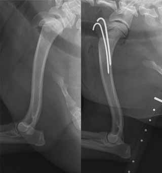 Humerus
fractures: The humeral head and greater tubercle have separate
centres of ossification, meaning they can fracture off alone or both
can separate off and remain attached together [See, photo at
right is of 12-month old cavalier King Charles spaniel
with S-H type one fracture of humerus] or separate completely,
leading to a three-piece fracture. The fractures of the proximal
humerus are usually Salter-Harris type one, although an additional
section of metaphyseal bone can remain attached to the humeral head,
converting it to a Salter-Harris type two. When the greater tubercle
is fractured (with or without the humeral head attached), fixation
is usually by two parallel K-wires so the articular surface of the
humeral head is not traumatised [see photo at far right].
An alternative in animals where the physis is close to fusing is a
lag screw, but this will prevent any remaining growth potential
across this physis. Fractures of the humeral head alone are
generally stabilised with two parallel K-wires inserted from the
cranial aspect of the proximal humerus, just distal to the greater
tubercle, so the tips of the wires reside in the humeral head. The
fractures of the proximal humerus are usually Salter-Harris type
one, although an additional section of metaphyseal bone can remain
attached to the humeral head, converting it to a Salter-Harris type
two." Radiograph photos above are of the humerus of a
12-month-old cavalier King Charles spaniel with a
Salter-Harris type one fracture of the proximal humeral physis
(left). This has been stabilised with parallel K-wires through the
greater tubercle into the humeral diaphysis (right).
Humerus
fractures: The humeral head and greater tubercle have separate
centres of ossification, meaning they can fracture off alone or both
can separate off and remain attached together [See, photo at
right is of 12-month old cavalier King Charles spaniel
with S-H type one fracture of humerus] or separate completely,
leading to a three-piece fracture. The fractures of the proximal
humerus are usually Salter-Harris type one, although an additional
section of metaphyseal bone can remain attached to the humeral head,
converting it to a Salter-Harris type two. When the greater tubercle
is fractured (with or without the humeral head attached), fixation
is usually by two parallel K-wires so the articular surface of the
humeral head is not traumatised [see photo at far right].
An alternative in animals where the physis is close to fusing is a
lag screw, but this will prevent any remaining growth potential
across this physis. Fractures of the humeral head alone are
generally stabilised with two parallel K-wires inserted from the
cranial aspect of the proximal humerus, just distal to the greater
tubercle, so the tips of the wires reside in the humeral head. The
fractures of the proximal humerus are usually Salter-Harris type
one, although an additional section of metaphyseal bone can remain
attached to the humeral head, converting it to a Salter-Harris type
two." Radiograph photos above are of the humerus of a
12-month-old cavalier King Charles spaniel with a
Salter-Harris type one fracture of the proximal humeral physis
(left). This has been stabilised with parallel K-wires through the
greater tubercle into the humeral diaphysis (right).
RETURN TO TOP
heatstroke:
Providing Care to Dogs with Heatstroke. Amy Newfield. Today's Vet. Nurse. May 2019. Quote: Thermoregulation is the ability to maintain body temperature within certain boundaries, even when the surrounding temperature is very different. This ability is a major aspect of homeostasis. Companion animals may experience large changes in environmental temperature, and although their bodies may initially be able to regulate the temperature to within normal range, at some point they may not be able to keep up with the demand. When this happens, and if the core body temperature reaches 41.1°C (106°F) or above, the animal is said to have heatstroke. Most commonly (although not the only cause), the increasing outside temperatures of summer can result in heatstroke in companion animals. As a veterinary nurse or assistant, you are likely to be the first person to deal with the client and the heatstroke patient and to perform the diagnostic testing, treatment, and nursing care for these critical patients. This article describes the physiology of normal thermoregulation; the pathophysiology of heatstroke; and how to recognize, treat, and care for the heatstroke patient.
Incidence and risk factors for heat-related illness (heatstroke) in UK dogs under primary veterinary care in 2016. Emily J. Hall, Anne J. Carter, Dan G. O’Neill. Nature: Sci. Repts. June 2020: doi: 10.1038/s41598-020-66015-8 Quote: This study aimed to report the incidence, fatality and canine risk factors of heat-related illness in UK dogs under primary veterinary care in 2016. The VetCompassTM programme collects de-identified electronic patient records from UK veterinary practices for research. From the clinical records of 905,543 dogs under veterinary care in 2016, 395 confirmed heat-related illness events were identified. The estimated 2016 incidence of heat-related illness was 0.04%, with an event fatality rate of 14.18%. ... Clinical signs: • panting excessively or continuously despite removal from heat/cessation of exercise, • collapse not subsequently attributed to another cause (e.g. heart failure, Addison’s), • stiffness, lethargy or reluctance to move, • gastrointestinal disturbance including hypersalivation, vomiting or diarrhoea, • neurological dysfunction including ataxia, seizures, coma or death, • haematological disturbances including petechiae or purpura. ... Multivariable analysis identified significant risk factors including breed (e.g. Chow Chow, Bulldog, French Bulldog, Dogue de Bordeaux, Greyhound, and Cavalier King Charles Spaniel), higher bodyweight relative to the breed/sex mean and being over two years of age. Dogs with a brachycephalic skull shape and dogs weighing over 50 kg were also at greater risk. Breeding for good respiratory function and maintaining a healthy bodyweight should be considered key welfare priorities for all dogs to limit the risk of heat-related illness.
RETURN TO TOP
icterus (jaundice):
Acute hepatic necrosis following xylitol ingestion in a 2-year-old dog. Hillary Wentworth. Cornell Univ. Lib. October 2009. Quote: A 2-year-old intact female Cavalier King Charles Spaniel presented to the triage service at the Cornell University Hospital for Animals (CUHA) with a 5-day history of icterus, severe lethargy, anorexia, and vomiting. The patient had a history of eating 2-3 pieces of xylitol-containing chewing gum 8 days before presentation to her referring veterinarian (48 hours before the onset of her clinical signs). Multiple laboratory abnormalities referable to liver damage were observed. Hepatic lymphadenopathy was detected on abdominal ultrasound. A presumptive diagnosis of xylitol toxicosis was made based on the patient's history and laboratory findings. Supportive and hepatoprotective therapy was instituted and the patient recovered after a treatment course of several weeks. Principals of the pathogenesis, diagnosis, treatment, and prognosis of canine xylitol toxicosis are discussed.
Icterus & Collapse in a Cavalier King Charles Spaniel. Leslie Sharkey. Clinicians Brief. July 2010. Quote: "A 7-year-old neutered male Cavalier King Charles spaniel is presented after an episode of collapse. The dog was presented to the referring veterinarian after an episode of collapse and weakness at a boarding kennel. The patient had been eating and drinking normally before the collapse episode but had been lethargic. The referring veterinarian noted evidence of hemolytic anemia, which was suspected to be immune-mediated. The dog was referred after initial treatment with vitamin K, gastroprotectants, and IV fluids. An untreated heart murmur was noted on the history. Physical Examination: On presentation to the referral hospital, the patient had icteric sclera and mucous membranes, a grade 3/6 systolic heart murmur, discomfort on abdominal palpation, nystagmus, and decreased pupillary light responses and mentation. Laboratory Results: A complete blood count revealed marked neutrophilic leukocytosis with a moderate regenerative left shift, including rare metamyelocytes, toxic change, and monocytosis. Other laboratory findings included a hematocrit of 9%, mild macrocytosis, normal mean corpuscular hemoglobin concentration, increased nucleated red blood cells, and normal platelet count. A review of the blood film revealed 50% to 75% Heinz bodies, numerous spherocytes, and red blood cell ghosts. Diagnosis: probable zinc toxicity. Abdominal radiography revealed metallic foreign bodies in the stomach.Multiple coins, including some zinc containing pennies minted after 1982, and a medallion were removed endoscopically."
Haemolytic anaemia and acute pancreatitis associated with zinc toxicosis in a dog. R. Blundell, F. Adam. Vety. Rec. July 2013;doi: 10.1136/vetreccr.100376rep. Quote: "We describe a case of zinc toxicity in a 14-month-old, female, neutered, Cavalier King Charles spaniel with a 48-hour history of haematochezia, icterus and collapse. Regenerative anaemia with a packed-cell volume of 7 per cent was seen. Prior to referral, radiography had revealed a gastric, metallic foreign body which was removed at exploratory laparotomy. On presentation, the dog was comatose, hypothermic and bradycardic – resuscitation was performed successfully, but the dog then displayed marked abdominal pain. The dog died 12 hours after presentation. At postmortem examination, the animal showed severe icterus. Both kidneys were diffusely dark red; the pancreas was diffusely pale and nodular. Histopathological examination revealed evidence of intravascular haemolysis with blood vessel lumens containing haemoglobin. The renal tubules also contained large amounts of intraluminal haemoglobin with haemoglobin crystals scattered throughout the cortex and medulla. The pancreas exhibited multifocal coagulative necrosis, surrounded by a neutrophil-dominated inflammatory infiltrate. Zinc levels were markedly increased above the normal reference range in both liver and kidney. This report describes the clinical and pathological findings of a case of acute zinc toxicity in a dog following ingestion of a metallic object which resulted in marked haemolytic anaemia and acute pancreatitis."
Retrospective evaluation of the clinical course and outcome of zinc toxicosis due to metallic foreign bodies in dogs (2005–2021): 55 cases. Cameron S. Henke, Matthew W. Beal, Rebecca A. L. Walton, Joanna B. Finstad, Brooke K. Newmans, Michael P. Sliman, Molly A. Racette, Nyssa A. Levy. Vet. Emerg. Crit. Care. October 2023; doi: 10.1111/vec.13330. Quote: Objective: To describe the overall clinical course of zinc toxicosis in dogs including source, time to source control, incidence of hemolytic anemia, acute liver injury (ALI), acute kidney injury (AKI), and pancreatitis. Design: Retrospective case series from 2005 to 2021. Setting: Six university veterinary teaching hospitals. Animals: Fifty-five client-owned dogs with known zinc toxicosis due to metallic foreign body (MFB) ingestion. Measurements and Main Results: The most common source of zinc was US pennies minted after 1982 (67.3%). Forty-five of 55 (81.8%) dogs survived and 10 of 55 (18.2%) died or were euthanized. Median length of hospitalization for survivors and nonsurvivors was 3 days. The most common clinical sequelae of zinc toxicosis were anemia (87%), ALI (82%), coagulopathy (71%), thrombocytopenia (30.5%), AKI (26.9%), and acute pancreatitis (5.5%). Most dogs (67.3%) required blood products and 83% of dogs achieved a stable HCT or PCV in a median of 24 hours after MFB removal. The median duration of illness prior to presentation was 48 hours for both survivors and nonsurvivors and there was no impact of time to presentation on the incidence of ALI, AKI, or pancreatitis. Conclusions: Zinc toxicosis secondary to MFB ingestion should be considered a differential diagnosis for dogs with gastrointestinal signs, hemolytic anemia, ALI, hemostatic abnormalities, AKI, and pancreatitis. AKI may be a more common sequela of zinc toxicosis than previously suspected. Acute pancreatitis is a rare but potentially serious sequela to zinc toxicosis.
RETURN TO TOP
insecticide poisoning reaction -- fipronil:
Efficiency of Levetiracetam and Gabapentin in the treatment of Epilepsy in a Cavalier King Charles Spaniel Dog with Myxomatous Mitral Valve Heart Disease, a Case Report. Sinan Kandır, Cengiz Yüce Kayabek, Bilge Kaan Tekelioğlu. 3d Int'l Mediterranean Sci. & Engr'g Conf. October 2018. Pgs. 1242-1245;paper ID456. Quote: Epilepsy and seizure have a complex physiopathological background and are most common idiopathic or primary neurologic disorders in dogs. Various anticonvulsant (ACDs) and antiepileptic drugs (AEDs) are used for the management of seizures and epilepsy. The use of levetiracetam is limited in the treatment of epilepsy and is used only with expert opinion because of its undesirable side effects and its low therapeutic effect, which are not chemically related to existing AEDs. The Cavalier King Charles Spaniels (CKCSs) are predisposed to numerous inherited neurologic and cardiologic problems due to their high predisposition to genetic pool originated consanguineous diseases. The dog was 3 years old, female, CKCSs breed stud and had been suffered from epileptic seizures almost 3 months. The dog was consulted at Veterinary Faulty Clinics and myxomatous mitral valve heart disease (MMVD) was diagnosed. Phenobarbital (2.5 mg/kg BID, PO) and enalapril (0.5 mg/kg BID, PO) were prescripted for the management of epileptic seizures and medication of the MMVD. However, the dog wasn’t reacted the treatment as expected and epileptic seizures were increased dramatically (~7 times per day) in addition with respiratory depression. The treatment protocol was changed to levetiracetam (15 mg/kg BID, PO). The dog was reacted well to levetiracetam treatment and epileptic seizures were disappeared with respiratory depression. Three months later a routine parasitic treatment was done with fipronil (0.067 ml/kg, spot-on). The epileptic attacks were dramatically increased and became more severe just after the fipronil treatment (~7 times per day). Thus, gabapentin (10 mg/kg, TID, PO) was decided to use together with levetiracetam (10 mg/kg BID, PO). Epileptic seizures disappeared with the use of gabapentin over again. In conclusion, the pharmacokinetics and antiepileptic effects of levetiracetam and gabapentin are not fully understood, but the combination of both drugs with a combined drug protocol may help to manage epileptic attacks without causing respiratory depression in CKCS. Fipronil, phenylpyrazole origin and other antiparasitic drugs should be used very carefully and under veterinary consultant supervision due to their side effects especially in epileptic dogs. Finally, gabapentin could be a good option for the alternative add-on therapy to control epileptic seizures in CKCSs.
Distribution of fipronil in humans, and adverse health outcomes of in utero fipronil sulfone exposure in newborns. Young Ah Kim, Yeong Sook Yoon, Hee Sun Kim, Se Jeong Jeon, Elizabeth Cole, Jeongsun Lee, Younglim Kho, Yoon Hee Cho. Internat'l J. Hygiene & Environ. Health. April 2019; doi: 10.1016/j.ijheh.2019.01.009. Quote: Fipronil is a highly effective insecticide with extensive usages; however, its distribution and toxic/health effects in the human population after chronic exposure have not yet been clearly identified. Our objectives were to determine the levels of serum fipronil and fipronil sulfone, a primary fipronil metabolite, in a general and sensitive human population using a birth cohort of parent-infant triads in Korea. We further investigated whether in utero exposure to fipronil and fipronil sulfone can affect health outcomes in newborn infants. Blood and umbilical cord blood from 169 participants, 59 mother-neonate pairs and 51 matching biological fathers, were collected; serum fipronil and fipronil sulfone (both blood and cord blood) and serum thyroid hormones (cord blood) were measured. Demographic, physiological, behavioral, clinical, and socioeconomic data for each participant were collected via a one-on-one interview and a questionnaire survey. Fipronil sulfone was detected in the serum of mothers, fathers, and infantile cord blood, while fipronil itself was not. Maternal fipronil sulfone levels were correlated to those of matched biological fathers and newborn infants. Adjusted analyses identified significant associations between parental fipronil sulfone levels and household income. Infantile fipronil sulfone levels were significantly associated with both maternal and paternal levels as well as maternal pre-pregnant BMI. Furthermore, infantile fipronil sulfone levels were inversely associated with cord blood T3 and free T3 levels as well as 5-min Apgar scores of newborn infants. Serum fipronil sulfone was detected in a specific population of mother-neonate pairs and their matched biological fathers in a manner suggestive of regular exposure to fipronil among urban residents. The findings also suggest that serum fipronil sulfone placentally transfers to the fetus and affects infantile adverse health outcomes. This is a first of its kind study; therefore, future studies are warranted.
RETURN TO TOP
insecticide poisoning reaction -- pyrethrins:
Tremor-Salivation Syndrome in Canine following Pyrethroid/Permethrin Intoxication. Sigal Klainbart, Yael Merbl, Efrat Kelmer, Olga Cuneah, Nir Edery, Jakob A Shimshon. Pharmaceutica Analytica Acta. September 2014;5:9. Quote: "A 17-month-old male King Charles cavalier was presented with acute onset of generalized body tremors, facial twitching and salivation after being exposed to 2 different classes of compounds of the pyrethrins/pyrethroids group as well as to imidacloprid. ... Biospotix spray (geraniol essential oils) and Advantage spot on (100 mg/mL imidaclopride and 25 mg/mL moxidectin) were applied on the dog’s skin. Furthermore, the owner’s household was sprayed against insects using a commercial pyrethroid preparation (Admiral) 24 hours before clinical signs appeared. The dog was otherwise healthy, fully vaccinated, lived in an apartment and leashwalked. ... Bifenthrin toxicity was confirmed by gas chromatography mass spectrometry. Pyrethroid toxicosis in dogs is to the best of our knowledge rarely reported in the literature. The dog displayed neurological signs highly characteristic of Tremor-Salivation-syndrome associated with pyrethroid toxicosis. The plasma half-life of bifenthrin in dogs was 7.6 hr). Initial therapy consisted of diazepam, methocarbamol and IV fluids, followed by general anesthesia with isofluran and diazepam CRI. Supportive nursing care was provided as needed. Twenty-four hours post admission, the dogs was no longer under general anesthesia. Seventy two hours post admission the dog was discharged had no menace response, was alert and responsive when stimulated, ataxic while walking and showed normal eating behavior." .
Bifenthrin Toxicity in a Dog. Sigal Klainbart, Yael Merbl, Efrat Kelmer, Olga Cuneah, Jakob A. Shimshoni. Annals Clin. Pathol. November 2014;2(4):1030. Quote: A 17-month-old male King Charles cavalier was presented with acute onset of generalized body tremors and facial twitching after being exposed to 2 different compounds of the pyrethrins/ pyrethroids group and imidacloprid. ... The dog presented in this case was exposed to 2 different compound of the pyrethrins/pyrethroids group, namely Admiral®, an insecticide comprised of 7.9 % bifenthrin and 1 % condensed naphthalene sulfonate andBiospotix®, an insecticide comprised of natural pyrethrum 0.2 %, geraniol 0,5 % (v/v) (containing természetespiretringeránium, lavender essential oils, citronella, aqua exipient, and alcohol qsp.) Concomitantly, Advantage® spot on (10% imidacloprid, 0.1% butylhydroxytoluene, benzyl alcohol) was applied. ... The dog exhibited clinical signs compatible with pyrethrin toxicity (swaying, hypersalivation, seizures, tremors, twitching of the facial muscles, four limb ataxia and paddling) consistent with the TS-syndrome [tremor associated with salivation]. ... Since pyrethroids and imidacloprid are metabolized via the liver by a conjugation pathway, we suspect this pathway was “overwhelmed” by the amount of compounds to be metabolized, resulting in enhanced toxicity. ... Bifenthrin toxicity was confirmed by gas chromatography mass spectrometry. Initial therapy consisted of diazepam, metacarbamol and IV fluids, followed by general anesthesia with isofloran and diazepam CRI. Blood specimens were collected for following bifebthrine blood levels over time. Supportive nursing care was provided as needed. Twenty-four hours post admission, the dog was no longer under general anesthesia. Seventy two hours post admission the dog was discharged had no menace response, was alert and responsive when stimulated, ataxic while walking and showed normal eating behavior. Pyretroid toxicosis in dogs was to our best knowledge never been reported before. ... Pyrethroids are reportedly eliminated in the first 12-24 hr after absorption. In the present case study, bifenthrin plasma levels dropped dramatically over a time period of 30 hr with a half-life of 7.6 hr. Several studies suggest that toxicity from pyrethroids occurs when central nervous system pyrethroid concentrations exceed threshold quantities. Since the clinical signs persisted long after the elimination of bifenthrin from the plasma compartment, cerebrospinal fluid levels might present a better predictor of bifenthrin toxicity. In conclusion, this is the first report of pyrethroid toxicity in a dog that was exposed to 2 types of pyrethroids and imidacloprid. The dog exhibited classic neurological signs of pyrethroid toxicity and was fully recovered following symptomatic and supportive care.
Management of presumptive canine permethrin toxicosis using intravenous lipid emulsion as an adjunctive therapy. Matthew A Kopke, Ivayla D Yozova. BMJ Vet. Rec. Case Rpts. April 2020; doi: 10.1136/vetreccr-2019-001041. Quote: A seven-month-old Cavalier King Charles spaniel presented with generalised, coarse muscle tremors that progressed to seizure activity, after observed ingestion (licking) of a cleaning product containing permethrin. At the time of presentation, the dog was receiving prednisone as management for masticatory muscle myositis. The dog was treated with symptomatic and supportive therapy in the form of midazolam, dexmedetomidine and intravenous lipid emulsion (ILE). This case details the first reported use of ILE as adjunctive therapy in the successful management of canine permethrin toxicosis. No further tremors or seizure activity, nor any adverse effects were observed following administration of ILE therapy.
RETURN TO TOP
meningoencephalitis:
Meningoencephalitis of unknown origin: investigation of prognostic factors and outcome using a standard treatment protocol. M. Lowrie, P. M. Smith, L. Garosi. Vet. Rec. May 2013; doi: 10.1136/vr.101431. Quote: Meningoencephalitis of unknown origin (MUO) is a common inflammatory CNS disease in dogs, with a variable and unpredictable outcome. MRI and cerebrospinal fluid (CSF) features were prospectively evaluated to establish their utility as prognostic markers for predicting mortality, relapse and long-term outcome in 39 dogs with MUO [including 2 cavalier King Charles spaniels (5%)]. MRI and CSF analysis were performed at initial diagnosis and three months into treatment with prednisolone and cytosine arabinoside. When possible, MRI was repeated every 12 months thereafter. Median survival time was 26 days. All deaths occurred within 52 days of diagnosis (22/39; 56 per cent). One-third (13/39) died within 72 hours of diagnosis. Outcome was good or excellent in 12/17 surviving dogs. Loss of the cerebral sulci and foramen magnum herniation on MRI were associated with increased risk of mortality. An abnormal CSF analysis at the three-month re-examination was associated with increased risk of relapse (P=0.04). The combination of MRI and CSF analysis provided a greater sensitivity for predicting relapse than one modality alone. Discontinuing treatment before MRI lesions resolved always resulted in relapse. The presence of certain MRI characteristics may indicate an increased risk of mortality. Dogs alive three months following diagnosis have a very low risk of death due to MUO.
Pasteurella Multocida meningoencephalomyelitis in a dog secondary to severe periodontal disease. Alexander E. Tun, Leontine Benedicenti, Evelyn M. Galban. Clinical Case Repts. April 2018; doi: 10.1002/ccr3.1561. Quote: Pasteurella multocida can cause meningoencephalomyelitis in canine patients with severe periodontal disease. Fever and neutrophilic pleocytosis in the cerebrospinal fluid are likely, and blood culture and/or empiric antibiotic therapy are indicated. A 5-year-old 5.14 kg (11.3-lb) spayed female Cavalier King Charles Spaniel was presented ... for evaluation of ataxia and cervical hyperesthesia. ... Physical examination revealed a rectal temperature of 40.2°C (104.4°F) and severe periodontal disease and was otherwise unremarkable. ... Consultation ... confirmed the severe periodontal disease with contact ulceration and possible necrotizing stomatitis. However, the changes were not considered to be to the degree that would cause lethargy and inappetence, and so dental cleaning with extractions was recommended once the patient condition had stabilized. ... Pasteurella multocida was isolated from blood and CSF cultures several days later. ... Pasteurella multocida is a small gram-negative coccobacillus. It is a facultative anaerobe that is part of the normal flora of the respiratory tract and oral cavity of many mammals. Carriage rates are reported to be highest among cats (70–90%) and dogs (20–50%). Pasteurella multocida meningitis is a rare clinical occurrence in humans and in veterinary medicine. ... [I]t is the hypothesis of the authors that this patient’s severe periodontal disease leads to a bacteremia causing hematogenous seeding of a bacterial meningitis originating at the disrupted blood–spinal cord barrier. ... Concluding remarks: The patient described exhibited clinical signs and laboratory findings consistent with previous reports of bacterial MEM [meningoencephalomyelitis] in dogs. Based on CSF and blood culture, as well as response to therapy, a diagnosis of P. multocida meningoencephalomyelitis was made. This is the first report of a canine patient with a P. multocida CNS infection without evidence of a direct inoculation. In addition, this is the first report where the source of the infection is suspected to be severe periodontal disease. Further support for this theory might have come from concurrent culture of the oral flora or any extracted teeth with matching susceptibility pattern. In the future, be aware that a fever with multifocal neurologic signs and severe periodontal disease in a canine patient may suggest a P. multocida infection and both CSF and blood cultures can be submitted for confirmation.
Serological prevalence of toxoplasmosis and neosporosis in dogs diagnosed with suspected meningoencephalitis in the UK. A. M. Coelho, G. Cherubini, A. De Stefani, A. Negrin, R. Gutierrez-Quintana, E. Bersan, J. Gueva. J. Sm. Anim. Pract. October 2018; doi: 10.1111/jsap.12937. Quote: Objectives: To assess the prevalence of antibodies to Toxoplasma gondii and Neospora caninum in a population of dogs with a diagnosis of suspected inflammatory meningoencephalitis. Materials and Methods: Medical records of three referral centres were reviewed from 2008 to 2016 to identify a cohort of dogs diagnosed and treated for suspected inflammatory meningoencephalitis after testing for evidence of exposure to these pathogens. Results: In our sample of 400 dogs [including 21 cavalier King Charles spaniels (5.25%)] the prevalence for exposure (IgG>1:50) to Toxoplasma gondii was 8/201 (3∙98%). Active infection (IgG titre >1:400 or/and an IgM titre >1:64 and/or positive PCR in CSF) was suspected in 1/400 (0∙25%). The prevalence for exposure [Indirect fluorescent antibody (IFA) titre >1:50] and active infection (IFA titres ≥⃒1:400 and/or positive PCR in CSF) with Neospora caninum were 14/201 (6∙96%) and 9/400 (2∙25%), respectively. Clinical Significance: In view of the low prevalence of protozoan infections, the risk associated with starting immunosuppressive medication in dogs with evidence of inflammatory meningitis or encephalitis in the UK appears low.
RETURN TO TOP
myoclonus:
Neurological diseases of the Cavalier King Charles spaniel. Rusbridge, C. J. Small Anim. Prac., June 2005, 46(6): 265-272. Quote: "Head nodding Occasionally CKCS may present with the complaint of head nodding. Face twitching may also be seen and the body may be observed to bounce up and down when the dog is standing stationary. Occasionally the dog may stagger or appear to lose balance. Episodes tend to last a few seconds and may be very frequent. This movement disorder can have a variety of causes and underlying CNS pathology such as GME and syringomyelia should be ruled out and treated. It is more common in geriatric CKCS. The episodes stop during sleep and when walking. Investigation into the exact aetiology is ongoing."
Unusual manifestation of bromide toxicity (bromism) in an idiopathic epileptic dog already treated with phenobarbital. Fabio Stabile, Alberta de Stefani, Luisa De Risio. Vet. Rec. March 2014; doi: 10.1136/vetreccr-2014-000043. Quote: A three-year seven-month-old female spayed Cavalier King Charles spaniel was diagnosed with idiopathic epilepsy. Because of poor seizure control, the dog was started on oral treatment with potassium bromide as adjunctive treatment to phenobarbital. The dog presented eight days following bromide loading, having developed sedation, general proprioceptive ataxia and generalised appendicular repetitive myoclonus. The serum bromide concentration was 15.9 mg/ml (target range 1 mg/ml to 2.5 mg/ml), which was suggestive of a bromide overdose. The dog improved after reduction of bromide dosing and no similar episodes were reported by the owners at a follow up of 26 months. To the authors’ knowledge this is the first report describing generalised repetitive myoclonus related to bromide toxicity.
Clinical and Breed Characteristics of Idiopathic Head Tremor Syndrome in 291 Dogs: A Retrospective Study. Linda G. Shell, John Berezowski, Mark Rishniw, Belle M. Nibblett, Patrick Kelly. Vet. Med. International. April 2015; doi: 10.1155/2015/165463. Quote: Objective: To establish signalment and phenomenology of canine idiopathic head tremor syndrome (IHTS), an episodic head movement disorder of undetermined pathogenesis. Design: Retrospective case series. Animals: 291 dogs with IHTS diagnosed between 1999 and 2013. Procedures: Clinical information was obtained from an online community of veterinary information aggregation and exchange (Veterinary Information Network, 777 W Covell Boulevard, Davis, CA 95616) and conducted with their approval. Information on breed, sex, age of onset, tremor description, mentation during the event, effect of distractions and drugs, diagnostics, presence of other problems, and outcome was analyzed. Results: IHTS was found in 24 pure breeds. Bulldogs, Labrador Retrievers, Boxers, and Doberman Pinschers comprised 69%; mixed breeds comprised 17%. Average onset age was 29 months (range: 3 months to 12 years). First episode occurred before 48 months of age in 88%. Vertical (35%), horizontal (50%), and rotational (15%) movements were documented. Possible trigger events were found in 21%. Mentation was normal in 93%. Distractions abated the tremor in 87%. Most dogs did not respond to antiepileptic drugs. Conclusions and Clinical Relevance: This retrospective study documents IHTS in many breeds including Labrador Retrievers, Boxers, and mixed breeds.
International Veterinary Epilepsy Task Force’s current understanding of idiopathic epilepsy of genetic or suspected genetic origin in purebred dogs. Velia-Isabel Hülsmeyer, Andrea Fischer, Paul J.J. Mandigers, Luisa DeRisio, Mette Berendt, Clare Rusbridge, Sofie F.M. Bhatti, Akos Pakozdy, Edward E. Patterson, Simon Platt, Rowena M.A. Packer, Holger A. Volk. BMC Vet. Res. September 2015;11:175. Quote: "Older Cavalier King Charles spaniels (>5 years old) have a high prevalence of myoclonus [spasmodic jerky contraction of groups of muscles], which manifests most commonly as a brief jerking of the head and forelimbs when the dog is standing or sitting. Initially the syndrome is relatively benign but can be progressive with affected dogs suffering frequent jerks which may interfere with function, for example cause the dog to fall or stumble. The syndrome can be confused with focal epileptic seizures but generally does not respond to AEDs [antiepileptic drugs] licenced for dogs although may respond to levetiracetam (personal communication Clare Rusbridge February 2015). The pathogenesis of the myoclonus is as yet undetermined."
Classification of Involuntary Movements in Dogs: Myoclonus and Myotonia. M. Lowrie, L. Garosi. J. Vet. Intern. Med. May 2017; doi: 10.1111/jvim.14771. Quote: Myoclonus is a sudden brief, involuntary muscle jerk. Of all the movement disorders, myoclonus is the most difficult to encapsulate into any simple framework. On the one hand, a classification system is required that is clinically useful to aid in guiding diagnosis and treatment. On the other hand, there is need for a system that organizes current knowledge regarding biological mechanisms to guide scientific research. These 2 needs are distinct, making it challenging to develop a robust classification system suitable for all purposes. We attempt to classify myoclonus as “epileptic” and “nonepileptic” based on its association with epileptic seizures. Myotonia in people may be divided into 2 clinically and molecularly defined forms: (1) nondystrophic myotonias and (2) myotonic dystrophies. The former are a group of skeletal muscle channelopathies characterized by delayed skeletal muscle relaxation. Many distinct clinical phenotypes are recognized in people, the majority relating to mutations in skeletal muscle voltage-gated chloride (CLCN1) and sodium channel (SCN4A) genes. In dogs, myotonia is associated with mutations in CLCN1. The myotonic dystrophies are considered a multisystem clinical syndrome in people encompassing 2 clinically and molecularly defined forms designated myotonic dystrophy types 1 and 2. No mutation has been linked to veterinary muscular dystrophies. We detail veterinary examples of myotonia and attempt classification according to guidelines used in humans. This more precise categorization of myoclonus and myotonia aims to promote the search for molecular markers contributing to the phenotypic spectrum of disease. Our work aimed to assist recognition for these 2 enigmatic conditions.
Myoclonus in older Cavalier King Charles Spaniel – clinical and histopathological findings. C. Rusbridge, P.J.Watson, C.J. Driver, A. Tauro, R.Fernandes, J.Rose, F. Constantino-Casas, F. Salguero-Bodes. 30th Symposium ESVN-ECVN, Abstract #03; J. Vet. Intern. Med. January 2020; doi: 10.1111/jvim.15681. Quote: Myoclonus has been reported in the Cavalier King Charles spaniel (CKCS) but its characteristics are not well described. Medical records were searched for CKCS observed to have myoclonic jerks. 40 dogs were identified (25 female). Most had been referred originally for another disease. The event was described as rapid eyelid blinking with head nodding / shuddering and variable extension down the limbs that could result in falling or stumbling. The movement lasted seconds, had no obvious trigger, was most likely when stationary in a sternal or sitting position and did not appear associated with loss of consciousness. Multiple daily episodes were seen and increased in frequency and intensity with time. The median age of onset was 9 years (interquartile range 7-10 years). 29 of 40 CKCS had brain and spinal MRI. All had chiari-like malformation, 23 of 29 had ventriculomegaly and 19 of 29 had syringomyelia. 11 of 40 dogs had epilepsy (generalised tonic clonic seizures) or other paroxysmal event (episodic falling genetic clear). Age of onset of seizures for 8 dogs with epilepsy was +/- 1 year from onset of the myoclonus (median 7 years old). 3 owners reported signs suggesting progressive cognitive dysfunction. Histopathology of the brains from 4 CKCS were unremarkable. Anecdotally myoclonus was improved with levetiracetum and possibly corticosteroids. Myoclonic jerks are common in older CKCS and should not be assumed to be a consequence of syringomyelia. More evidence is required as to whether there is an association with generalised tonic‐clonic seizures and mental deterioration.
Myoclonus in older Cavalier King Charles Spaniels. Carina Rotter, Danielle Whittaker, Clare Rusbridge. J. Vet. Intern. Med. March 2022; doi: 10.1111/jvim.16404. Quote: Background: Myoclonus is observed in older Cavalier King Charles Spaniels (CKCS) but a full description is lacking. Objectives: The presence, age of onset, characteristics and treatment of myoclonic episodes were retrospectively evaluated in a cohort of CKCS which presented to 1 board-certified neurologist. Clinical data, imaging studies, presence of seizures and their management, as well as other comorbidities were noted. Animals: Thirty-nine CKCS that were presented to 2 institutions between 2001 and 2018 with signs consistent with myoclonus. Clinical examination, blood sampling, advanced diagnostic imaging, cerebrospinal fluid analysis, and record keeping of other comorbidities was performed. ... Twenty of 30 cases showed signs of SM [syringomyelia], 3 a presyrinx and 4 a central canal dilatation. OME [otitis media with effusion (primary secretory otitis media -- PSOM)] was visible in 13 dogs. ... The myoclonic event was a mixture of positive and negative myoclonus and described as rapid eyelid blinking with head nodding/shuddering and variable extension down the thoracic limbs, that could result in falling or stumbling of the thoracic limbs. The movement lasted seconds had no obvious trigger and occurred mostly when stationary in a sternal or sitting position. It did not seem to be associated with loss of consciousness. Multiple daily episodes were seen and increased in frequency and intensity over time. Owners whose dogs would frequently fall, often disrupting them while eating, reported the myoclonus to interfere with the dog's quality of life. The mean age of onset of myoclonus was 8.38 years (SD ±1.96). ... Methods: This is a retrospective case series, describing the presence of myoclonus in CKCS. Results: Clinical signs reported were spontaneous in onset, lasted a few seconds and consisted of rapid blinking with head nodding and variable extension down the thoracic limbs. Myoclonus occasionally led to stumbling of the thoracic limbs or collapse. Mean age of onset was 8.38 years (SD ±1.96). Thirteen of 39 dogs with myoclonus had paroxysmal events, such as generalized seizures (9/13). ... Twenty cases showed signs of CM-associated pain (CM-P) [Chiari-like malformation associated pain] and 9 dogs had SM specific signs in including phantom scratching (SM-S) [syringomyelia associated phantom scratching]. Two dogs displayed signs of cognitive dysfunction, with 1 dog described as tending to stare blankly into space, showing less interaction with the owner, wandering aimlessly when left alone and signs suggesting anxiety. Both dogs had seizures which were managed with imepitoin. Four dogs had pelvic limb orthostatic tremor, 1/4 involved 1 of the thoracic limbs as well. The tremor was present while standing, disappeared when walking. ... Twenty-three of 39 dogs were prescribed gabapentin/pregabalin due to CM-P and/or SM-S. It is questionable if gabapentin or pregabalin worsened the myoclonus; however, no deterioration of myoclonus after start of gabapentinoids was documented. Two of the cases were prescribed gabapentin after the myoclonus occurred and the myoclonus did not worsen but the nature of this study does not help to draw a conclusion and it remains questionable if myoclonus was drug-induced in these cases. ... Conclusions and Clinical Importance: Myoclonus occurs in middle-aged to older CKCS and seems to be another epiphenomena of this breed. A link to epilepsy might be present. ... Myoclonic jerks are reported in middle-aged to older CKCS and should not be assumed to be a consequence of SM. ... 100% of dogs that underwent MRI showed signs of CM and 66% SM, this resembles the overall reported breed prevalence in the general CKCS population. In other words, a link between CM and SM was neither proven nor disproven. The frequency of CM and SM was that expected for the age of the population and dogs could have myoclonus without CM or SM. ... There might be a link with generalized tonic-clonic seizures. It seems to be another epiphenomena for this breed Although the number of CKCS treated successfully with levetiracetam was low this could be a possible treatment for myoclonus in this breed. Further research to substantiate this is needed. (See video #1 and video #2.)
Use of levetiracetam for the successful treatment of suspected myoclonic seizures: five dogs (2016-2022). J. Linder, J. Mehra, S. Miller, M. J. Lewis, R. T. Bentley, S. Thomovsky. J. Sm. Anim. Pract. April 2024; doi: 10.1111/jsap.13719. Quote: Objectives: Myoclonic seizures are considered a type of generalised seizure characterised by brief, jerking movements of the body. The aim of this study is to describe cases of suspected canine myoclonic seizure of idiopathic aetiology and to discuss the successful use of the anticonvulsant levetiracetam as treatment in each of these cases. ... Most recently, myoclonus has also been reported in older Cavalier King Charles Spaniels – approximately 25% of this population of Cavaliers also experienced at least one generalised tonic–clonic seizure and 10% experienced another type of paroxysmal episode (Rotter et al., 2022). ... The cases included in this case series suggest that myoclonic seizures in dogs can be idiopathic in origin and, in some instances, be secondary to auditory stimuli in dogs. No breeds previously reported to have myoclonic seizures (Rhodesian Ridgebacks, Beagles with Lafora disease, Cavalier King Charles Spaniels) were included in the current study. There is the possibility that the myoclonic seizures were a result of a DIRAS1 mutation or Lafora disease; genetic testing was not performed in any cases. Meanwhile, the origin of myoclonus in Cavalier King Charles Spaniels is unknown and could originate from a cortical or subcortical aetiology. In the most recent case series published in this breed, 30 cases had MRI performed, all which had caudal occipital malformation syndrome, 13 had lateral ventriculomegaly, 20 had syringomyelia and 13 had otitis media (Rotter et al., 2022). ... Materials and Methods: Dogs with epileptic myoclonus suspected to be idiopathic in aetiology were considered for inclusion. Medical records were reviewed for physical and neurologic examination findings, clinicopathologic results, and diagnostic imaging results. All included dogs were treated with levetiracetam, and their response was reported. Results: Five dogs were included, all of which had suspected myoclonic seizures either observed in-person or on video recording by a board-certified veterinary neurologist. The duration of myoclonic seizures preceding treatment ranged from one day to one year. One dog also experienced a generalised tonic–clonic seizure. All dogs were treated with levetiracetam. Two dogs experienced long-term myoclonic seizure freedom (duration seizure-free of at least 1 year), and two dogs experienced marked decreased myoclonic seizure frequency. One dog experienced immediate abatement of myoclonic seizures, although levetiracetam was only utilised for 1 month following onset of myoclonic seizures in this patient. Clinical Significance: Myoclonic seizures can be idiopathic in aetiology. Levetiracetam can be used effectively to rapidly stop myoclonic seizures and to decrease the frequency of myoclonic seizures.
RETURN TO TOP
necrotizing fasciitis (NF):
Early and aggressive surgical debridement and negative pressure wound therapy to treat necrotizing fasciitis in three dogs. Alicia Mastrocco, Jennifer Prittie. Vet. Surg. March 2021; doi: 10.1111/vsu.13576. Quote: Objective: To report the management and outcomes of dogs with necrotizing fasciitis (NF) treated with early, aggressive surgical debridement and negative pressure wound therapy (NPWT). Study design: Short case series. Animals: Three dogs surgically treated for NF [including a 4.5 year old female cavalier King Charles spaniel]. Methods: Clinical signs in the three dogs included an identified wound, severe pain, fever, and progressive erythema. A tentative diagnosis of NF was based on the presence of suppurative inflammation and intracellular bacteria seen in fine needle aspirates and clinical progression in each case. Each dog was treated with surgical debridement within 6 hours of clinical suspicion for NF. Necrosis affected multiple tissue layers was noted surgical exploration. Systemic supportive care and antibiotherapy were also provided for 3 to 4 weeks postoperatively. Results: Three surgical debridements were required in two dogs, and four surgical debridements were required in one dog. All of the surgical sites were managed with NPWT until final primary closure was possible at 4, 5, and 6 days after initial surgery. Results of histopathology and culture of the surgical sites were consistent with NF as described in each case. All dogs survived to discharge and long-term follow up. Conclusion: Management with early surgery, multiple debridements, and the use of NPWT led to resolution of NF in three dogs.
RETURN TO TOP
neutering:
Gonadectomy effects on the risk of immune disorders in the dog: a retrospective study. Crystal R. Sundburg, Janelle M. Belanger, Danika L. Bannasch, Thomas R. Famula, Anita M. Oberbauer. Vet. Res. December 2016;12:278. Quote: Background: Gonadectomy (spay or neuter or castration) is one of the most common procedures performed on dogs in the United States. Neutering has been shown to reduce the risk for some diseases although recent reports suggest increased prevalence for structural disorders and some neoplasias. The relation between neuter status and autoimmune diseases has not been explored. This study evaluated the prevalence and risk of atopic dermatitis (ATOP), autoimmune hemolytic anemia (AIHA), canine myasthenia gravis (CMG), colitis (COL), hypoadrenocorticism (ADD), hypothyroidism (HYPO), immune-mediated polyarthritis (IMPA), immune-mediated thrombocytopenia (ITP), inflammatory bowel disease (IBD), lupus erythematosus (LUP), and pemphigus complex (PEMC), for intact females, intact males, neutered females, and neutered males. Pyometra (PYO) was evaluated as a control condition. Results: Patient records (90,090) from the William R. Pritchard Veterinary Medical Teaching Hospital at the University of California, Davis from 1995 to 2010 were analyzed in order to determine the risk of immune-mediated disease relative to neuter status in dogs. Neutered dogs had a significantly greater risk of ATOP, AIHA, ADD, HYPO, ITP, and IBD than intact dogs with neutered females being at greater risk than neutered males for all but AIHA and ADD. Neutered females, but not males, had a significantly greater risk of LUP than intact females. Pyometra was a greater risk for intact females. Conclusions: The data underscore the importance of sex steroids on immune function emphasizing a role of these hormones on tissue self-recognition. Neutering is critically important for population control, reduction of reproductive disorders, and offers convenience for owners. Despite these advantages, the analyses of the present study suggest that neutering is associated with increased risk for certain autoimmune disorders and underscore the need for owners to consult with their veterinary practitioner prior to neutering to evaluate possible benefits and risks associated with such a procedure. ... Six of the autoimmune diseases evaluated showed an increased prevalence in neutered dogs, supporting the concept that neutering may have a detrimental effect on a canine’s health and immune function. Lupus was the only disease that did not show increased prevalence in neutered dogs of both sexes compared to intact dogs. These findings indicate the importance of sex steroids on immune function and suggest a role of these hormones on self-recognition.
Assisting Decision-Making on Age of Neutering for 35 Breeds of Dogs: Associated Joint Disorders, Cancers, and Urinary Incontinence. Benjamin L. Hart, Lynette A. Hart, Abigail P. Thigpen, Neil H. Willits. Front. Vet. Sci. July 2020; doi: 10.3389/fvets.2020.00388. Quote: Neutering (including spaying) of male and female dogs in the first year after birth has become routine in the U.S. and much of Europe, but recent research reveals that for some dog breeds, neutering may be associated with increased risks of debilitating joint disorders and some cancers, complicating pet owners' decisions on neutering. The joint disorders include hip dysplasia, cranial cruciate ligament tear or rupture, and elbow dysplasia. The cancers include lymphoma, mast cell tumor, hemangiosarcoma, and osteosarcoma. In previous studies on the Golden Retriever, Labrador Retriever and German Shepherd Dog, neutering before a year of age was associated with increased risks of one or more joint disorders, 2–4 times that of intact dogs. The increase was particularly seen with dogs neutered by 6 months of age. In female Golden Retrievers, there was an increase in one or more of the cancers followed to about 2–4 times that of intact females with neutering at any age. The goal of the present study was to expand and use the same data collection and analyses to cover an additional 29 breeds [including cavalier King Charles spaniels], plus three varieties of Poodles. There were major breed differences in vulnerability to neutering, both with regard to joint disorders and cancers. In most cases, the caregiver can choose the age of neutering without increasing the risks of these joint disorders or cancers. Small-dog breeds seemed to have no increased risks of joint disorders associated with neutering, and in only two small breeds (Boston Terrier and Shih Tzu) was there a significant increase in cancers. To assist pet owners and veterinarians in deciding on the age of neutering a specific dog, guidelines that avoid increasing the risks of a dog acquiring these joint disorders or cancers are laid out for neutering ages on a breed-by-breed and sex basis. ... The present study examined the occurrence in both sexes of the joint disorders: HD [hip dysplasia], CCL [cranial cruciate ligament tears or ruptures] and ED [elbow dysplasia]. Also examined in both sexes were the cancers LSA [lymphoma/lymphosarcoma], HSA [hemangiosarcoma], MCT [mast cell tumors], and OSA [osteosarcoma], because these had been shown in some multi-breed studies to be increased in risks with neutering. In addition, mammary cancer (MC), pyometra (PYO), and urinary incontinence (UI) were examined in female dogs. Of interest was the possible association of early neutering and the occurrence of intervertebral disc disorders (IDD) in the Corgi and Dachshund, two breeds known to be at risk for these diseases. All of the above diseases were examined with regard to dogs neutered in one of the age periods of: <6 mo., 6–11 mo., 1 year (12 to <24 mo.) or 2–8 years, or left intact. The diseases were tracked until the dogs were last seen at the hospital, or through 11 years of age, if seen past their 12th birthday. ... Cavalier King Charles Spaniel: The study population was 51 intact males, 72 neutered males, 87 intact females, and 76 spayed females, for a sample size of 286 cases. Joint disorders: hip dysplasia (HD), cranial cruciate ligament rupture (CCL), and elbow dysplasia (ED). For males and females left intact, the occurrences of one or more joint disorders were just 1-4 percent. For males and females neutered at any age, there was no noteworthy increase of joint disorder occurrence, undoubtedly reflecting the small body size. Cancers: lymphoma (LSA), mast cell tumor (MCT), hemangiosarcoma (HSA), and osteosarcoma (OSA). In intact males, the occurrence of cancers was 2 percent and zero in intact females. For both sexes, neutering was not associated with any increase in this measure. Mammary Cancer (MC), Pyometra (PYO) and Urinary Incontinence (UI) in Females. The occurrence of MC in females left intact was zero. There was no occurrence of UI in spayed females. PYO was diagnosed in 2 percent of intact females. Bottom line. Lacking a noticeable occurrence of increased joint disorders or cancers with neutering males or females, those wishing to neuter should decide on the appropriate age.
Gonadectomy in Dogs: Considerations & Review. Karen M. Tobias. Clinician's Brief. January 2022. Quote: In the United States, canine gonadectomy has traditionally been performed at ≈4 to 6 months of age, after puppies complete their vaccine series. From a technical perspective, surgery at this age or younger is easy, fast, and safe because of small patient size, lack of body fat, and rapid recovery; however, recent studies suggest gonadectomy before skeletal maturity can have adverse effects, particularly in certain dog breeds. More owners and clinicians are thus reconsidering their opinions on timing of gonadectomy and whether it should be performed. There are no randomized, controlled, lifetime studies to provide unequivocal evidence on appropriate timing of gonadectomies3; therefore, the decision as to when and if a patient should be neutered should be based on the individual patient, owner, and available data. ... Dogs should be allowed to reach musculoskeletal maturity (ie, be fully grown, with a fully functional urethral sphincter) before being neutered. Breed type and temperament should also be considered. ... Multiple factors, including breed and age of the dog and inclinations and needs of the owner, should be considered when determining whether to perform gonadectomy, as well as the appropriate timing. Whether a dog is neutered or left intact, owners should be informed of common breed and sex conditions, and the dog should be examined for those conditions on subsequent physical examinations. There is no single or definitive source of information on effects of gonadectomy for each breed, and most current articles have some bias. Current evidence should be evaluated and the positives and negatives should be considered for each patient and owner before a recommendation is made.
Vasectomy and ovary-sparing spay in dogs: comparison of health and behavior outcomes with gonadectomized and sexually intact dogs. Chris Zink, , Mikel M. Delgado, Judith L. Stella. J. Amer. Vet. Med. Assn. January 2023; doi: 10.2460/javma.22.08.0382. Quote: Objective: To compare health and behavior outcomes for dogs that underwent vasectomy or ovary-sparing spay (hysterectomy) with sexually intact dogs or dogs that had undergone traditional castration or spay. Sample: 6,018 dog owners responded to a web-based survey between November 3, 2021, and January 7, 2022. Procedures: Participants were asked demographic questions and to provide information about 1 or more dogs (living or deceased). Options for reproductive status were as follows: sexually intact, castrated, spayed (ovariohysterectomy or ovariectomy), vasectomy, or ovary-sparing spay (hysterectomy). Participants were asked questions about orthopedic and other health problems, cancer, and problematic behavior. Logistic regression models, survival analyses, and descriptive statistics were used to assess relationships between reproductive status and outcomes. Resuts: Owners provided valid surveys for 6,018 dogs, including 1,056 sexually intact, 1,672 castrated, and 58 vasectomized male dogs and 792 sexually intact, 2,281 spayed, and 159 female dogs that had undergone ovary-sparing spay. Longer exposure to gonadal hormones, regardless of reproductive status, was associated with reduced odds of general health problems and both problematic and nuisance behaviors. Clinical Relevance: TTo our knowledge, this study provides the first data on health and behavior outcomes of vasectomy and ovary-sparing spay in dogs and is the first to compare these outcomes to sexually intact and gonadectomized dogs. It adds to accumulating data on the mixed benefits and risks of removing the gonads to prevent reproduction and emphasizes the importance of developing an informed, case-by-case assessment of each patient, taking into consideration the potential risks and benefits of spaying or neutering and alternative reproductive surgeries.
Understanding the long-term risks of neutering dogs. Michelle Kutzler, et al. Vet. Rec. June 2023; doi: 10.1002/vetr.3195. Quote: Despite the limitations of a survey-based and retrospective study, the findings are clear: longer exposure to gonadal hormones is associated with fewer general health problems, longer lifespan, lower odds of orthopaedic problems, cancer and obesity, and fewer problematic and nuisance behaviours. ... Numerous peer-reviewed research papers now describe how routine neutering of dogs has been linked to increased incidence of health problems such as cruciate ligament rupture, hip dysplasia, osetoarthritis, hypothyroidim and many cancers (including haemangiosarcoma, mast cell tumours, lymphosarcoma, osteosarcoma, transitional cell carcinoma of the bladder), and the assertino that gondadectomised dogs live longer has largely been discredited. ... Kutzler concludes: "Gonads should no longer be considered mere gamete-producing or ancillary sex organs but rather necessary endocrine glands for normal metabolic, endocrinologic, musculoskeletal, and anti-neoplastic health. Reproductive sterillisation with gonad removal should not be considered a routine procedure."
RETURN TO TOP
orofacial clefts:
Incidence patterns of orofacial clefts in purebred dogs. Nicholas Roman, Patrick C. Carney, Nadine Fiani, Santiago Peralta. PLos ONE November 2019; doi: 10.1371/journal.pone.0224574. Quote: Cleft lip (CL), cleft palate (CP) and cleft lip and palate (CLP) are the most common types of orofacial clefts in dogs. Orofacial clefts in dogs are clinically relevant because of the associated morbidity and high newborn mortality rate and are of interest as comparative models of disease. However, the incidence of CL, CP and CLP has not been investigated in purebred dogs, and the financial impact on breeders is unknown. The aims of this study were to document the incidence patterns of CL, CP and CLP in different breeds of dogs, determine whether defect phenotype is associated with skull type, genetic cluster and geographic location, and estimate the financial impact in breeding programs in the United States by means of an anonymous online survey. A total of 228 orofacial clefts [10 in cavalier King Charles spaniels -- 7.5% of all reported live births] were reported among 7,429 puppies [134 CKCSs] whelped in the 12 preceding months. Breeds in the mastiff/terrier genetic cluster and brachycephalic breeds were predisposed to orofacial clefts. Certain breeds in the ancient genetic cluster were at increased odds of orofacial clefts. ... Breeds identified as having significantly increased odds of CL ... were the Boston Terrier, French Bulldog, Cavalier King Charles Spaniel, and English Bulldog. ...[T]he Boston Terrier, French Bulldog, Cavalier King Charles Spaniel and Papillon had increased odds of any orofacial cleft. ... Male purebred dogs were at increased odds of CPs. Results confirm that brachycephalic breeds are overrepresented among cases of orofacial clefts. Furthermore, geographic region appeared to be a relevant risk factor and orofacial clefts represented a considerable financial loss to breeders. Improved understanding of the epidemiology of orofacial clefts (frequency, causes, predictors and risk factors) may help in identifying ways to minimize their occurrence. Information gained may potentially help veterinarians and researchers to diagnose, treat and prevent orofacial clefts.
RETURN TO TOP
parasites:
Facultative myiasis caused by rat-tailed maggots in a dog in Belgium
(Facultatieve myiasis veroorzaakt door rattenstaartlarven bij een
hond in België). H. De Bosschere, A.-S. Platteeuw, A.
Verstraeten. Vlaams Diergeneeskundig Tijdschrift. February 2023;
doi: 10.21825/vdt.85858. Quote: A two-year-old cavalier King
Charles spaniel was presented with accidentally observed
larvae in fresh feces, which were identified as rat-tailed larvae of
the common drone fly (Eristalis tenax). This common drone fly can
cause facultative myiasis in both humans and animals. ... Myiasis is
the term ... to define the presence of fly larvae (Insecta: Diptera)
in the body of humans and animals. This term was more precisely
defined ... as the parasitic infestation of organs or tissues of
humans and live vertebrates by dipteran larvae, which feed on a
host’s tissue, liquid body substances, or ingested food. ...
[F]acultative myiasis means that the species does not necessarily
need a host for its development. ... The owner had observed several
(2-3) larvae in fresh feces of the dog three times with more than
one week in between. They were still alive after defecation. Since a
period of
seven weeks before these larvae were found, the dog’s
feces had been dark (consistent with melena [dark black, tarry feces
that are associated with upper gastrointestinal bleeding]) and with
a weak consistency. The dog showed signs of abdominal discomfort.
During that period, the dog’s feces had been examined three times
for occult blood, Giardia sp. and other intestinal parasites. All
occult blood tests were positive while the dog tested negative for
Giardia and other intestinal parasites. Nevertheless, the dog was
treated for intestinal parasites with Milbemax and Panacur. One
month after presentation, the dog’s feces tested negative for occult
blood; no more larvae were found during that period and clinical
symptoms were absent. The owner had a fishpond in the garden, where
the dog used to drink. The dog also had the habit of digging and
chewing plants in the garden. The larvae were 1.5 to 2 cm long,
white-beige colored. They express as a fly maggot-like appearance
with a telescopic extendable tube at their posterior end. This organ
gives the larva its common name of rat-tailed larva/maggot. ... The
drone fly larvae does not penetrate the epithelium of the digestive
tract, and therefore does not infiltrate any body tissues. They are
spontaneously expelled from the host organism after a period of
partial maturation. Thereafter, the transformation of mature larvae
into pupae occurs externally. This implies that no physical symptoms
or limited anal discomfort and pruritus are experienced. ... The dog
of the present case showed melena and abdominal discomfort. To the
authors’ knowledge, the symptom of melaena hasn’t been previously
described in combination with a rat-tailed maggot infestation;
however, the authors seriously question whether melena in the
present case was attributed to the rat-tailed maggots, as they are
believed to inhabit the rectum. In addition, fresh blood in the
feces could have been expected in case the maggots had caused
intestinal hemorrhage. If it is assumed that myiases involving
syrphid flies are due to the accidental ingestion of eggs or first
instar larvae in contaminated food or drink, they should be
classified as accidental myiasis. However, since expulsed maggots
are 2.5 to 3 cm long and close to pupation, larval development must
have been completed inside the digestive system of vertebrates.
Therefore, drone fly larvae should be considered as true facultative
myiasis agents instead of accidental agents or pseudomyiasis, as
previously reported. In case of pseudomyiasis, the larvae are not
able to continue their development. Nonetheless, as the parasitic
stage is not obligatory and as the bowel constitutes an accidental
site for their development, the infestation by E. tenax larvae
should only be described as facultative myiasis. In conclusion,
discovery of living drone fly larvae in pet’s feces may be
frightening for the owner, but in most cases, facultative myiasis
does not involve pathology. ... Although facultative myasis has
already been described in humans, to the authors’ knowledge, this is
the first description of such a case in an animal in Belgium.
RETURN TO TOP
plants -- toxic, poisonous:
The 10 most toxic plants. Eric Dunayer. Clinician's Brief. March 2005;11-14. Quote: The plants discussed in this article [lilies, castor bean, cycad palms (such as sago), yews, autumn crocuses, foxglove, lily of the valley, oleander, and yesterday, today, and tomorrow] are considered to be among the most toxic for dogs and cats; serious illness and death can result from consumption of relatively small amounts. These plants are commonly found in the home or yard. Identification of a plant may be difficult. Plants have common names that may differ between various regions of the country. In some cases, plants of different species may share common names, making determination of a genus or species important for proper identification. If necessary, nursery personnel or a florist can be consulted to help identify the plant. General Treatment Considerations: All cases of toxic plant ingestion are best treated by early, aggressive decontamination. Attempts at emesis should be made in asymptomatic patients, followed by administration of activated charcoal. Charcoal administration can be repeated every 4 to 6 hours while plant matter remains in the gastrointestinal tract. An enema given about 6 to 12 hours after ingestion may further clear plant matter from the gastrointestinal tract. Other treatments are aimed at controlling such signs as vomiting, diarrhea, and arrhythmias.
Cycad Palm Toxicosis in 14 Dogs from Texas. Carolyn Clarke, Derek Burney. JAAHA. May 2017;53(3):1-8. Quote: The goal of this study is to report clinical information, diagnostic findings, and treatment modalities; assess variables that may help distinguish survivors from nonsurvivors; and review the outcome of cycad palm toxicosis in dogs. Fourteen client-owned dogs [including two cavalier King Charles spaniels] with confirmed cycad palm ingestion were identified by reviewing the medical record database at Gulf Coast Veterinary Specialists. Information on signalment, time of ingestion to presentation, clinical signs, physical examination findings, initial and peak/nadir laboratory abnormalities, radiographic and ultrasonographic findings, treatment modalities, liver histopathology, and clinical outcome was retrieved. Of the 14 dogs, nine (64%) died as a direct result of cycad palm intoxication, and three survivors had persistently elevated liver enzymes, signifying residual liver damage. Despite decontamination, patients continued to display evidence of illness, indicating rapid absorption of toxins. When evaluating initial and peak/nadir laboratory values, nadir serum albumin levels and nadir platelet counts were significantly lower in nonsurvivors compared to survivors (1.25 g/dL [0.4–2.1 g/dL] versus 2.6 g/dL [1.7–3.4 g/dL] and 21 × 103 [0–64 × 103] versus 62 × 103 [6–144 × 103], respectively). In this cohort of dogs, the case fatality rate was higher than previously reported. Nadir serum albumin levels and nadir platelet counts may help distinguish potential survivors from nonsurvivors.
RETURN TO TOP
polyglandular deficiency syndrome:
A Case of Canine Polyglandular Deficiency Syndrome with Diabetes Mellitus and Hypoadrenocorticism. Sho Furukawa, Natsuko Meguri, Kazue Koura, Hiroyuki Koura, Akira Matsuda. Vet. Sci. March 2021; doI: 10.3390/vetsci8030043. Quote: This report describes the first clinical case, to our knowledge, of a dog with polyglandular deficiency syndrome with diabetes mellitus and hypoadrenocorticism. A six-year-old female Cavalier King Charles Spaniel presented with a history of lethargy and appetite loss. ... Polyglandular deficiency syndrome (PDS), also described as autoimmune polyglandular syndrome (APS), is characterized by multiple endocrine end-organ failure. ... To date, although the combination of PDS and APS is clinically recognized frequently today, only a few cases of PDS with hypoadrenocorticism and hypothyroidism in canines have been reported in the literature. ... Primary hypoadrenocorticism is an uncommon disease in dogs. Commonly, the zonae glomerulosa, fasciculata, and reticularis in the adrenal cortex are destroyed, resulting in a lack of both cortisol and aldosterone. Its etiology is not fully understood, but in some animals, an immune-mediated adrenal cortical destruction has been observed. Most of the clinical signs, including lethargy, anorexia, decreased appetite, vomiting, and weight loss are intermittent in dogs with primary hypoadrenocorticism; however, some dogs present with hypovolemic shock with severe hyperkalemia, needing acute management with aggressive intravenous fluid resuscitation. Lifelong glucocorticoid and mineralocorticoid supplementation is warranted in chronic therapy for canine primary hypoadrenocorticism. Either daily fludrocortisone oral administration or monthly desoxycorticosterone pivalate (DOCP) injection is used to supplement the mineralocorticoids. Fludrocortisone is a shorter-acting oral mineralocorticoid with some glucocorticoid activity, whereas DOCP has no glucocorticoid activity; thus, all dogs receiving DOCP require additional glucocorticoid administration. Prednisolone is most frequently used for supplementing glucocorticoid at the physiological dose. ... The dog was diagnosed with diabetic ketoacidosis based on hyperglycemia and renal glucose and ketone body loss. The dog’s condition improved on intensive treatment of diabetes mellitus; daily subcutaneous insulin detemir injection maintained an appropriate blood glucose level over half a year. However, the dog’s body weight gradually decreased from day 207, and on day 501, it presented with a decreased appetite; the precise cause could not be determined. Based on mild hyponatremia and hyperkalemia, hypoadrenocorticism was suggested; the diagnosis was made using an adrenocorticotropic hormone stimulation test. Daily fludrocortisone with low-dose prednisolone oral administration resulted in poor recovery of the blood chemistry abnormalities; however, monthly desoxycorticosterone pivalate (DOCP) subcutaneous injection with daily low-dose prednisolone oral administration helped in the significant recovery of the abnormalities. Therefore, clinicians should consider the possibility of coexistence of hypoadrenocorticism in dogs with diabetes mellitus presenting with undifferentiated weight loss. Additionally, DOCP (not fludrocortisone) may be useful in treating dogs with diabetes mellitus complicated with hypoadrenocorticism.
RETURN TO TOP
porencephaly:
Porencephaly in dogs and cats: relationships between magnetic
resonance imaging (MRI) features and hippocampal atrophy.
![]() Ai
Hori, Kiwamu Hanazono, Kenjirou Miyoshi, Tetsuya Nakade. J. Vet.
Med. Sci. July 2015;77(7):889–892. Quote: "Porencephaly is the
congenital cerebral defect and a rare malformation and described few
MRI reports in veterinary medicine. MRI features of porencephaly are
recognized the coexistence with the unilateral/bilateral hippocampal
atrophy, caused by the seizure symptoms in human medicine. ... [T]he
purpose of this study was to characterize the clinical signs and MRI
features of porencephaly in dogs and cats, and to discuss the
associated MRI with hippocampal atrophy. ... We studied 2 dogs
[including one cavalier King Charles spaniel -- 9
month old female] and 1 cat with congenital porencephaly to
characterize the clinical signs and MRI, and to discuss the
associated MRI with hippocampal atrophy. The main clinical sign was
the seizure symptoms, and all had hippocampal atrophy at the lesion
side or the larger defect side. ... [CKCS] seizure
symptoms: Secondary generalized seizures; neurological findings:
Chewing, excitement, fly-biting. ... [CKCS] had
abnormal behavior, such as chewing and excitement, before
generalized seizures, and the dog showed 'fly-biting' after the
seizures for 5–6 min. [At right, see MRI scan of CKCS' lesion at
arrow.] ... All cases had the less hippocampal volume or
hippocampal loss at the lesion side or the larger defect side.
Furthermore, the severity of seizure symptoms was attributed to cyst
ratio and asymmetric ratio. ... [B]oth cyst ratio and asymmetric
ratio had correlation with the seizure symptoms in this study. ...
In conclusion, it is suggest that porencephaly coexists with the
hippocampal atrophy as well as humans. We should evaluate carefully
the hippocampal volume and asymmetry in MRI, because the atrophy may
have relationships with porencephaly-related seizures."
Ai
Hori, Kiwamu Hanazono, Kenjirou Miyoshi, Tetsuya Nakade. J. Vet.
Med. Sci. July 2015;77(7):889–892. Quote: "Porencephaly is the
congenital cerebral defect and a rare malformation and described few
MRI reports in veterinary medicine. MRI features of porencephaly are
recognized the coexistence with the unilateral/bilateral hippocampal
atrophy, caused by the seizure symptoms in human medicine. ... [T]he
purpose of this study was to characterize the clinical signs and MRI
features of porencephaly in dogs and cats, and to discuss the
associated MRI with hippocampal atrophy. ... We studied 2 dogs
[including one cavalier King Charles spaniel -- 9
month old female] and 1 cat with congenital porencephaly to
characterize the clinical signs and MRI, and to discuss the
associated MRI with hippocampal atrophy. The main clinical sign was
the seizure symptoms, and all had hippocampal atrophy at the lesion
side or the larger defect side. ... [CKCS] seizure
symptoms: Secondary generalized seizures; neurological findings:
Chewing, excitement, fly-biting. ... [CKCS] had
abnormal behavior, such as chewing and excitement, before
generalized seizures, and the dog showed 'fly-biting' after the
seizures for 5–6 min. [At right, see MRI scan of CKCS' lesion at
arrow.] ... All cases had the less hippocampal volume or
hippocampal loss at the lesion side or the larger defect side.
Furthermore, the severity of seizure symptoms was attributed to cyst
ratio and asymmetric ratio. ... [B]oth cyst ratio and asymmetric
ratio had correlation with the seizure symptoms in this study. ...
In conclusion, it is suggest that porencephaly coexists with the
hippocampal atrophy as well as humans. We should evaluate carefully
the hippocampal volume and asymmetry in MRI, because the atrophy may
have relationships with porencephaly-related seizures."
RETURN TO TOP
pregnancy:
Estimated pregnancy length from ovulation to parturition in the bitch and its influencing factors: a retrospective study in 162 pregnancies. F Mir, C Billault, E Fontaine, J Sendra. Reprod. Domest. Anim. December 2011;46(6):994-8. Quote: An accurate timing of parturition is very useful for managing canine parturition. It is generally accepted that parturition in bitches occurs between 64 and 66 days after the luteinizing hormone peak. In this retrospective study, we determined pregnancy length in different breeds and its influencing factors dating it from the estimated day of ovulation (EDO), defined as the day when peripheral plasma level of progesterone (P4) reaches 6 ng/ml. From January 2001 to December 2006, 162 pregnancies in 151 bitches of 53 different breeds were followed [including 25 cavalier King Charles spaniels]. Different parameters concerning the bitch, the litter, the type of semen and the type of reproduction were studied. The mean estimated pregnancy length in the bitch from EDO to parturition was 63.1 ± 2.1 days. [Cavaliers' was 61 ± 1.5 days.] The main influencing factors for the pregnancies studied were the breed, the size of the bitch and the number of puppies within the litter.
Changes in serum progesterone concentrations in Bernese mountain dogs and Cavalier King Charles Spaniels during pregnancy. K. Thejll Kirchhoff, S. Goericke-Pesch. Theriogenology. June 2016. Quote: Progesterone (P4) concentrations during canine pregnancy follow a specific pattern. Although the general pattern is similar, it is likely that breed-specific differences exist. Detailed knowledge about the physiological range of P4 concentrations may be helpful in cases of suspected hypoluteoidism. The aim of this study was to investigate P4 changes during pregnancy in a small and a large breed, to obtain reference values for specific intervals during pregnancy and to test for breed- or body weight–specific differences. We studied P4 concentrations in pregnancies from healthy Bernese mountain dogs (BMDs, n = 6) and Cavalier King Charles Spaniels (CKCSs, n = 6) with a normal reproductive history. Blood samples for P4 were taken to determine the day of ovulation and after confirmation of pregnancy in regular intervals from Days 23 to 29 to Days 60 to 64. Bernese mountain dogs delivered 6.2 ± 2.6 puppies (range: 3–9) 63.4 ± 1.5 (range: 61–65) days after ovulation (excluding data from one BMD with elective c-section) and CKCS delivered 3.3 ± 1.9 puppies (range: 1–5) 63.5 ± 1.1 (range: 62–65) days after ovulation. In general, the P4 concentrations of individual dogs continuously decreased from the first to the last sampling during pregnancy. Respective mean concentrations were Days 23 to 29: 19.2 ± 4.3/22.2 ± 3.9 ng/mL (BMD/CKCS), Days 30 to 34: 15.6 ± 2.3/17.7 ± 5.8 ng/mL, Days 35 to 39: 12.5 ± 2.8/14.1 ± 3.4 ng/mL, Days 40 to 44: 8.9 ± 1.4/11.8 ± 3.7 ng/mL, Days 45 to 49: 7.7 ± 1.6/8.9 ± 1.9 ng/mL, Days 50 to 54: 6.0 ± 1.3/8.7 ± 7.1 ng/mL, Days 55 to 59: 4.7 ± 1.2/5.3 ± 2.8 ng/mL, and Days 60 to 64: 3.69 ± 1.86/2.62 ± 0.42 ng/mL. ANOVA indicated significant differences over time within each breed when considered individually (P < 0.0001 each), but not between breeds although mean P4 was slightly lower in BMD until Days 55 to 59. The present data clearly confirm the previously described P4 pattern during canine pregnancy with highest P4 concentrations obtained in the first interval (Days 23–29) and a subsequent decrease of P4. The lack of a significant rapid prepartal P4 drop might be related to methodological issues (time of last collection in regards to parturition). Other than expected, we failed to proof significant differences in P4 between CKCS and BMD. Further studies are required to confirm the results on a larger population of both breeds, but also other large-sized breeds to test for the hypothesis if BMD might have lower P4 concentrations and smaller litter size compared to other large breeds with larger litter size.
Short gestation length in Cavalier King Charles Spaniels. K. S. Baltutis, T. M. Beachler, S. K. Lyle, C. S. Bailey. Therio 2016 annual conference. July 2016. Quote: Background: Anecdotal evidence from breeders suggests that Cavaliers regularly whelp earlier than their due date. The purpose of this case series was to evaluate whether the gestation length of Cavaliers is shorter than the average canine gestation length. Methods: 17 client-owned CKCS and 17 client-owned bitches of various breeds as control group. Gestation length evaluated on timing of LH [luteinizing hormone] surge. LH surge estimated as the date on which progesterone measured between 2.0 and 3.0 ng/ml. All bitches went into labor naturally. Results: CKCS Gestation Length: 62.8 ± 2.0 days. Range: 60 to 66 days. Non-CKCS: Gestation Length: 64.5 ± 1.4 days. Range: 62 to 68 days. Gestation length of CKCS is statistically shorter than gestation length of control group (p<0.05). Conclusions: Gestation length of Cavalier King Charles bitches was 3.2% shorter than the reported canine gestation length (65 days from the LH surge). Gestation length of control group was not significantly shorter than reported canine gestation length. Difference between CKCS group and control group was statistically significant (p<0.05). Clinical implications of this research for pregnancy management of CKCS bitches include: • Recommendations for scheduling a timed Caesarean section. • Approaches to managing late-term complications.
Reproductive performance and pre-weaning mortality: Preliminary
analysis of 27,221 purebred female dogs and 204,537 puppies in
France. Sylvie Chastant-Maillard, C Guillemot, A Feugier, C Mariani,
A Grellet, H Mila. Reprod. in Domestic Anim. October 2016;51(Suppl.
3):1-5. Quote: The objective of this study was to describe
efficiency of reproduction of purebred dogs in field breeding
conditions, from mating to weaning in France. Data were collected
between 2010 and 2014 in 5,667 French breeding kennels. ... Data on
45,913 heats (all with mating), from 27,221 bitches from 248 breeds,
were analysed. Fifty-six per cent of the heats were from mini
breeds, 17.4% from medium, 20.5% from maxi and 6.1% from giant. The
most represented breeds were Chihuahua (7.8% of the heats, 2,132
bitches), Yorkshire Terrier (6.2%; 1,698), Cavalier King
Charles Spaniel (6.1%; 1,668) and 10 breeds had more than
1,000 heats included. ...
Effect
of breed size (Mini: adult body weight <10 kg; Medium: 10–25 kg;
Maxi: 25–40 kg; Giant: >40 kg), age of dam and male on pregnancy
rate, abortion rate and litter size were evaluated by multivariable
models. Data on 45,913 heats (all with mating), from 27,221 bitches
from 248 breeds, were analysed. At mating, mean age (±SD) was 3.1 ±
1.8 years for bitches and 3.3 ± 2.0 for males. Males originated from
the same kennel as the females in 88.5% of the matings. Based on
breeder’s evaluation of the pregnancy status, pregnancy rate (number
of pregnant females based on breeders declaration/number of heats)
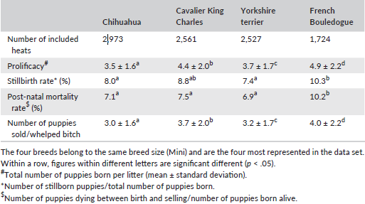 was 87.8% and abortion rate was 6.8%. Finally, 81.9% of the mated
females gave birth to a litter. On 37,946 litters (204,537 puppies),
mean litter size was 5.4 ± 2.8 puppies (range 1–24), which was
influenced by breed size and dam age (p < .0001). Stillbirth rate
was 7.4% and puppy mortality rate (stillbirth + mortality until 2
months of age) was 13.4%. Prolificacy and puppy mortality rates were
affected by breed size and within a breed size, by breed. Despite
probable approximations (as data originate from breeders
declaration), this large-scale analysis provides reference values on
reproductive performance in dogs.
was 87.8% and abortion rate was 6.8%. Finally, 81.9% of the mated
females gave birth to a litter. On 37,946 litters (204,537 puppies),
mean litter size was 5.4 ± 2.8 puppies (range 1–24), which was
influenced by breed size and dam age (p < .0001). Stillbirth rate
was 7.4% and puppy mortality rate (stillbirth + mortality until 2
months of age) was 13.4%. Prolificacy and puppy mortality rates were
affected by breed size and within a breed size, by breed. Despite
probable approximations (as data originate from breeders
declaration), this large-scale analysis provides reference values on
reproductive performance in dogs.
Duration of pregnancy is shorter in Cavalier King Charles spaniels. Kristina Baltutis, Katherine Settle, Theresa Beachler, Sara Lyle, C. Scott Bailey. Clin. Theriogenology. December 2020;12:475–480. Quote: Duration of pregnancy was evaluated in 17 [litters from 10] Cavalier King Charles spaniel bitches [in central North Carolina], based on timing of LH surge (progesterone concentrations between 2.0 and 3.0 ng/ml, with continued rise in subsequent 48 hours) and compared to 17 bitches of other breeds [Lucas terrier, Tibetan terrier, French bulldog, English springer spaniel, Border collie, Airedale terrier, Nova Scotia duck tolling retriever, Labrador retriever, Polish lowland sheepdog, German shorthaired pointer, Briard, black and tan Coonhound, German shepherd dog, Gordon setter, Doberman pinscher, Rottweiler, and Great Dane]. Duration of pregnancy was shorter in Cavalier King Charles spaniel bitches compared to others (mean ± SD, 62.8 ± 2.0 days [range; 60 - 66 days] versus 64.5 ± 1.4 days [range; 62 - 68 days]). This observation has clinical implications for pregnancy management of this breed, including recommendations for scheduling a timed caesarean section and approaches to managing late-term complications.
Ultrasonography for the evaluation of pregnancy in the female canine. Cheryl Lopate. Reproduction in Domestic Anim. September 2023; doi: 10.1111/rda.14446. Quote: Ultrasonography can be used for canine pregnancy diagnosis, determination of gestational age, assessing foetal maturation and readiness for birth, monitoring high-risk pregnancies, assessing foetal distress, evaluating bitches in dystocia and foetal gender determination. As the quality and resolution of ultrasound machines have improved, the clinician's abilities to utilize ultrasound as an integral part of reproductive evaluation of all aspects of pregnancy have exponentially increased. This paper reviews the use of ultrasonography throughout pregnancy and the advances made in the interpretation of captured images. ... In the author's experience, Cavalier King Charles Spaniels also have shorter gestation length. In another study using Drever bitches, it was shown that once litter size exceeds the average for the breed, that each additional puppy will reduce gestation length by 0.25 days.
RETURN TO TOP
pulmonic stenosis:
Percutaneous balloon valvuloplasty in four dogs with pulmonic stenosis. S. E. Brownlie, M. A. Cobb, J. Chambers, G. Jackson, S. Thomas. J. Sm. Anim. Pract. April 1991; doi: 10.1111/j.1748-5827.1991.tb00537.x. Quote: Four young dogs [2 cavalier King Charles spaniels (50%)] with severe pulmonic stenosis were treated by percutaneous balloon valvuloplasty. Pressure gradients between the right ventricle and pulmonary artery were markedly reduced by the procedure resulting in dramatic improvements in growth rate and clinical condition. No postoperative problems were encountered. ... Case 2 was a five-month-old male Cavalier King Charles spaniel. ... The dog had a grade IV/V harsh pansystolic murmur in the pulmonic area. Right axis deviation was present on ECG examination and echocardiography showed right ventricular hypertrophy and an enlarged right atrium. Chest radiographs revealed right-sided enlargement and hypovascular lung fields. A non-selective angiogram was performed under sedation with acetylpromazine/pethidine. This demonstrated very severe subvalvular pulmonic stenosis and a large post stenotic dilation. ... Case 3 was also a male five-month-old Cavalier King Charles Spaniel puppy which had a grade V/V harsh pansystolic murmur and a palpable thrill in the pulmonic area on the left side of the thorax. The murmur was also clearly audible on the right side. The puppy was bright, with good exercise tolerance, but very stunted, weighing only 3 kg. ECG, chest radiographs and ultrasound results were similar to the previous case. A nonselective angiogram showed severe pulmonic stenosis and a large post stenotic dilatation. During catheterisation for valvuloplasty, the catheter slipped from the right ventricle into the left demonstrating the presence of a ventricular septa1 defect.
Pulmonary artery lesions in Cavalier King Charles Spaniels. Karlstam E, Häggström J, Kvart C, Jönsson L. Michaelsson, M. Vet. Rec. August 2000;147:166–167. Quote: Abstract Postmortem samples from 7 Cavalier King Charles Spaniels (aged 8-9 years) that died or been killed because of problems associated with chronic valvular disease (CVD) were examined. The findings included mitral CVD and dilation of the left atrium and ventricle in all dogs, and left atrial rupture in two and severe pulmonary artery thrombosis in one. all dogs also showed wrinkling and irregularity of the endothelial surface of the main pulmonary artery and its branches. The pulmonary artery wall showed sever intimal thickening due to subendothelial fibrosis and abundant vacuolated, Alcian blue-PAS-positive intercellular substances compatible with glycosaminoglycans (GAGS). GAGS were also found in the tunica media. In more sever cases the internal elastic lamina, separating the tunica intima from the tunica media was fragmented and poorly discernible. It is suggested that CVD is part of a more generalised connective tissue diseases. ... In cavalier King Charles spaniels, intimal thickening and breaks in the internal elastic lamina of the femoral artery have been reported (Buchanan and others 1997), and these lesions were associated with thrombosis and vascular occlusion. ... The cases presented here were seven consecutive, nonselected spaniels that underwent postmortem examination over a comparably short period of time. The observations reported indicate that pulmonary vascular lesions are common in middle-aged to old cavalier King Charles spaniels. However, the study does not allow a conclusion as to the true prevalence.
Retrospective review of congenital heart disease in 976 dogs. P. Oliveira, O. Domenech, J. Silva, S. Vannini, R. Bussadori, C. Bussadori. J. Vet. Intern. Med. March 2011; doi: 10.1111/j.1939-1676.2011.0711.x. Quote: Background: Knowledge of epidemiology is important for recognition of cardiovascular malformations. Objective: Review the incidence of congenital heart defects in dogs in Italy and assess breed and sex predispositions. Animals: Nine hundred and seventy-six dogs diagnosed with congenital heart disease (CHD) of 4,480 dogs presented to Clinica Veterinaria Gran Sasso for cardiovascular examination from 1997 to 2010. Methods: A retrospective analysis of medical records regarding signalment, history, clinical examination, radiography, electrocardiography, echocardiography, angiography, and postmortem examination was performed. Breed and sex predisposition were assessed with the odds ratio test. Results: CHD was observed in 21.7% of cases. A total of 1,132 defects were observed with single defects in 832 cases (85%), 2 concurrent defects in 132 cases (14%), and 3 concurrent defects in 12 cases (1%). The most common defects were pulmonic stenosis (PS; 32.1%) [6 cavalier King Charles spaniels (1.6%)], subaortic stenosis (SAS; 21.3%), and patent ductus arteriosus (20.9%), followed by ventricular septal defect (VSD; 7.5%), valvular aortic stenosis (AS; 5.7%), and tricuspid dysplasia (3.1%). SAS, PS, and VSD frequently were associated with other defects. Several breed and sex predispositions were identified. Conclusions and Clinical Relevance: The results of this study are in accordance with previous studies, with slight differences. The breed and sex predilections identified may be of value for the diagnosis and screening of CHD in dogs. Additionally, the relatively high percentage of concurrent heart defects emphasizes the importance of accurate and complete examinations for identification. Because these data are from a cardiology referral center, a bias may exist.
Diagnosis and treatment of pulmonic stenosis. Emily Dutton. Vet. Times. June 2015;22-24. Quote: "This article will review the importance of early detection of heart murmurs, including the diagnosis and management of one of the most common congenital cardiac defects – pulmonic stenosis. Early recognition of congenital heart disease is important to achieve appropriate medical or surgical management, improve outcome and provide an accurate prognosis. The four most common congenital cardiac defects reported in prevalence studies are pulmonic stenosis; patent ductus arteriosus; sub-aortic stenosis; and ventricular septal defects. When severe, these can all result in clinical signs and premature death. However, not every dog with one of these conditions will develop clinical signs or die prematurely. For example, cardiologists grade pulmonic stenosis into one of three categories – mild, moderate or severe. We know dogs with moderate pulmonic stenosis may not have a discernibly worse prognosis than those with mild disease. Pulmonic stenosis: Maldevelopment of the distal bulbus cordis may lead to pulmonary valve stenosis. Breeds at increased risk for pulmonic stenosis include English bulldog, boxer, beagle, Samoyed, French bulldog, West Highland white terrier, Labrador retriever, cavalier King Charles spaniel and bullmastiff. Pulmonic stenosis can be classified as sub-valvular, valvular or supra-valvular according to the location of the lesion. Valvular stenosis is the most common form. There are two main types – A and B. Type A is typified by fusion of the peripheral edges of the semilunar valves (commissural fusion) and poststenotic dilatation of the pulmonary trunk. Type B may involve hypoplasia of the pulmonary annulus and pulmonary trunk, with the cusps appearing thickened and immobile6. Type A is more prevalent. The two forms are not mutually exclusive."
Outcome of valvuloplasty in three dogs with pulmonic stenosis. Chollada Buranakarl, Anusak Kijtawornrat, Wasan Udayachalerm, Sirilak Disatian Surachetpong, Saikaew Sutayatram, Pasakorn Briksawan, Sumit Durongphongtorn, Rampaipat Tungjitpeanpong, Nardtiwa Chaivoravitsakul. 9th Vet. Practitioners Assn. of Thailand (VPAT) Regional Vet. Cong. July 2016. Quote: Valvuloplasty is the interventional technique for correction of pulmonic stenosis (PS), one of the most common congenital heart disease in dogs. The procedure was less invasive and the results are commonly satisfied. However, the success may depend on many factors such as the severity of stenosis, type of PS, the presence of aberrant coronary artery and other concurrent cardiac diseases. The procedure includes insertion of special catheter with balloon at the tip and advanced into the area of stenosis. The balloon is inflated under fluoroscopy to the disappearance of the waist of stenotic area. We report the first 3 cases of PS. All dogs had stenosis at valvular area. However, the second dog [a cavalier King Charles spaniel] had additional subvalvular stenosis while the third dog also had ventricular septal defect (VSD). (See also a more extensive report on this CKCS, below.)
Special surgical procedure by UF vets saved Rumple's life. Gainesville Sun. July 2016.
Balloon valvuloplasty for pulmonic stenosis in two dogs: case report. Chollada Buranakarl, Anusak Kijtawornrat, Wasan Udayachalerm, Sirilak Disatian Surachetpong, Saikaew Sutayatram, Pasakorn Briksawan, Sumit Durongpongtorn, Rampaipat Tungjitpeanpong, Nardtiwa Chaivoravitsakul. Thai J. Vet. Med. December 2016;46(4):713-721. Quote: Balloon valvuloplasty for correction of congenital pulmonic stenosis (PS) in two dogs by intervention technique was first reported in Thailand. ... In the present study, dog no.1 was Chihuahua and dog no.2 was Cavalier King Charles Spaniel. Chihuahua is one of the most popular breeds of pet dogs in Thailand, whereas Cavalier King Charles Spaniel is uncommon. ... Both dogs did not show any clinical signs related to congestive heart failure. Physical examination in both dogs revealed heart murmur. Electrocardiographs of both dogs were recorded and showed normal sinus rhythm with deep S wave. In the first dog, thoracic radiograph revealed cardiomegaly with enlargement of the main pulmonary artery and left deviation of the apex due to right ventricular enlargement. Definitive diagnosis of PS was performed by echocardiography. Computed tomography (CT) of both dogs demonstrated stenosis of the pulmonic valve with immediate post-stenotic dilatation of the pulmonary trunk. ... In the current report, both dogs were type A with severe PS (systolic pressure gradient > 80 mmHg). These dogs had no concurrence of cardiac disease. By performing CT, no aberrant coronary artery was observed. Thus, cardiac intervention with BV was highly recommended. ... Both left and right coronary arteries originated from their aortic sinuses. In addition, in dog no.2 (Case 2: A 15-month-old Cavalier King Charles Spaniel weighing 6 kg was referred from another veterinary hospital for echocardiography. Neither history of syncope nor exercise intolerance was found. Physical examination revealed that the dog was bright and alert. Heart rate and respiratory rate were 120 beats/min and 32 breath/min, respectively. Systolic murmur grade V/VI was presented along with precordial thrill from chest palpitation.) the CT scan showed slight narrowing of the subvalvular area, corresponding to that seen during echocardiography. Both dogs received PS correction by balloon valvuloplasty under fluoroscopic guidance at the Small Animal Teaching Hospital, Faculty of Veterinary Science, Chulalongkorn University. The procedures were successful as suggested by reduced pressure gradient without major complications. Seven days after the operation the pressure gradient reduced as assessed by echocardiography and both dogs remained healthy. ... In conclusion, this study is the first to report the procedure for BV performed in Thailand in two dogs with type A pulmonic stenosis, of which one dog had additional subvalvular stenosis. During the follow-up there were improvements in clinical signs in both dogs associated with enhanced pulmonary outflow due to the reduction in right ventricular afterload. (See a briefer report on the same CKCS, above.)
Anaesthetic management and complications of balloon valvuloplasty for pulmonic stenosis in dogs. J. Viscasillas, S. Sanchis-Mora, C. Palacios, A. Mathis, H. Alibhai, D. C. Brodbelt. Vet. Rec. October 2015; doi: 10.1136/vr.103146. Quote: Pulmonic stenosis (PS) is a common congenital cardiac defect in dogs (Buchanan 1992). Depending on the type of stenosis and its severity, surgical management is required. Balloon valvuloplasty (BV) is a non-invasive surgical technique that has become commonly performed in veterinary medicine (Bussadori and others 2001). Several protocols to anaesthetise patients undergoing this procedure have been described. However, only one retrospective study (Ramos and others 2014) assessing and reporting the perioperative complications during BV has been published recently. The aim of this study was to describe the anaesthetic management used over the past years in a large referring European veterinary institution and to explore which major factors are associated with complications in dogs undergoing BV. Forty-six dogs in which PS was managed with BV were included in this study. ... The most commonly represented breed was the cocker spaniel (eight patients, 17 per cent), followed by the boxer and Cavalier King Charles (three patients, 6 per cent each of them). The median age was 11.5 months (range 2–85 months) and median weight was 10.3 kg (range 2.5–65.5 kg).
Breed does not affect the association between murmur intensity and disease severity in dogs with pulmonic or subaortic stenosis. M. Rishniw, D. Caivano, D. Dickson, S. Swift, C. Rouben, S. Dennis, C. Sammarco, J. Lustgarten, I. Ljungvall. J. Sm. Anim. Pract. April 2019; doi: 10.1111/jsap.13015. Quote: Objectives: To determine whether breed affects the ability of murmur intensity to predict the severity of stenosis in dogs with pulmonic stenosis or subaortic stenosis. Materials and Methods: Retrospective multi-investigator study of dogs with pulmonic stenosis or subaortic stenosis. Murmur intensity, assessed by a four-level classification scheme, was compared with echocardiographically-determined pressure gradient across the affected valve. Breeds represented by at least 10 dogs at any murmur intensity were compared to determine the effect, if any, of breed. Results: A total of 1088 dogs (520 with pulmonic stenosis and 568 with subaortic stenosis, representing 106 breeds and the mixed breed group) were included; 208 dogs had soft, 210 had moderate, 283 had loud and 387 had palpable murmurs. Fifteen breeds were represented by at least 10 dogs: five breeds with at least 10 dogs had soft murmurs (132 dogs), nine breeds had moderate murmurs (149 dogs), 10 breeds had loud murmurs (188 dogs), and 11 breeds had palpable murmurs (286 dogs). No breeds differed in stenosis severity from any other breeds within any murmur grade. Post hoc power calculations suggested that we would have been able to detect at least a moderate or large effect size, had one existed. Several dogs with soft murmurs had more-than-mild disease severity. ... We found only one breed with a large number of individuals that expressed only one disease – the cavalier King Charles spaniel, in which investigators only diagnosed pulmonic stenosis. While this breed has been reported to have pulmonic stenosis (Oliveira et al. 2011, Viscasillas et al. 2015), a previous study found no increased odds in this breed (Oliveira et al. 2011). These previous studies reported only nine cases of cavalier King Charles spaniels with pulmonic stenosis, whereas our study reported 26 cases, identified by six of seven investigators, all of whom practice in different parts of the world. In addition, most cavalier King Charles spaniels in our study had severe disease (Table 2). Therefore, our data suggest that cavalier King Charles spaniels might be predisposed to pulmonic stenosis, although the popularity of this breed might account for the high number of observed cases. ... Clinical Significance: Despite anecdotally perceived differences in the detection of heart murmurs between breeds, which have been proposed to potentially affect the interpretation of stenosis severity, we found no obvious breed effect in the ability to predict severity of stenosis.
Epidemiological study of congenital heart diseases in dogs: Prevalence, popularity, and volatility throughout twenty years of clinical practice. Paola Giuseppina Brambilla, Michele Polli, Danitza Pradelli, Melissa Papa, Rita Rizzi, Mara Bagardi, Claudio Bussadori. PLoS One. July 2020;15(7):e0230160. Quote: The epidemiology of Congenital Heart Diseases (CHDs) has changed over the past twenty years. This study aimed to evaluate the prevalence of CHDs in the population of dogs recruited in a single referral center (RC); compare the epidemiological features of CHDs in screened breeds (Boxers) versus non-screened (French and English Bulldogs, German Shepherds); investigate the association of breeds with the prevalence of CHDs; determine the popularity and volatility of breeds over a 20-year period; analysed the trends of the most popular breeds in the overall population of new-born dogs registered in the Italian Kennel Club (IKC) from 1st January 1997 to 31st December 2017. The RC's cardiological database was analysed, and 1,779 clinical records were included in a retrospective observation study. Descriptive statistics and frequencies regarding the most representative breeds and CHDs were generated. A logistic regression model was used to analyse the trends of the most common CHDs found in single and in cluster of breeds. The relationship between breed popularity and presence of CHDs was studied. The most common CHDs were Pulmonic Stenosis (PS), Patent Ductus Arteriosus (PDA), Subaortic Stenosis (SAS), Ventricular Septal Defect (VSD), Aortic Stenosis (AS), Tricuspid Dysplasia (TD), Atrial Septal Defect (ASD), Double Chamber Right Ventricle (DCRV), Mitral Dysplasia (MD), and others less frequent. The most represented pure breeds were Boxer, German Shepherd, French Bulldog, English Bulldog, Maltese, Newfoundland, Rottweiler, Golden Retriever, Chihuahua, [Poodle, Labrador Retriever, Cavalier King Charles Spaniel] and others in lower percentage. Chihuahuas, American Staffordshire Terriers, Border Collies, French Bulldogs, and Cavalier King Charles Spaniel were the most appreciated all of which showed a high value of volatility. ... The probability of detecting a CHDi at first presentation is significantly elevated in French Bulldogs, Cavalier King Charles Spaniels, English Bulldogs and Miniature Pinschers. ... This study found evidence for the value of the screening program implemented in Boxers; fashions and trends influence dog owners' choices more than the worries of health problems in a breed. Effective breeding programs are needed in order to control the diffusion of CHDs without impoverishing the genetic pool. ... The owners are not often fully aware of the potential problems their dog may face prior to acquisition of a dog. It is also possible that owners do not perceive the clinical signs of some inherited cardiac disorders as problems, but rather as normal, breed-specific characteristics (e.g., murmur in CKCS). ... [Among the CKCSs, 40% had PS and 60% had PDA.]
RETURN TO TOP
quadricuspid aortic valve:
Quadricuspid aortic valve and associated abnormalities in the dog: Report of six cases. François Serres, Valérie Chetboul, Carolina Carlos Sampedrano, Vassiliki Gouni, Jean-Louis Pouchelon. J. Vet. Cardiol. June 2008; doi: 10.1016/j.jvc.2008.03.003. Quote: Objectives: To describe the clinical and echocardiographic findings in dogs with quadricuspid aortic valve (QAV). Background: QAV is a rare canine congenital heart disease which has been reported only three times in the young dog. Animals, materials and methods: Six dogs (0.3- to 13-year-old) with QAV diagnosed by two-dimensional echocardiography were retrospectively evaluated [including 2 cavalier King Charles spaniels (33.3%)]. Medical records, echocardiograms, and follow-ups were reviewed. Results: According to aortic cusp morphology, QAV was classified as type A [four equally or nearly equally sized cusps] (n = 1), type B [three equal large cusps and one smaller cusp] (n = 4) or type C [two equal large cusps and two equal smaller cusps] (n = 1). QAV was associated with at least one other heart disease in all of the dogs including, ventricular septal defect (n = 1) [CKCS Case #5], enlarged left coronary ostium (n = 4), degenerative mitral valve disease (MVD, n = 1) [CKCS Case #4] and patent ductus arteriosus (PDA, n = 3). Mild to moderate aortic regurgitation was also detected in all dogs by continuous-wave and color-flow Doppler echocardiography. QAV was diagnosed in four asymptomatic dogs referred for evaluation of a heart murmur. The remaining two dogs had QAV and PDA with evidence of mild exercise intolerance and moderately retarded growth. The PDA was surgically corrected in both dogs and at the time of writing, 1–2.5 years after the initial diagnosis, none of the six animals shows evidence of clinical signs. Conclusion: QAV is a cause of aortic insufficiency. It may incidentally be found by two-dimensional echocardiography in dogs of various ages in association with other congenital or acquired cardiac abnormalities.
RETURN TO TOP
salivary glands disorders:
• hypersialism:
Phenobarbitone-responsive hypersialism in two dogs. B. L. Chapman, R. Malik. J. Sm. Anim. Pract. November 1992; doi: 10.1111/j.1748-5827.1992.tb01051.x. Quote: Two dogs [both cavalier King Charles spaniels] were examined for hypersialism associated with non-painful, symmetrical enlargement of both mandibular salivary glands. These signs were accompanied by inappetence and weight loss. ... The cause of hypersialism in the two dogs of this report could not be determined despite extensive investigations. It is not known why the condition responded to anticonvulsant medication. ... The increased size of the mandibular salivary glands and absence of structural or functional abnormalities within the oropharynx suggests that hypersialism was attributable to increased production of saliva and, or, a reluctance to swallow, rather than an inability to swallow. ... The underlying cause of the problem could not be determined despite extensive investigations including fine needle aspiration biopsy of affected salivary glands. Hypersialism and mandibular salivary gland enlargement resolved completely during phenobarbitone administration. ... This paper describes hypersialism associated with mandibular salivary gland enlargement in two female cavalier King Charles spaniels which resolved completely with phenobarbitone dosing. ... Severe hypersialism and mandibular salivary gland enlargement comprise a serious and potentially life-threatening syndrome. Affected dogs may be inappetent and lose weight, they are at risk of aspiration and constant drooling is a major management problem for owners who may consider euthanasia. Extensive investigations are required to rule out the various causes of hypersialism, including procedures such as salivary gland biopsy, cerebrospinal fluid analysis and electroencephalography which were not carried out in the present cases. In dogs where hypersialism is associated with non-painful enlargement of both mandibular salivary glands, and investigations have ruled out sialadenitis and oropharyngeal disease, it may be prudent to consider a therapeutic trial with phenobarbitone before proceeding with more invasive diagnostic procedures.
• sialadenitis:
Phenobarbital-Responsive Bilateral Zygomatic Sialadenitis following an Enterotomy in a Cavalier King Charles Spaniel. Paoul S. Martinez, Renee Carter, Lorrie Gaschen, Kirk Ryan. Res. J. Vet. Practioners. August 2018; doi: 10.17582/journal.rjvp/2018/6.2.14.19. Quote: This report describes a case of acute onset phenobarbital-responsive zygomatic sialadenitis in a Cavalier King Charles Spaniel diagnosed by computed tomography of the skull. A 6 year old male castrated Cavalier King Charles Spaniel presented ... for a three day history of bilateral acute trismus, exophthalmos, and blindness. These signs developed after an exploratory laparotomy and gastrointestinal foreign body removal was performed. On presentation, the ophthalmic examination revealed bilateral decreased retropulsion, absent bilateral direct and consensual pupillary light responses, bilateral blindness, and a superficial ulcer in right eye. Ocular ultrasound and computed tomography of the skull were performed and identified a bilateral retrobulbar mass effect originating from the zygomatic salivary glands. Ultrasound guided fine needle aspirate cytology revealed neutrophilic inflammation. Fungal testing and bacterial cultures were performed which were negative for identification of an infectious organism. The patient was placed on a 5-day course of antibiotic therapy and the response was poor. Phenobarbital was added to the treatment regimen. Two days following phenobarbital administration, the exophthalmos and trismus began to resolve. Following phenobarbital therapy, ocular position normalized, trismus completely resolved, and patient returned to normal behavior although blindness remained in both eyes. No other neurologic condition occurred. This paper highlights the early use of phenobarbital for the rapid resolution of zygomatic sialadenitis.
• sialoceles:
Clinical and CT sialography findings in 22 dogs with surgically confirmed sialoceles. Yi Lin Tan, Ana Marques, Tobias Schwarz, Jordan Mitchell, Tiziana Liuti. Vet. Rad. & Ultrasound. May 2022; doi: 10.1111/vru.13104. Quote: Sialoceles are an uncommon canine salivary gland disease, and complete surgical resection is important for a positive outcome. ... In dogs, the most commonly affected gland is the monostomatic sublingual gland, giving rise to the characteristic ventral neck swelling and ranula formation. Underlying causes of sialoceles have been suggested to include trauma, foreign bodies, sialolithiasis, and obstruction of the duct secondary to inflammation. Idiopathic sialocele formation, where an underlying cause has not been identified, has been more commonly reported. ... A total of 51 confirmed cases of sialoceles were retrieved from the HfSA database, with 22 dogs meeting the inclusion criteria [including one cavalier King Charles spaniel]. ... The most common clinical sign was a mass or swelling in the mandibular region (11/22, 50.0%), and three of the 22 dogs (13.6%) displayed a combination of two or more of [these clinical signs: mandibular swelling/mass; neck swelling/mass; sublingual swelling; exopthalmos (abnormal protrusion of the eyeball from its socket).]. ... Radiographic sialography has been described as a diagnostic test for presurgical planning; however, superimposition artifacts may limit the diagnosis and detection of all affected glands. Computed tomographic (CT) sialography is a promising technique for delineating the salivary gland apparatus. The aims of this retrospective, observational study were to describe clinical and CT sialographic findings in a group of dogs with confirmed sialoceles, to determine the sensitivity of CT sialography for detecting affected salivary glands using surgery as the reference standard and to determine interobserver agreement for CT sialographic assessments. Dogs were included if they underwent a CT sialography study followed by surgical resection of the diseased gland(s) and histopathological analysis. Computed tomography sialography studies of dogs with surgically confirmed sialoceles (n = 22) [including the cavalier King Charles spaniel] were reviewed by a European College of Veterinary Diagnostic Imaging (ECVDI)-certified radiologist and an ECVDI resident. Interobserver agreement was calculated using Cohen's kappa statistics. CT sialography results were compared to surgical findings to determine sensitivity. Contrast leakage was detected in 12 of 22 dogs (54.5%) [including the cavalier], with intrasialocele leakage being most frequently observed (7/12, 58.3%) [including the cavalier]. There was substantial agreement (κ = 0.70) between reviewers identifying diseased glands, substantial agreement (κ = 0.62) on the diagnostic quality, and no to slight agreement (к = 0.13) in the detection of contrast leakage. The overall sensitivity of CT sialography to detect surgically confirmed diseased glands was 66.7% (95% confidence interval: 48.8-80.8). In conclusion, these findings support the use of CT sialography as an adjunct diagnostic test for treatment planning in dogs with sialoceles.
• sialolithiasis:
Parotid
sialolithiasis in a dog. D. A. Jeffreys, A. Stasiw, R.
Dennis. J. Sm. Anim. Pract. June 1996; doi:
10.1111/j.1748-5827.1996.tb02385.x. Quote: A case of parotid
sialolithiasis is reported. Rupture of the [parotid] duct followed
and this was successfully treated by surgical drainage through
anatomical reconstruction of the duct. Subcutaneous swelling ventral
to the ear canal can be due to enlargement of the parotid salivary
gland or to pathology of the surrounding tissues. ... An
eight-week-old neutered female Cavalier King Charles spaniel
presented with a tense, unilateral subcutaneous swelling ventral to
the right ear canal in the region of the parotid salivary gland. ...
The radiology report was as follows. The ventrodorsal view of the
skull showed a linear, calcified structure approximately 15 mm long
and 2 mm wide within the soft tissues of the right side of the face,
rostral to the ear canal. This was confirmed to be ventral to the
ear canal on the orthogonal view. Transverse lines suggested that
this structure consisted of multiple small particles stacking up
behind each other. A diagnosis of parotid sialolithiasis was made.
The patient was re-examined under general anaesthesia. ...
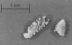 The
whole of the right side of the face was prepared for surgery and an
initial incision was made over the expected path of the duct. The
tissues were dissected to identify the duct. ... Gross examination
revealed that the [parotid] duct had ruptured approximately
two-thirds of its length proximally and a copious exudate was
released from the soft tissues at this level. ... [T]wo sialoliths
[were] identified and removed (Fig 2 at right). These were
dissimilar in length and showed transverse grooves, which had given
rise to the radiographic appearance of multiple, small sialoliths.
... In this case, once the sialoliths had been removed a large
defect remained in the parotid duct. Ligation of the parotid duct
proximal to such a defect has been described as a simple and
effective way of dealing with such a lesion (Harvey 1977). In this
case, however, anatomical reconstruction of the duct produced a
successful outcome. This approach was chosen because of concern
about infection in the salivary gland. This case illustrates that,
while rare in dogs, blockage of the parotid duct by a sialolith,
followed by rupture of the duct, can be successfully managed by
anatomical reconstruction following removal of the sialolith.
The
whole of the right side of the face was prepared for surgery and an
initial incision was made over the expected path of the duct. The
tissues were dissected to identify the duct. ... Gross examination
revealed that the [parotid] duct had ruptured approximately
two-thirds of its length proximally and a copious exudate was
released from the soft tissues at this level. ... [T]wo sialoliths
[were] identified and removed (Fig 2 at right). These were
dissimilar in length and showed transverse grooves, which had given
rise to the radiographic appearance of multiple, small sialoliths.
... In this case, once the sialoliths had been removed a large
defect remained in the parotid duct. Ligation of the parotid duct
proximal to such a defect has been described as a simple and
effective way of dealing with such a lesion (Harvey 1977). In this
case, however, anatomical reconstruction of the duct produced a
successful outcome. This approach was chosen because of concern
about infection in the salivary gland. This case illustrates that,
while rare in dogs, blockage of the parotid duct by a sialolith,
followed by rupture of the duct, can be successfully managed by
anatomical reconstruction following removal of the sialolith.
Retropharyngeal pain and swelling in a dog. Geraldine B. Hunt, K. R. Youmans, D. J. Simpson, R. J. Steel. July 1997; Aust. Vet. J.;75(7):476, 491. Quote: A 7-year-old desexed male Cavalier King Charles Spaniel was referred to The University of Sydney Veterinary Teaching Hospital with a history of three episodes of acute swelling of the left retropharyngeal region over an 8-week period. These episodes were characterised by severe pain on palpation of the affected area, pain on opening the mouth and oedema of the surrounding tissues. ... It had a painful soft tissue swelling ventral to the left ear, with oedema extending into the intermandibular area. Tenacious, turbid saliva was present at the cornmissure of the mouth. ... The radiograph revealed a radiodense foreign body (sialolith) rostrolatera1 to the zygomatic arch. An open-mouth radiograph shows the foreign body ventral to the zygomatic arch. This location corresponds to the intraoral portion of the parotid salivary duct. ... A 1-cm incision through the oral mucosa revealed a single large sialolith in the distal portion of the parotid salivary duct. Chemical analysis showed it to be composed mainly of calcium carbonate with some magnesium ammonium phosphate. Pain and swelling resolved promptly after removal of the sialolith, but recurred 4 months later, at which time the parotid salivary gland and its duct were removed surgically. ... Ultrasonographic examination early in the course of the present case failed to demonstrate a sialolith, however, this may have been because the sialolith was located in the terminal, intraoral portion of the salivary duct, rather than the region of maximum swelling. If the condition is identified early, before dilatation and distortion of the duct occur, removal or reduction of the sialolith using minimally invasive techniques has been shown to be effective in relieving signs in humans. In the present case, the chronic changes within the salivary duct presumably predisposed the dog to infection even after removal of the sialolith, necessitating surgical extirpation of the gland and duct.
RETURN TO TOP
sand impaction:
Sand impaction of the small intestine in eight dogs. A. D. Moles, J. A. McGhie, O. R. Schaaf, R. Read. J. Sm. Anim. Pract. December 2009; doi: 10.1111/j.1748-5827.2009.00847.x. Quote: Objective: To describe signalment, clinical fi ndings, imaging and treatment of intestinal sand impaction in the dog. Methods: Medical records of dogs with radiographic evidence of small intestinal sand impaction were reviewed. Results: Sand impaction resulting in small intestinal obstruction was diagnosed in eight dogs ... [including] a cavalier King Charles spaniel. All dogs presented with signs of vomiting. Other clinical signs included anorexia, lethargy and abdominal pain. Radiographs confi rmed the presence of radio-opaque material consistent with sand causing distension of the terminal small intestine in all dogs. Four dogs were treated surgically for their impaction and four dogs were managed medically. Seven of the eight dogs survived. ... One dog (dog 8) [the cavalier King Charles spaniel] died, suffering respiratory and cardiac arrest immediately following surgery. This dog had previously been diagnosed with cardiac disease by the referring veterinarian and was being medically managed with pimobendan (Vetmedin; Boehringer Ingelheim) and frusemide (Frudix; Jurox). The dog became increasingly acidotic (pH 7·065) and hyperlactataemic (5·6 mmol/l (reference range 0 to 2·5 mmol/l)) following presentation. It is likely that the combination of the cardiac disease, acidosis, gastrointestinal disease and surgery all contributed to this dog’s death. ... Clinical Significance: Both medical and surgical management of intestinal sand impaction in the dog can be effective and both afford a good prognosis for recovery.
RETURN TO TOP
shadow chasing:
A Cavalier King Charles dog with shadow chasing: Clinical recovery and normalization of the dopamine transporter binding after clomipramine treatment. Simon Vermeire, Kurt Audenaert, Andre Dobbeleir, Eva Vandermeulen, Tim Waelbers, Kathelijne Peremans. J.Vet.Behavior: Clinical Applications & Research. Nov. 2010;5(6):345-349. Quote: A 30-month-old female Cavalier King Charles dog was presented with a history of worsening compulsive behavior (shadow chasing). In vivo brain imaging using single-photon emission computed tomography and the dopamine transporter (DAT)-specific radiopharmaceutical 123I-FP-CIT revealed a significantly higher DAT striatal-to-brain ratio. Treatment was started with the tricyclic antidepressant clomipramine 2.5 mg/kg PO, q. 12 hours. After 2 months of medication that resulted in clinical improvement, the DAT binding regained normal values. ... In our case, clinical improvement was already noticeable after 7 days of medication. ... After 2 months of treatment, a post-therapeutic DAT scan was performed. The new DAT ratios were 10.82 and 12.49 for left and right striatum, respectively, both ratios located closely to or in the normal range. To minimize the gluttony, an attempt was made to reduce the dose of clomipramine without renewal of shadow chasing. A dose of 2.5 mg PO, q. 24 hours was daily alternated with a dose of 2.5 mg PO, q. 12 hours, with success. However, not giving the medication dose resulted in relapse of shadow chasing already after 2 days.
RETURN TO TOP
temporomandibular joint morphology:
The effect of obliquity on the radiographic appearance of the temporomandibular joint in dogs. Alison M. Dickie, Martin Sullivan. Vet. Radiology & Ultrasound. May 2001;42(3):205-217. Quote: The temporomandibular joint is formed between the condyloid process of the mandible and the mandibular fossa of the temporal bone. The basic anatomy of this joint was assessed and described in a series of skulls including dolichocephalic, mesaticephalic and brachycephalic breeds. The facial index and rotational angles were measured with the facial index providing a useful method of classifying skull types but the rotational angle being of limited use in assessment of the temporomandibular joint until normal breed values are established. ... Breed size within the brachycephalic group appeared to influence facial index with large brachycephalic breeds (boxer and mastiff) producing lower facial indices (159 to 190, mean 172) and small breeds (cavalier king Charles spaniel, Pekingese, Chihuahua) producing larger indices (2 15 to 480, mean 231). ... The temporomandibular joints of the cavalier king Charles spaniel and Pekingese skulls in the anatomical collection were considered to be grossly abnormal. They were therefore excluded from further consideration in the present study and their rotational angles omitted from Table 2. ... Equipment was designed to allow repeatable positioning of the temporomandibular joint for radiography at a variety of lateral and long axis rotational angles relative to the central x-ray beam. The regions of the joint and anatomic features visualized in each view are demonstrated. 10° rotation was required in either axis to project the joints independently of each other. Lateral rotational angles of 10 to 30° in mesaticephalic and dolichocephalic breeds and 20 to 30° in brachycephalics and long axis rotational views of 10 to 30° depending on the region of interest were considered to be the most useful.
Temporomandibular joint morphology in cavalier king charles spaniels. Alison M. Dickie, Tobias Schwarz, Martin Sullivan. Veterinary Radiology & Ultrasound. May 2002;43:260. Quote: “Temporomandibular joint dysplasia has been reported in several breeds of dog. Three Cavalier King Charles Spaniel (CKCS) skulls were examined and radiographed during the course of a separate study and all demonstrated changes consistent with bilateral temporomandibular joint dysplasia. Subsequently, skull radiographs from all CKCS dogs examined at the University of Glasgow Veterinary School between 1991 and 2001 were reviewed (n = 26). Only two of these dogs were radiographed specifically for investigation of the temporomandibular joint, although varying degrees of dysplasia were identified in all dogs where the joints were adequately visualized (n = 20). The head of four CKCS cadavers was also radiographed, and similar changes were found. This finding suggests that temporomandibular joint dysplasia is a widespread asymptomatic condition in the CKCS and should be regarded as a normal morphologic variation rather than a pathologic anomaly. Subtle changes are best seen on lateral oblique radiographs, although marked changes are also visible on dorsoventral views. The rotational angle or angle of articulation of each of the dysplastic mandibular condyles was measured and was related to the severity of the dysplastic changes. However, there was overlap between the values calculated for these abnormal joints and normal ones in other breeds, suggesting this measurement was of limited significance and the shape of the components of the temporomandibular joint are more relevant when assessing this joint for the presence of dysplastic changes.”
Morphologic and Morphometric Description of the Temporomandibular Joint in the Domestic Dog Using Computed Tomography. Lenin A. Villamizar-Martinez, Cristian M. Villegas, Marco A. Gioso, Alexander M. Reiter, Geni C. Patricio, Ana C. Pinto. J. Vet. Dentistry. August 2016;33(2):75-82. Quote: The temporomandibular joint (TMJ) in the domestic dog is a synovial joint with 2 articular surfaces, the mandibular fossa of the squamous portion of the temporal bone and the articular head of the condylar process of the mandible. Although different diagnostic imaging techniques have been used to study the TMJ in dogs, morphologic and morphometric studies based on computed tomography (CT) are scarce. The purpose of the present study was to describe the morphologic and morphometric features of the TMJ in domestic dogs using CT. Width and depth of the mandibular fossa and 2 different angles between the mandibular fossa and the condylar process were measured in 96 TMJs of 48 dogs of different breeds (Labrador retriever, German shepherd, cocker spaniel, boxer, English bulldog, pug, shih tzu, and Cavalier King Charles spaniel [11 CKCSs]). Temporomandibular joint conformation differed between breeds. Mid- and small-sized dogs had mandibular fossae that were more shallow, less developed retroarticular processes, and irregularly shaped condylar processes. The TMJs were more congruent in large dogs, presenting with deeper mandibular fossae, prominent retroarticular processes, and more uniform condylar processes. The measurements proposed in this study demonstrated 3 different morphologic conformations for the TMJ in the dogs of this study. ... The TMJs of Cavalier King Charles spaniels showed remarkably lower values associated with shallow mandibular fossae and incongruent articular heads. ... Cavalier King Charles spaniels had the shallowest mandibular fossae of all breeds. The retroarticular process appeared as a bony extension on the medioventral aspect of the mandibular fossa, articulating with the caudomedial aspect of the condylar process. This bony extension was prominent in Labrador retrievers, German shepherds, boxers, and English bulldogs. The retroarticular process was less developed in cocker spaniels, shih tzus, and pug. In Cavalier King Charles spaniels, it was small or absent. ... The wide angles documented in Labrador retrievers, German shepherds, boxers, and English bulldogs were consistent with long and well-defined retroarticular processes, whereas the narrow angles found in cocker spaniels, shih tzus, Cavalier King Charles spaniels, and a pug were due to less developed or absent retroarticular processes. A dramatically reduced ventral extension of the retroarticular process, as observed in cocker spaniels and Cavaliers King Charles spaniels in this study, may permit slight caudal dislocation of the head of the condylar process and predispose to TMJ instability. ... German shepherds, boxers, and English bulldogswere consistent with deeper (more concave) mandibular fossae and more prominent retroarticular processes, whereas the narrow angles found in cocker spaniels, shih tzus, CavalierKing Charles spaniels, and the pug were due to more shallower (less concave) mandibular fossae and less prominent or absent retroarticular processes. ... The present study agrees with the asymptomatic dysplastic condition of the TMJ previously reported in the Cavalier King Charles spaniel and suggests that this morphological features may exist in certain dog breeds such as cocker spaniels and small breeds such as shih tzus and pugs.
Morphological Assessment of the Temporomandibular Joint in Asymptomatic Brachycephalic Dogs Using Computed Tomography. Emilie Paran, Sarah Bouyssou, Alison King. J. Vet. Dentistry. April 2023; doi: 10.1177/08987564231171529. Quote: Temporomandibular joint (TMJ) incongruity and morphological variations can result in clinical signs but have also been reported in asymptomatic brachycephalic dogs. The purpose of this study was to assess TMJ morphology in a group of brachycephalic dogs using computed tomography (CT). French Bulldogs, English Bulldogs, Boxers, Cavalier King Charles Spaniels (CKCS), Chihuahuas, Lhasa Apsos, Pugs, Shih Tzus, and Staffordshire Bull Terriers were retrospectively enrolled. The severity of the TMJ morphological changes was determined using a modified 5-grade classification system. The intra- and inter-observer agreements were calculated. One hundred fifty-three dogs were included. When evaluating the medial aspect of the TMJ in the sagittal plane, there was a spectrum of variations in the shape of the head of the condylar process of the mandible, the mandibular fossa and the retroarticular process ranging from a rounded concave TMJ with a long retroarticular process to a flattened TMJ with an absent process. Variations in the articular surface of the head of the condyle in the transverse plane ranged from flat, through curved and trapezoid to sigmoid. The prevalence of severe TMJ dysplasia (grades B3 and C) in the CKCS and French Bulldog was high (69.2% and 53.8%, respectively). The intra- and inter-observer agreements were moderate. Variations in TMJ morphology exist in asymptomatic brachycephalic dogs. Marked changes seem to be highly prevalent in the French Bulldog and CKCS and should be considered a breed variation. The TMJ classification described in this study could be used to standardize assessment of canine TMJ morphology. However, further research is needed to determine its clinical application.
RETURN TO TOP
tonsillitis:
Reagan Dog Has Surgery. UPI. January 1986. Quote: President Reagan's spaniel Rex successfully underwent a tonsillectomy today, the White House reported. Rex, the year-old King Charles spaniel that replaced another dog, Lucky, in December, checked into a veterinary hospital at an undisclosed location on Monday.
RETURN TO TOP
vaccines:
Long-lived immunity to canine core vaccine antigens in UK dogs as assessed by an in-practice test kit. R. Killey, C. Mynors, R. Pearce, A. Nell, A. Prentis, M. J. Day. J. Sm. Anim. Pract. January 2018;59(1):27-31. Quote: Objectives: To determine the utility of an in-practice test kit to detect protective serum antibody against canine distemper virus, canine adenovirus and canine parvovirus type 2 in a sample of the UK dog population. Materials and Methods: Serum samples from 486 dogs, last vaccinated between less than 1 month and 124 months previously, were tested with the VacciCheck™ test kit for protective antibodies against distemper, adenovirus and parvovirus type 2. Results: A high proportion of the dogs tested (93·6%) had protective antibody against all three of the core vaccine antigens: 95·7% of the dogs were seropositive against canine distemper virus, 97·3% against canine adenovirus and 98·5% against canine parvovirus type 2. The small number of dogs that were seronegative for one or more of the antigens (n = 31) may have had waning of previous serum antibody or may have been rare genetic non-responders to that specific antigen. Clinical Significance: UK veterinarians can be reassured that triennial revaccination of adult dogs with core vaccines provides long-lived protective immunity. In-practice serological test kits are a valuable tool for informing decision-making about canine core revaccination.
RETURN TO TOP
vasculitis:
Histologic and clinical features of primary and secondary vasculitis: a retrospective study of 42 dogs (2004–2011). James W. Swann, Simon L. Priestnall, Charlotte Dawson, Yu-Mei Chang, Oliver A. Garden. J.Vet. Diagnostic Investigation. June 2015. Quote: "Inflammation of the blood vessel wall has been reported infrequently in dogs, and it may occur without apparent cause (primary vasculitis) or as a pathologic reaction to a range of initiating insults (secondary vasculitis). The aims of our study were to report histologic, clinical, and survival data from a large series of cases with primary and secondary vasculitis, and to compare the clinical parameters and outcome data between groups. Clinical data was collected retrospectively from the medical records of 42 client-owned dogs [Cavalier King Charles Spaniel (6)] with a histologic diagnosis of primary or secondary vasculitis, and follow-up information was obtained. Cases were grouped according to clinical and histologic descriptors, and biochemical, hematologic, and survival data was compared between groups. Several forms of primary vasculitis were observed, and vascular inflammation was observed in conjunction with numerous other diseases. Female dogs were more likely to develop primary vasculitis, and serum globulin concentration was greater in dogs with primary vasculitis compared to those with underlying disease. All dogs with primary vasculitis of the central nervous system died or were euthanized shortly after presentation, but other forms of primary vasculitis could be managed effectively. In conclusion, presentation of clinical cases in this series was variable, and there did not appear to be well-defined vasculitic syndromes as described in people."
RETURN TO TOP
ventricular septal defect:
Quadricuspid aortic valve and associated abnormalities in the dog: Report of six cases. François Serres, Valérie Chetboul, Carolina Carlos Sampedrano, Vassiliki Gouni, Jean-Louis Pouchelon. J. Vet. Cardiol. June 2008; doi: 10.1016/j.jvc.2008.03.003. Quote: Objectives: To describe the clinical and echocardiographic findings in dogs with quadricuspid aortic valve (QAV). Background: QAV is a rare canine congenital heart disease which has been reported only three times in the young dog. Animals, materials and methods: Six dogs (0.3- to 13-year-old) with QAV diagnosed by two-dimensional echocardiography were retrospectively evaluated [including 2 cavalier King Charles spaniels (33.3%)]. Medical records, echocardiograms, and follow-ups were reviewed. Results: According to aortic cusp morphology, QAV was classified as type A [four equally or nearly equally sized cusps] (n = 1), type B [three equal large cusps and one smaller cusp] (n = 4) or type C [two equal large cusps and two equal smaller cusps] (n = 1). QAV was associated with at least one other heart disease in all of the dogs including, ventricular septal defect (n = 1) [CKCS Case #5], enlarged left coronary ostium (n = 4), degenerative mitral valve disease (MVD, n = 1) [CKCS Case #4] and patent ductus arteriosus (PDA, n = 3). Mild to moderate aortic regurgitation was also detected in all dogs by continuous-wave and color-flow Doppler echocardiography. QAV was diagnosed in four asymptomatic dogs referred for evaluation of a heart murmur. The remaining two dogs had QAV and PDA with evidence of mild exercise intolerance and moderately retarded growth. The PDA was surgically corrected in both dogs and at the time of writing, 1–2.5 years after the initial diagnosis, none of the six animals shows evidence of clinical signs. Conclusion: QAV is a cause of aortic insufficiency. It may incidentally be found by two-dimensional echocardiography in dogs of various ages in association with other congenital or acquired cardiac abnormalities.
Signalment, clinical features, echocardiographic findings, and
outcome of dogs and cats with ventricular septal defects: 109 cases
(1992–2013). Eric Bomassi, Charlotte Misbach, Renaud
Tissier, Vassiliki Gouni, Emilie Trehiou-Sechi, Amandine M. Petit,
Aude Desmyter, Cécile Damoiseaux, Jean-Louis Pouchelon, Valérie
Chetboul. JAVMA. July 2015; doi: 10.2460/javma.247.2.166. Quote:
Objective: To determine the signalment, clinical features,
echocardiographic findings, and outcome of dogs and cats with
ventricular septal defects (VSDs). Design: Retrospective case
series. Animals: 56 dogs [including 2 cavalier King Charles
spaniels (4%)] and 53 cats with VSDs. Procedures: Medical
records of dogs and cats with VSDs diagnosed by means of
conventional and Doppler echocardiography were reviewed. Signalment,
clinical status, echocardiographic findings, and outcome data were
recorded. Variables of interest were analyzed for the study
population and subgroups according to species and clinical status.
Results: VSDs were isolated (ie, solitary defects) in 53 of 109
(48.6%) patients. Most (82/109 [75.2%]) VSDs were membranous or
perimembranous. Terriers and French Bulldogs were commonly
represented canine breeds. Most isolated VSDs were subclinical
(43/53 [81%]) and had a pulmonary-to-systemic flow ratio < 1. 5
(24/32 [75%]). The VSD diameter and VSD-to-aortic diameter ratio
were significantly correlated with pulmonary-to-systemic flow ratio
in dogs (r = 0.529 and r = 0.689, respectively) and in cats (r =
0.713 and r = 0.829, respectively). One dog underwent open surgical
repair for an isolated VSD and was excluded from survival analysis.
Of the remaining animals with isolated VSDs for which data were
available (37/52 [71%]), no subclinically affected animals developed
signs after initial diagnosis, and median age at death from all
causes was 12 years. Conclusions and Clinical Relevance: Most dogs
and cats with isolated VSDs had a long survival time; few had
clinical signs at diagnosis, and none with follow-up developed
clinical signs after diagnosis.
Transcatheter closure of aneurysmal perimembranous ventricular septal defect with the canine duct occluder in two dogs. I.-J.B. Chi, B.A. Scansen, B.M. Potter, .V. Pierce, A.L. Gagnon, C.Q. Sloan. J. Vet. Cardiol. October 2022; doi: 10.1016/j.jvc.2022.07.003. Quote: Congenital membranous ventricular septal aneurysm has been reported in dogs and can be associated with a perimembranous ventricular septal defect (VSD). The windsock-like ventricular septal aneurysm is formed by tissue of the membranous ventricular septum and portions of the septal leaflet of the tricuspid valve. We report two dogs that underwent transcatheter closure of perimembranous VSD associated with membranous ventricular septal aneurysm using a commercial device marketed for transcatheter closure of patent ductus arteriosus, the canine duct occluder. Partial closure was achieved in the first dog with reduction in left heart dimensions documented on echocardiography both at one day and nine months after procedure. In the second dog, three-dimensional transesophageal echocardiography, cardiac computed tomography, and a three-dimensionally printed whole heart model were used to evaluate feasibility for transcatheter device closure. Complete closure of the VSD was subsequently achieved. Both cases had good short- to medium-term outcomes, no perioperative complications were observed, and both dogs are apparently healthy and receiving no cardiac medications at 34 months and 17 months after procedure. Transcatheter attenuation of perimembranous VSD with membranous ventricular septal aneurysm is clinically feasible using the canine duct occluder, and multimodal cardiac imaging allows accurate assessment and planning prior to transcatheter intervention for structural heart disease in dogs.
zoonotic diseases:
Detection of a novel clone of Acinetobacter baumannii isolated from a dog with otitis externa. Francesca Paola Nocera, Luciana Addante, Loredana Capozzi, Angelica Bianco, Filomena Fiorito, Luisa De Martino, Antonio Parisi. Comp. Immun., Microbiol., & Infectious Diseases. June 2020; doi: 10.1016/j.cimid.2020.101471. Quote: In this study, the isolation of Acinetobacter baumannii in a dog ... a 10-years-old Cavalier King Charles Spaniel male ... with clinical bilateral otitis externa is described. Moreover, to investigate the zoonotic potential of the isolate, microbiological examinations on the family members were performed. An A. baumannii strain was isolated from nasal swab in one of the dog owners. The identity of bacterial strains, either from dog and owner, was confirmed by phenotypic and molecular typing (wgMLST). Furthermore, to assess the pathogenic potential of the isolates a deep characterization of virulence and antibiotic resistance genes was done by Whole Genome Sequencing (WGS). Finally, the susceptibility towards a wide panel of antimicrobials was investigated. In our knowledge, this is the first recorded case of A. baumannii isolation from canine auricular swabs in Italy. And interestingly, this study underlines the possible spread of this microorganism from human to animal. Highlights: • We detected A. baumannii, a nosocomial bacterial agent, from the external auditory canal of a dog with otitis externa. • Could be the pet owner or dog a potential reservoir of A. baumannii? • Whole genome sequencing has been used to assess the identity of human and animal A. baumanni isolates.
CONNECT WITH US