
Hip Dysplasia in Cavalier King Charles Spaniels
-
 Symptoms
- Diagnosis
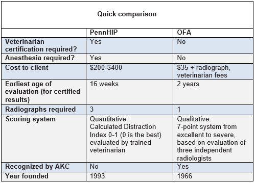
- - OFA Evaluation
- - PennHIP Method
- - Cornell's DLS Test
- - BVA/KC Scheme
- Treatment
- Breeders' Responsibilities
- What You Can Do
- Research News
- Related Links
- Veterinary Resources
Hip dysplasia (HD) is a heritable disease in cavalier King Charles spaniels which can cause the dogs terrible pain and debilitation.* HD is the abnormal development of the hip, which can produce various degrees of arthritis (which also may be called degenerative joint disease, arthrosis, or osteoarthrosis). Estimates of affected CKCSs range from 15.5%** to more than double*** that amount.
* See
Canine Inherited
Disorders Database.
**15.5%
is the current percentage of dysplastic CKCSs on the
OFA database.
*** In the April 1985 issue of Cornell University's Animal Health newsletter, it
estimated that OFA's statistics under-state the true extent of hip dysplasia in
a given breed by 1.5 times to 2.5 times, due to non-submissions to OFA of x-rays
of obviously affected dogs.
However, hip dysplasia can be affected by environmental factors upon young puppies, including body weight, floor surfaces, and types of exercise. For example, a 2006 study showed:
"[T]hat increased early postnatal body weight, particularly birth weight, was associated with reduced femoral head coverage at 4 mo and a greater probability of degenerative changes at 8 mo. Increased birth weight was negatively associated with age at ossification onset for the femoral head; however, the effect was likely due to differences in skeletal maturity rather than to weight per se. These results support the hypothesis that increased body weight during the critical early postnatal period was sufficient to alter the course of hip development and result in measurable degenerative changes at adulthood."
 Courtesy of The Dog Channel
RETURN TO TOP
 Symptoms
Early symptoms of hip dysplasia include bunny hopping or swiveling the hips when running, having a difficult time rising from a down position or when climbing stairs. It is not predictable as to when or even if a dog with HD will start showing signs of lameness due to pain. There are many environmental factors, including caloric intake and the level of exercise, which can affect the severity of pain and lameness. Many dysplastic dogs with severe HD can run, jump, and play as if nothing is wrong, and other dogs with barely any HD x-ray indications are severely lame.
RETURN TO TOP
Diagnosis
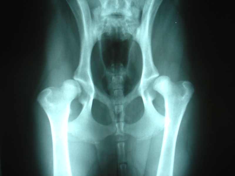
The only way to accurately diagnose HD is by radiography ( x-ray). Hip dysplasia is a developmental disease, meaning that it is not present at birth, but develops with age; it normally does not appear at all in x-rays of puppies or young cavaliers.
-- OFA evaluation
 The
Orthopedic Foundation for Animals
(OFA) provides x-ray evaluations by panels of board certified veterinary
radiologists. The PennHip
method of hip joint analysis is an alternative means of diagnosing HD, also by
x-ray.
The
Orthopedic Foundation for Animals
(OFA) provides x-ray evaluations by panels of board certified veterinary
radiologists. The PennHip
method of hip joint analysis is an alternative means of diagnosing HD, also by
x-ray.
According to OFA's statistics, 15.5% of cavaliers are afflicted with hip dysplasia. However, the vast majority of cavalier breeders do not submit to OFA any x-rays of their breeding stock which show obviously dysplastic hips.* Therefore, OFA's statistics do not accurately reflect the incidence of HD in cavaliers. For example, only 7,715 cavaliers have been evaluated by OFA in the 37 years from 1974 through 2016. Considering that in 2008 alone, the AKC registered over 8,000 cavaliers, the OFA data base is woefully short. This indicates a major lack of interest by most all CKCS breeders in obtaining objective, expert evaluations of their dogs' hips.
*The Cavalier King Charles Spaniel Club,USA makes this comment on its website about the lack of submission of x-rays to OFA: "Since x-ray results are not always sent to OFA by breeders whose vets consider them dysplastic, this number is not exact and one can assume that 12.6% [the 2001 statistics] is a low figure." In The Orthopedic Foundation for Animals Hip Dysplasia Database: A Review, published in 2013, the author states: "It is assumed that radiographs submitted to OFA are generally prescreened by the veterinarian and the more obvious cases of hip dysplasia are probably not submitted. Therefore, the actual frequency of hip dysplasia in the general population is unknown but has been approximated by Corley and Rettenmaier to be higher than reported by OFA."
 OFA requires that dogs be at least 24 months
old when x-rayed in order to qualify for a permanent OFA evaluation. OFA
evaluates the x-rays by assigning a classification to each dog, from excellent,
good, fair, borderline, mild, moderate, to severe. Ideally, breeders should
consider x-raying their breeding stock again annually thereafter, as hip
dysplasia has been found to be a progressive genetic disorder which results in
OFA requires that dogs be at least 24 months
old when x-rayed in order to qualify for a permanent OFA evaluation. OFA
evaluates the x-rays by assigning a classification to each dog, from excellent,
good, fair, borderline, mild, moderate, to severe. Ideally, breeders should
consider x-raying their breeding stock again annually thereafter, as hip
dysplasia has been found to be a progressive genetic disorder which results in
![]() deterioration of the hip joints after age two years.
Here is a YouTube link to a video discussion on the proper method to x-ray
dogs' hips for OFA.
deterioration of the hip joints after age two years.
Here is a YouTube link to a video discussion on the proper method to x-ray
dogs' hips for OFA.
It is believed by veterinary specialists in the field of hip dysplasia that the true incidence of HD in the cavalier King Charles spaniel probably is at least twice as high as those statistics would indicate, meaning 25% or more of all cavaliers.* Even higher percentages of cavaliers have been reported in recent studies of dogs requiring surgical hip procedures. See Veterinary Resources below. By comparison, OFA's current statistics show that 66% of pugs had dysplastic hips, 44.8% of Clumber spaniels, 30.4% of French bulldogs, and 19.7% of golden retrievers.
* A 2010 study of the PennHIP method, described below, also suggests that OFA scoring can underestimate the true incidence of hip dysplasia in dogs. In the April 1985 issue of Cornell University's Animal Health newsletter, it estimated that OFA's statistics under-state the true extent of hip dysplasia in a given breed by 1.5 times to 2.5 times, due to non-submissions to OFA of x-rays of obviously affected dogs.

RETURN TO TOP
-- PennHIP method
 The
PennHIP method was developed in 1983 by Dr. Gail
K. Smith (right), an orthopedic
surgeon at the University of Pennsylvania School of Veterinary Medicine.
It measures hip joint laxity (looseness), which is the primary cause of
degenerative joint disease. The Distraction Index (DI) used in the PennHip
method is a measurement of passive hip laxity, the degree of looseness of the
hip joint when the dog's hips are completely relaxed. Dogs with a DI of 0.3 have
tighter hips and are less likely to develop hip dysplasia, and dogs with looser
hips and DI values approaching 0.7 or more are at greater risk.
The
PennHIP method was developed in 1983 by Dr. Gail
K. Smith (right), an orthopedic
surgeon at the University of Pennsylvania School of Veterinary Medicine.
It measures hip joint laxity (looseness), which is the primary cause of
degenerative joint disease. The Distraction Index (DI) used in the PennHip
method is a measurement of passive hip laxity, the degree of looseness of the
hip joint when the dog's hips are completely relaxed. Dogs with a DI of 0.3 have
tighter hips and are less likely to develop hip dysplasia, and dogs with looser
hips and DI values approaching 0.7 or more are at greater risk.
 In a 2010 study
conducted by Dr. Smith and others, they compared OFA and PennHIP evaluation
results for 439 dogs from 1987 through 2008. The results showed that high
percentages of dogs judged by OFA to be phenotypically normal nevertheless had
clinically important passive hip joint laxity as determined by PennHIP's
distraction radiography. The study concluded that dogs judged as "normal" by OFA
can have clinically important passive hip joint laxity as determined by the
PennHIP method. The results suggest that OFA scoring can underestimate
susceptibility to osteoarthritis in dogs, which may impede progress in reducing
or eliminating hip dysplasia through breeding.
In a 2010 study
conducted by Dr. Smith and others, they compared OFA and PennHIP evaluation
results for 439 dogs from 1987 through 2008. The results showed that high
percentages of dogs judged by OFA to be phenotypically normal nevertheless had
clinically important passive hip joint laxity as determined by PennHIP's
distraction radiography. The study concluded that dogs judged as "normal" by OFA
can have clinically important passive hip joint laxity as determined by the
PennHIP method. The results suggest that OFA scoring can underestimate
susceptibility to osteoarthritis in dogs, which may impede progress in reducing
or eliminating hip dysplasia through breeding.
You may contact Dr. Gail Smith at email smithgk@vet.upenn.edu
RETURN TO TOP
-- Cornell's dorsolateral subluxation (DLS) test
Researchers at Cornell University's College of Veterinary
Medicine have developed a new test for hip
dysplasia, called the dorsolateral
 subluxation (DLS) test. The report that this
procedure improves upon the OFA protocol by being more accurate at a younger age
than the OFA test. Studies have shown the DLS test to be accurate as early as 8
months of age. The main difference between the DLS test and the OFA method is
the way in which the dog is positioned while being x-rayed. While the OFA test
places the hind limbs in a position that is not natural and may hide symptoms of
hip dysplasia, the DLS test relies on a position much more similar to
positioning normally found in a standing dog.
subluxation (DLS) test. The report that this
procedure improves upon the OFA protocol by being more accurate at a younger age
than the OFA test. Studies have shown the DLS test to be accurate as early as 8
months of age. The main difference between the DLS test and the OFA method is
the way in which the dog is positioned while being x-rayed. While the OFA test
places the hind limbs in a position that is not natural and may hide symptoms of
hip dysplasia, the DLS test relies on a position much more similar to
positioning normally found in a standing dog.
Before the DLS x-ray examination may take place, the dog must be anesthetized or deeply sedated. It is then placed on its stomach on a foam rubber pad. There is a hole cut in the pad for the dog's hind legs. The stifles (corresponding to a human knee joint) make contact with the x-ray table, and the dog's femurs are nearly perpendicular to the table. Arranging the dog in a position that mimics its natural posture allows the x-ray to show with a high level of accuracy what the position of the hip joints is.
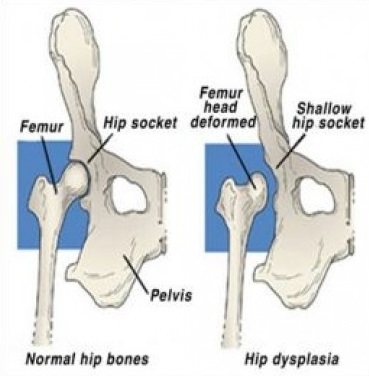
Cornell's specialists explain that, in a normal hip joint, the head of the femur fits snugly into the joint socket, or acetabulum, but in the dysplastic joint, the femoral head conforms poorly to the acetabulum. More space is evident between the bones. Displacement of the femoral head is the hallmark of the disease. Joints are evaluated using the DLS score. This measurement, expressed as a percent, is calculated from the radiograph and represents the percent of the femoral head covered by the acetabular rim. The greater the coverage, the higher the DLS score, and the healthier the hip joint. The DLS test may be carried out by the dog's regular veterinarian. See details here.
RETURN TO TOP
-- BVC/KC hip dysplasia scheme
 The British Veterinary Association (BVA) and the UK's Kennel Club
(KC) have
instituted a hip dysplasia x-ray scheme for all breeds, which is similar to
those of OFA and PennHIP. Details may be found at
BVA's
website here. Statistics for cavaliers under the BVA/KC hip scheme indicate
that, as of October 2010, x-rays of only 270 CKCSs (compared to, e.g., over
70,000 Labrador retrievers) have been submitted for
review under the scheme. Analysis of those x-rays show that the ratings of the
UK
cavaliers' hips ranged from "0" (no dysplasia) to "93" (severe dysplasia).
See the
BVA/KC statistics here.
The British Veterinary Association (BVA) and the UK's Kennel Club
(KC) have
instituted a hip dysplasia x-ray scheme for all breeds, which is similar to
those of OFA and PennHIP. Details may be found at
BVA's
website here. Statistics for cavaliers under the BVA/KC hip scheme indicate
that, as of October 2010, x-rays of only 270 CKCSs (compared to, e.g., over
70,000 Labrador retrievers) have been submitted for
review under the scheme. Analysis of those x-rays show that the ratings of the
UK
cavaliers' hips ranged from "0" (no dysplasia) to "93" (severe dysplasia).
See the
BVA/KC statistics here.
In a February 2017 article, it is reported plans to introduce a new hip testing scheme in the UK.
RETURN TO TOP
Treatment
 Initial treatment is aimed at relieving pain and improving function with
medication for the treatment of degenerative joint disease. Typical medications
include non-steroidal anti-inflammatory drugs (NSAIDs), such as carprofen*
(Rimadyl, Quellin), meloxicam (Metacam,
Loxicom), firocoxib (Previcox), mavacoxib (Trocoxil),
grapiprant (Galliprant), and
aspirin.
Initial treatment is aimed at relieving pain and improving function with
medication for the treatment of degenerative joint disease. Typical medications
include non-steroidal anti-inflammatory drugs (NSAIDs), such as carprofen*
(Rimadyl, Quellin), meloxicam (Metacam,
Loxicom), firocoxib (Previcox), mavacoxib (Trocoxil),
grapiprant (Galliprant), and
aspirin.
The anticonvulsant (antiepileptic) gabapentin (Neurontin, Gabarone), has been successful in relieving neuropathic pain. Gabapentin works through a receptor on the membranes of brain and peripheral nerve cells. It binds to calcium channels and modulates calcium influx as well as influences GABergic neurotransmission. Its effect is to deaden the irritated nerve impulses in the dog's neck. In humans, gabapentin reportedly does not interact with any other medications, and it is not metabolized, so it is fully excreted in the urine and has no affect upon the liver. However, in dogs, gabapentin is partially metabolized in the liver, and therefore the prescribing neurologist may be expected to order periodic blood tests to check the liver enzymes.
In an August 2020 article, researchers studied the treatment records of 240 dogs suffering chronic pain due to a variety of diagnosed causes, including osteoarthritis (84.6%), generalized, nonspecific back pain (9.2%), intervertebral disk disease (7.5%), luxating patellas (6.67%), and 56.67% not having a definitive anatomical cause for their pain. They report finding that:
"The results from this case series suggest that gabapentin is well-tolerated at much higher doses than what is typically prescribed. Side effects were uncommon, with no clear pattern based on dose, or dog size or age. Therefore, like many other analgesic medications, the efficacy of gabapentin appears patient-specific and should be dosed to effect until side effects are noted or analgesia is achieved.
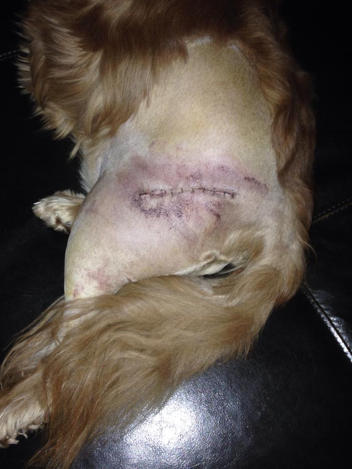
If pain cannot be controlled there are surgical procedures (right, above) which may relieve pain and improve function in some dogs. They include hip replacement, removing the femoral head (ball of the hip joint) and the acetabulum of the pelvis (hip socket) and replacing them with metal proteses. (See x-rays below.)

Hip "resurfacing" is an alternative to total hip replacement. Also called "coxofemoral hemi-arthroplasty", involves implanting a hollow cap*, shaped like a mushroom, over the head of the femur, and a matching metal cup in the pelvis socket.
* The cap may be made of metal; polycaprolactone (PCL), a biodegradable polyester material, ceramics, or a combination of them.
Stem cell therapy is being used on cavaliers by some practitioners, with reportedly successful results. See, e.g., this 2010 clinical report and this 2012 Paw Prints magazine article about a border collie, using Vet-Stem Regenerative Cell technology. In a 2007 study, in which dogs suffering from chronic osteoarthritis of the hip were treated with their own stems cells, the treated dogs "had significantly improved scores for lameness and the compiled scores for lameness pain, and range of motion compared with control dogs."
In a 2011 report, a team of Irish veterinary surgeons repaired the hips of 70 dogs with hip dysplasia (seven were CKCSs), using a "modified transarticular pinning technique". In a 2012 report, US surgeons, including Dr. Dominic Marino, reported on micro total hip prosthesis of 41 dogs, including six cavalier King Charles spaniels. In another 2012 report, the same US surgical team reviewed the outcomes of nano total hip replacement (NanoTHR) in twelve dogs, including a cavalier.
Nutraceuticals are nutrients necessary for supporting or improving normal structure and function of the hip joint. They have been found to:
• Support or enhance metabolism of cartilage cells (chondrocyte) and joint membranes (synoviocyte) -- an anabolic effect;
• Inhibit damaging enzymes within synovial fluid and cartilage -- a catabolic effect;
• Inhibit formation of thrombi in small blood vessels supplying the joint (antithrombolic).
The most common nutraceuticals are glucosamine and chondroitin.
Glucosamine regulates the synthesis of
 collagen
in cartilage, and may provide mild anit-inflammatory effects.
Crystalline glucosamine sulphate has the greatest efficacy and
bioavailablity for osteoarthritis. Chondroitin sulfate inhibits
destructive enzymes in joint fluid and cartilage. Both of them also
contribute to the building blocks synthesis of glycoaminoglycans and
proteoglycans) for the formation and repair of cartilage.
collagen
in cartilage, and may provide mild anit-inflammatory effects.
Crystalline glucosamine sulphate has the greatest efficacy and
bioavailablity for osteoarthritis. Chondroitin sulfate inhibits
destructive enzymes in joint fluid and cartilage. Both of them also
contribute to the building blocks synthesis of glycoaminoglycans and
proteoglycans) for the formation and repair of cartilage.
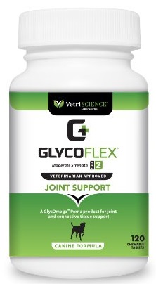
Owners may provide their affected dogs with supplements such as glucoamine HCI, methylsulfonylmethane (MSM), N,N-Dimethylglycine HCI (DMG), and manganese. Retail combinations of these supplements include Vetri-Science GlycoFlex Joint Support.
RETURN TO TOP
Breeders' Responsibilities
 The
Canine Inherited Disorders Database
recommends that breeders breed only dogs that have disease-free joints and that
come from ancestors with disease-free joints. The Canine Inherited Disorders
Database further recommends not breeding any cavaliers whose offspring have hip
dysplasia (dogs with hip dysplasia can produce normal offspring, and
normal-appearing dogs can produce offspring with hip dysplasia).
The
Canine Inherited Disorders Database
recommends that breeders breed only dogs that have disease-free joints and that
come from ancestors with disease-free joints. The Canine Inherited Disorders
Database further recommends not breeding any cavaliers whose offspring have hip
dysplasia (dogs with hip dysplasia can produce normal offspring, and
normal-appearing dogs can produce offspring with hip dysplasia).
The general principles recommended by OFA for breeding away from HD are:
1) Breed only normal dogs (meaning, not dysplastic) to normal dogs.
2) The normal dogs should come from normal parents and grandparents.
3) The normal dogs should have over seventy-five percent normal siblings.
4) A dog with excellent hips from a litter having more than twenty-five percent dysplastic pups is a worse breeding choice than a dog with fair hips from a litter experiencing less than twenty-five percent dysplasia.
5) Choose replacement dams that have better hips than their parents and the breed average.
Breeders should have their breeding cavaliers' hips x-rayed after age two years and have the x-rays evaluated by OFA before considering the dogs for breeding. Ideally, breeders should consider x-raying their breeding stock again annually thereafter, as hip dysplasia has been found to be a progressive genetic disorder which results in deterioration of the hip joints after age two years.
The Cavalier King Charles Spaniel Club, USA recommends that, prior to breeding any cavalier, the dog have a passing grade from an x-ray for hip dysplasia submitted to the OFA.
The Canine Health Information Center (CHIC) is a centralized canine health database sponsored by the AKC/Canine Health Foundation (AKC/CHF) and OFA. The CHIC, working with participating parent clubs, provides a resource for breeders and owners of purebred dogs to research and maintain information on the health issues prevalent in specific breeds.
AKC's national breed clubs establish the breed specific testing protocols. Dogs complying with the breed specific testing requirements are issued CHIC numbers. The ACKCSC requires that, to qualify for CHIC certification, cavaliers must be screened for hip dysplasia, using the OFA, PennHIP, or OVC protocol.
Since the most critical period of canine hip development is from birth to age 12 weeks, breeders should take care regarding body weight, as well as the floor and ground surfaces on which the young puppy is raised. In a June 2012 article, researchers reported that:
"Results indicated that puppies ≤ 3 months old should not be allowed access to stairs, but should be allowed outdoor exercise on soft ground in moderately rough terrain to decrease the risk for developing radiographically detectable HD."
RETURN TO TOP
What You Can Do
Protective factors:
• Limit puppies' food consumption.
• Provide puppies with early off-leash exercise to strengthen the hip joints.
To a certain extent, in this case, the "What You Can Do" section really should be labeled "What You Should Not Do":
• Do not over-feed your cavalier. Do not allow your cavalier to become obese. Follow the Body Condition Scoring Charts on our Diets webpage.
• Do not allow cavalier puppies to gain weight rapidly.
• Do not over-supplement your cavalier with calcium.
• Do not neuter your cavaliers before they reach physical maturity. The reproductive hormones have a great influence on bone development. They initiate growth plate closure in long bones. They are responsible for maintaining tendon, ligament, and muscle integrity. There is a current theory that their abrupt removal may lead to weakening of joints and musculature as well as changes in joint conformation.
RETURN TO TOP
Research News
August 2020:
Colorado State Univ. researchers find gabapentin is
well-tolerated at high dosages for dogs in chronic pain.
 In
an
August 2020 article, a team of Colorado State University researchers
(Lily V Davis, Peter W Hellyer, Robin A Downing, Lori R Kogan [right])
studied the treatment records of 240 dogs suffering chronic pain due to
a variety of diagnosed causes, including osteoarthritis (84.6%),
generalized, nonspecific back pain (9.2%), intervertebral disk disease
(7.5%), hip dysplasia (3%), and 56.67% not having a
definitive anatomical cause for their pain. They
report finding that:
In
an
August 2020 article, a team of Colorado State University researchers
(Lily V Davis, Peter W Hellyer, Robin A Downing, Lori R Kogan [right])
studied the treatment records of 240 dogs suffering chronic pain due to
a variety of diagnosed causes, including osteoarthritis (84.6%),
generalized, nonspecific back pain (9.2%), intervertebral disk disease
(7.5%), hip dysplasia (3%), and 56.67% not having a
definitive anatomical cause for their pain. They
report finding that:
"The results from this case series suggest that gabapentin is well-tolerated at much higher doses than what is typically prescribed. Side effects were uncommon, with no clear pattern based on dose, or dog size or age. Therefore, like many other analgesic medications, the efficacy of gabapentin appears patient-specific and should be dosed to effect until side effects are noted or analgesia is achieved.
June 2020:
Swedish hip dysplasia (HD) screening and breeding protocol has
resulted in less than 10% moderate and severe HD.
 In
a
May 2020 article, Swedish veterinary geneticist Åke Hedhammar
(right) reports that the Swedish Kennel Club's mandatory screening
and open registry data on seven breeds has resulted in the reduction of
moderate and severe hip dysplasia (grades D and E) in those breeds to
less than 10% of current offspring. Specifically, the prevalance of HD
is: German shepherd dogs 7%, Labrador retrievers 4%, Golden retrievers
5%, Bernese mountain dogs 6%, and Rottweilers 6%). Only in a few giant
breeds with a few registered dogs and no restrictions on breeding stock
is the prevalence of grades D and E above 10%.
In
a
May 2020 article, Swedish veterinary geneticist Åke Hedhammar
(right) reports that the Swedish Kennel Club's mandatory screening
and open registry data on seven breeds has resulted in the reduction of
moderate and severe hip dysplasia (grades D and E) in those breeds to
less than 10% of current offspring. Specifically, the prevalance of HD
is: German shepherd dogs 7%, Labrador retrievers 4%, Golden retrievers
5%, Bernese mountain dogs 6%, and Rottweilers 6%). Only in a few giant
breeds with a few registered dogs and no restrictions on breeding stock
is the prevalence of grades D and E above 10%.
June 2017:
OFA statistics show that hip dysplasia is on a stunning rise in the
cavalier King Charles spaniel breed.
 A comparison between
statistical records maintained by the Orthopedic Foundation for Animals
(OFA) of cavalier King Charles spaniels between 2006 and 2016 show that
the percentage of CKCSs' hips judged to be dysplastic has jumped from
11.8% in the 2006-2010 years to 15.5% in the 2011-2015 years. The
current 15.5% is the highest percentage of dysplastic cavaliers since
1981. This amounts to a surprising jump of nearly 4% between 2010 and
2015. The full statistical record for hip dysplasia in the CKCS between
1981 and 2015:
A comparison between
statistical records maintained by the Orthopedic Foundation for Animals
(OFA) of cavalier King Charles spaniels between 2006 and 2016 show that
the percentage of CKCSs' hips judged to be dysplastic has jumped from
11.8% in the 2006-2010 years to 15.5% in the 2011-2015 years. The
current 15.5% is the highest percentage of dysplastic cavaliers since
1981. This amounts to a surprising jump of nearly 4% between 2010 and
2015. The full statistical record for hip dysplasia in the CKCS between
1981 and 2015:
• 1981 to 1985: 12.4%
• 1986 to 1990: 8.9%
• 1991 to 1995: 10.8%
• 1996 to 2000: 9.9%
• 2001 to 2005: 11.7%
• 2006 to 2010: 11.8%
• 2011 to 2015: 15.5%
See this September 2013 article and the current OFA website for more details.
February 2013: Early neutering may affect male dogs' risk for developing hip dysplasia. A University of California at Davis study of golden retrievers has found that early castration was associated with an increase in the occurrence of hip dysplasia. The study showed a surprising 100 percent increase, or doubling, of the incidence of hip dysplasia among early-neutered males. Earlier studies had reported a 17 percent increase among all neutered dogs compared to all non-neutered dogs, indicating the importance of the new study in making gender and age-of-neutering comparisons.
September 2010:
PennHIP's Dr. Gail Smith finds "OFA Normal" dogs have hip joint laxity.
In a 2010 study, Dr. Gail
K. Smith (right), an orthopedic surgeon at the University of Pennsylvania
School of Veterinary Medicine who developed the PennHIP method of testing dogs'
for hip dysplasia, and others compared OFA and PennHIP evaluation
results for 439 dogs from 1987 through 2008. The results showed that high
percentages of dogs judged by OFA to be phenotypically normal nevertheless had
clinically important passive hip joint laxity as determined by PennHIP's
distraction radiography. The study concluded that dogs judged as "normal" by OFA
can have clinically important passive hip joint laxity as determined by the
PennHIP method. The results suggest that OFA scoring can underestimate
susceptibility to osteoarthritis in dogs, which may impede progress in reducing
or eliminating hip dysplasia through breeding. You may contact Dr. Gail Smith at email
smithgk@vet.upenn.edu
RETURN TO TOP
Related Links
RETURN TO TOP
Veterinary Resources
Hip Dysplasia, Chapter 83, Textbook of Small Animal Orthopedics. Wayne H. Riser, W. Harker Rhodes, and Charles D. Newton. 1982; U. Penn CAL small animal orthopedics.
Prevalence and inheritance of and slection for hip dysplasia in seven breeds of dogs in Sweden and benefit:cost analysis of a screening and control program. Swenson, L., Audell, L. & Hedhammar, Å. JAVMA. 1997;210(2):207-214. Quote: "Objective: To determine the prevalence and changes over time in the prevalence of hip dysplasia; to ascertain whether prevalence or severity of hip dysplasia was associated with sex of the dogs, age at which coxofemoral joint status was evaluated, or ancestral background; to determine the effects of selective breeding; and to conduct an economic evaluation of the hip dysplasia program operated by the Swedish Kennel Club. Design: Analysis of radiographic evaluations of coxofemoral joint conformity. Animals: 83,229 dogs from 7 breeds registered by the Swedish Kennel Club. Procedure: All radiographs were scrutinized by a single radiologist (LA), and coxofemoral joint conformation was classified as normal or dysplastic, with the degree of dysplasia classified as 1,2,3, or 4. Results: Decreasing prevalence of hip dysplasia corresponding to selection of breeding stock and high heritabilities was found. Sex differences were documented in 3 of the breeds. This was interpreted as breed differences in the distribution of genes related to hip dysplasia. Economic analyses showed that costs of screening and registration of coxofemoral joints was less than the value of dogs estimated to have been saved from moderate, severe, or very severe hip dysplasia in 6 of the breeds. Clinical Implications: Documented effects of age suggest that all dogs should be screened at the same age, rather than screening a few dogs at an older, more revealing age. In screening and control programs based on an open registry with access to family records, decreasing prevalence of hip dysplasia can be expected, and related to selection of breeding stock."
Control of Canine Genetic Diseases. Padgett, G.A., Howell Book House 1998, pp. 198-199, 245.
Hip dysplasia. Canine Inherited Disorders Database.
Effect of early postnatal body weight on femoral head ossification onset and hip osteoarthritis in a canine model of developmental dysplasia of the hip. Wendy S. Vanden Berg-Foels, Rory J. Todhunter, Steven J. Schwager, Anthony P. Reeves. Pediatric Res. September 2006;60(5):549-554. Quote: Developmental dysplasia of the hip (DDH) is a well-known precipitator of hip osteoarthritis. An increase in body weight during the critical early postnatal growth period may alter joint contact, and thus alter hip development and influence joint health in adulthood. The objective of this study was to determine whether early postnatal body weight affected the course of hip development and the onset of osteoarthritis in a canine model of DDH. A longitudinal study, from birth to skeletal maturity, was conducted. Serial body weight, age at femoral head ossification onset, and femoral head coverage at 4 mo were measured. Presence and severity of degeneration at 8 mo were determined using necropsy and cartilage biochemistry. There was a negative association between birth weight and age at femoral head ossification onset; however, the association was likely due to skeletal maturity level rather than body weight per se. Lower birth weight subjects had greater femoral head coverage at 4 mo. Greater birth weight was associated with greater probability of moderate degenerative changes or macroscopic lesions at 8 mo. These results support the hypothesis that increased birth weight is sufficient to alter the course of hip development and result in measurable degenerative changes at adulthood.
Lifelong diet restriction and radiographic evidence of osteoarthritis of the hip joint in dogs. Gail K. Smith, Erin R. Paster, Michelle Y. Powers, Dennis F. Lawler, Darryl N. Biery, Frances S. Shofer, Pamela J. McKelvie, Richard D. Kealy. JAVMA. Sept. 2006;229(5):690-693, Quote: "Objective: To evaluate the effects of diet restriction on development of radiographic evidence of hip joint osteoarthritis in dogs. Design: Longitudinal cohort study. Animals: 48 Labrador Retrievers from 7 litters. Procedures: Forty-eight 6-week-old puppies from 7 litters were paired with littermates by sex and weight, and each pairmate was randomly assigned to 1 of 2 groups of 24 dogs each. Starting at 8 weeks of age, 1 group was fed ad libitum (control fed) and the other was fed 25% less (restricted fed) of the same diet for life on a pairwise basis. The dogs' hip joints were radiographed in the standard ventrodorsal hip-extended view at multiple intervals prior to 1 year of age and at annual intervals thereafter on the basis of birth anniversary. A board-certified radiologist unaware of group assignment scored the radiographs for evidence of osteoarthritis. Results: Prevalence of radiographic evidence of hip joint osteoarthritis in all dogs increased linearly throughout the study, from an overall prevalence of 15% at 2 years to 67% by 14 years. Restricted-fed dogs had lower prevalence and later onset of hip joint osteoarthritis. Median age at first identification of radiographic evidence of hip joint osteoarthritis was significantly lower in the control-fed group (6 years), compared with the restricted-fed group (12 years). Conclusions and Clinical Relevance: Restricted feeding delayed or prevented development of radiographic signs of hip joint osteoarthritis in this cohort of Labrador Retrievers. Lifetime maintenance of 25% diet restriction delayed onset and reduced severity of hip joint osteoarthritis, thus favorably affecting both duration and quality of life. In addition, the data indicated that development of hip joint osteoarthritis was not bimodal in these dogs but occurred as a continuum throughout life."
Impact of sedation method on the diagnosis of hip and elbow dysplasia in Swedish dogs. Sofia Malm, Erling Strandberg, Birgitta Danell, Lars Audell, Lennart Swenson, Ake Hedhammar. Preventive Vet. Med. 2007;78:196-209. Quote: "Our objective was to investigate the effect of sedation method on the screening result for hip and elbow dysplasia. The study was based on a questionnaire survey of routines for hip and elbow screening at Swedish veterinary clinics and results of hip and elbow status, for eight breeds (Bernese Mountain Dog, Boxer, German Shepherd Dog, Golden Retriever, Labrador Retriever, Newfoundland, Rottweiler, and Saint Bernard) recorded by the Swedish Kennel Club. In total 5877 and 5406 dogs examined for hip and elbow dysplasia, respectively, from January 2002 through March 2003 were included. We used logistic regression to examine whether the type of chemical restraint used for sedation affected the screening result for hip and elbow dysplasia. In addition to sedation method, the effects of veterinary clinic, sex, breed, and age at screening were studied. The type of chemical restraint used for sedation affected the screening result for hip but not for elbow dysplasia. Acepromazine gave less than half the odds of hip dysplasia compared with medetomidine and butorphanol (the most common method), medetomidine alone or xylazine. Females had about 25% higher odds for developing hip dysplasia whereas males had almost 40% higher odds for developing elbow dysplasia. Saint Bernard, Newfoundland and German Shepherd Dog had the highest odds of developing hip dysplasia, whereas Rottweiler and Labrador Retriever had the lowest odds. Boxer had the lowest risk for elbow dysplasia, followed by Labrador Retriever. Saint Bernard and Rottweiler had the highest odds of elbow dysplasia. Increasing age increased the odds of both hip and elbow dysplasia, by about 2.5% per month. Following the results in this study, recording of the type of chemical restraint used for sedation during hip screening has now become mandatory in Sweden. This makes it possible to account for the effect of sedation method in a model for prediction of breeding values for hip dysplasia."
Effect of Adipose-Derived Mesenchymal Stem and Regenerative Cells on Lameness in Dogs with Chronic Osteoarthritis of the Coxofemoral Joints: A Randomized, Double-Blinded, Multicenter, Controlled Trial. Linda L. Black, James Gaynor, Dean Gahring, Cheryl Adams, Dennis Aron, Susan Harmon, Daniel A. Gingerich, and Robert Harman. Vety. Therapeutics. 2007; 8(4). Quote: "Autologous stem cell therapy in the field of regenerative veterinary mediciine involves harvesting tissue, such as fat, from the patient, isolating the stem and regenerative cells, and administering the cells back to the patient. Autologous adipose-derived stem cell therapy has been commercially available since 2003, and the current study evaluated such therapy in dogs with chronic osteoarthritis of the hip. Dogs treated with adipose-derived stem cell therapy had significantly improved scores for lameness and the compiled scores for lameness pain, and range of motion compared with control dogs. This is the first randomized, blinded, placebo-controlled clinical trial reporting on the effectiveness of stem cell therapy in dogs."
Evaluation of the relationship between Orthopedic Foundation for Animals' hip joint scores and PennHIP distraction index values in dogs. Michelle Y. Powers, Georga T. Karbe, Thomas P. Gregor, Pamela McKelvie, William T. N. Culp, Hilary H. Fordyce, and Gail K. Smith. JAVMA Sept 2010; 237(5):532-541. Quote: "Objective--To compare 2 screening methods for detecting evidence of hip dysplasia (Orthopedic Foundation for Animals [OFA] and PennHIP) in dogs. 439 dogs > 24 months of age [Cavalier King Charles spaniel included] that received routine hip joint screening from June 1987 through July 2008. Dogs were sedated, and PennHIP radiography was performed (hip joint- extended [HE], compression, and distraction radiographic views). The HE radiographic view was submitted for OFA evaluation. A copy of the HE radiographic view plus the compression and distraction radiographic views were submitted for routine PennHIP evaluation, including quantification of hip joint laxity via the distraction index (DI). Results: 14% (60/439) of dogs had hip joints scored as excellent by OFA standards; however, 52% (31/60) of those had a DI > 0.30 (range, 0.14 to 0.61). Eighty-two percent of (183/223) dogs with OFA-rated good hip joints had a DI > 0.30 (range, 0.10 to 0.77), and 94% (79/84) of dogs with OFA-rated fair hip joints had a DI > 0.30 (range, 0.14 to 0.77). Of all dogs with fair to excellent hip joints by OFA standards, 80% (293/367) had a DI > 0.30. All dogs with OFA-rated borderline hip joints or mild, moderate, or severe hip dysplasia had a DI > 0.30 (range, 0.30 to 0.83). Conclusion and Clinical Relevance: Dogs judged as phenotypically normal by the OFA harbored clinically important passive hip joint laxity as determined via distraction radiography. Results suggested that OFA scoring of HE radiographs underestimated susceptibility to osteoarthritis in dogs, which may impede progress in reducing or eliminating hip dysplasia through breeding."
Breeding for Improved Hip and Elbow Health in Swedish Dogs. Sofia Malm. Uppsala Univ. 2010. Quote: "Decades of selective breeding to reduce the prevalence of hip and elbow dysplasia (HD and ED) in Swedish dogs, based on phenotypic assessment of radiographic hip status, has had limited success. The prevalence of dysplastic dogs is still high in many large-sized and giant breeds and among the most common causes for euthanasia and costly veterinary care in numerous breeds are joint-related problems. The aim of this thesis was to investigate the possibilities of more efficient breeding strategies for improved hip and elbow health in Swedish dogs by improving the genetic evaluation of HD and ED, and by evaluating the clinical significance of radiographic hip status. Data from several different sources were combined in the analyses: pedigree information and screening records for HD and ED from the Swedish Kennel Club, questionnaire data about routines for hip and elbow screening from Swedish veterinary clinics and insurance data on hip-related veterinary care and mortality from Agria Insurance Company. Moreover, simulated data were used to evaluate selection strategies. Radiographic hip status in young adult dogs was shown to be strongly associated with subsequent incidence of veterinary care and mortality related to the hip joint. Furthermore, the genetic analyses of screening records for HD and ED showed considerable genetic variation and moderate heritability of both traits. Taken together, these findings support the use of screening records for HD and ED in selection to reduce prevalence of clinical problems related to hip and elbow joints. However, the impact of systematic environmental factors, such as sedation method, on the phenotypic expression of HD and ED implies that the individual's screening result alone is an imprecise estimate of its breeding value. Simulation of selection strategies against HD showed that selection based on BLUP breeding values was superior to phenotypic selection, leading to faster genetic progress and more rapid reduction of dysplastic dogs. Based on the studies included in this thesis, it is concluded that implementation of breeding schemes based on BLUP breeding values, instead of phenotypic records, should be prioritised to enable a more efficient breeding for improved hip and elbow health in Swedish dogs."
Genetic Evaluation of Hip Score in UK Labrador Retrievers. Thomas W. Lewis, Sarah C. Blott, John A. Woolliams. PLoS One. October 2010;5(10). Quote: "Hip dysplasia is an important and complex genetic disease in dogs with both genetic and environmental influences. Since the osteoarthritis that develops is irreversible the only way to improve welfare, through reducing the prevalence, is through genetic selection. This study aimed to evaluate the progress of selection against hip dysplasia, to quantify potential improvements in the response to selection via use of genetic information and increases in selection intensity, and to prepare for public provision of estimated breeding values (EBV) for hip dysplasia in the UK. Data consisted of 25,243 single records of hip scores of Labrador Retrievers between one and four years old, from radiographs evaluated between 2000 and 2007 as part of the British Veterinary Association (BVA) hip score scheme. A natural logarithm transformation was applied to improve normality and linear mixed models were evaluated using ASREML. Genetic correlations between left and right scores, and total hip scores at one, two and three years of age were found to be close to one, endorsing analysis of total hip score in dogs aged one to three as an appropriate approach. A heritability of 0.35±0.016 and small but significant litter effect (0.07±0.009) were estimated. The observed trends in both mean hip score and mean EBV over year of birth indicate that a small genetic improvement has been taking place, approximately equivalent to avoiding those dogs with the worst 15% of scores. Deterministic analysis supported by simulations showed that a 19% greater response could be achieved using EBV compared to phenotype through increases in accuracy alone. This study establishes that consistent but slow genetic improvement in the hip score of UK Labrador Retrievers has been achieved over the previous decade, and demonstrates that progress may be easily enhanced through the use of EBVs and more intense selection."
Treatment of 70 dogs with traumatic hip luxation using a modified transarticular pinning technique. W. McCartney, K. Kiss, F. McGovern. Vet.Rec. Mar 2011; 168(13):355. "Luxation of the hip is the most common joint luxation in dogs. Treatment options are based on closed reduction, or surgical reduction and stabilisation using intra-articular or extra-articular methods. The intra-articular techniques include toggle pin variations or transarticular pinning, whereas the extra-articular techniques include de Vita pinning and extracapsular prosthesis. This short communication describes an analysis of 70 cases of traumatic hip luxation in dogs treated with a modified transarticular pinning technique. Seventy dogs were operated on for hip luxation between 2003 and 2009. The predominant breeds were labrador retriever (13), collie (10), golden retriever (nine), German shepherd dog (eight), Cavalier King Charles spaniel (CKCS) (seven) and mixed breed (12). The remaining 11 dogs were poodles (two), Staffordshire bull terriers... A retrospective analysis of hip luxation case details was undertaken in conjunction with a telephone questionnaire survey of the owners; all 70 dogs had sufficient data for analysis. Dogs with underlying hip dysplasia or osteoarthritis did not have transarticular pinning performed as a treatment for hip luxation. Only cases in which an outcome assessment had been made using the owner questionnaire more than six months postoperatively were included. Two questions were asked in the questionnaire. For the first, 'Is your dog stiff or lame at any time and if so can you grade it as frequent, intermittent or never?', frequent was described as more than three times a day, intermittent as up to three times a week and never as no lameness or stiffness at anytime. For the second, 'Are you satisfied with the outcome?', there were two possible answers - 'satisfied' and 'not satisfied'. On the basis of the owner's repsonses, the outcome was graded as failed, poor, good or excellent. The outcome grading was classified as excellent if a dog had no complications and was not stiff or lame at any time, good if the dog was only intermittently stiff or lame up to three times a week, poor if there was frequent (daily) lameness or stiffness, and failed if there were any serious complications. There were 33 females and 37 males, and the age range was six months to 11 years."
Treatment of hip dysplasia. A. Anderson. J.Sm.Anim.Prac. April 2011;52(4):182-189. Quote: "Hip dysplasia is a common orthopaedic developmental disorder of dogs. This paper reviews the treatment options available for management of the condition in the skeletally immature and adult dog."
Canine Hip Resurfacing. Fred S. Pike. ACVS. 2011.
Micro Total Hip Replacement in Dogs and Cats. Dominic J Marino, Shadi J Ireifej DVM, Catherine A Loughin. Vet. Sur. Jan 2012; 41(1):121-129. Quote: "Objective: To describe the surgical technique using power-assisted femoral preparation and clinical outcome in 41 dogs and 2 cats surgically treated with the micro total hip prosthesis. Animals: Dogs (n = 41[cavalier King Charles spaniels = 6]) and 2 cats. Results: Six dogs had staged bilateral MicroTHR, 35 dogs and 2 cats had unilateral MicroTHR. Median body weight was 8.2 kg (range, 2.1-14.2 kg) whereas for those that had complications (fracture or luxation) it was 8.6 kg (range, 6.6-14.1 kg). Median operative time was 71 minutes (range, 55-105 minutes). Complications included luxations (5 of 49; 10%) and femoral fracture (1; 2%). Lameness grades assigned at the 3-month recheck examination: 39 (80%) animals were grade 1, 6 (12%) were grade 2, and 4 (8%) were grade 3. Forty-five animals (92%) had good or excellent results. Conclusions: MicroTHR is a practical and effective surgery in small breed dogs and cats with coxofemoral disease."
Nano Total Hip Replacement in 12 Dogs. Shadi Ireifej, Dominic Marino, Catherine Loughin. Vet. Sur. Jan 2012; 41(1):130-135. Quote: "Objective: To determine the short-term clinical outcome of nano total hip replacement (NanoTHR) in dogs. Results: Breeds were Yorkshire Terriers (n = 6), Toy Poodles (2), with 1 each of Maltese, Pomeranian, Cavalier King Charles Spaniel, and Shih-Tzu. Median body was 4.87 kg (range, 2.5-5.90 kg) and median age, 35.75 months (range, 12-144 months). Radiographs were taken in 4 dogs at 12 days (n = 2), 14 days (1), and 30 days (1) after surgery because of presentation for an acute grade 5 lameness. Three dogs had femoral fractures distal to the femoral implant tip and 1 dog displaced the acetabular implant medially. After revision surgery, all femoral fractures were assessed as healed with intact plate fixation. The dog with the medially displaced acetabular component responded to conservative management including strict confinement and analgesic administration. Eight dogs (58%) were assigned a grade 1 lameness and 4 dogs were grade 2 (33%) at 12-week examination. The 3 dogs with grade 5 lameness scores found to have femoral fractures within 1 month after surgery, subsequently improved to grade 1 (n = 1) and 2 (2) 12 weeks after revision surgery. The dog with medial acetabular displacement improved to a grade 2 lameness 12 weeks after conservative management. Conclusions: Although all 12 dogs had good-to-excellent outcomes, 33% experienced significant complications associated with the technique. As improvements in instrumentation and refinements in the technique are developed, NanoTHR can be considered an alternative to the femoral head and neck ostectomy (FHO) or medical management of coxofemoral disease for toy breed dogs. Further studies with a larger number of dogs and longer follow-up times are required."
British Veterinary Association/Kennel Club Hip Dysplasia Scheme - Procedure Notes. January 2017. Quote: These procedure notes are intended to explain the BVA/Kennel Club Hip Dysplasia Scheme and to provide helpful instruction to those using the Scheme. They are due to be effective from 1 January 2017 and replace all previous documents in relation to the scheme. These notes may be modified from time to time; please consult the BVA website for the latest version. Introduction Hip dysplasia (HD) is a genetically-transmitted condition but environmental factors may influence the final score achieved. The score does not therefore absolutely reflect the potential for transmission of HD of an individual animal but should be regarded only as an indicator of possible transmission of the condition. For the Scheme to be meaningful and successful it is important that images from EVERY dog radiographed be submitted for scoring, whether or not the animal is required for breeding and whatever the state of the hips, in order to provide the widest possible information for use by a geneticist and for generation of estimated breeding values (EBVs). Further information about hip dysplasia and the use of the scoring scheme is available in the Canine Health Schemes section of the BVA website: www.bva.co.uk/chs or by contacting the Canine Health Schemes office at the BVA.
Interpretation and use of BVA/KC hip scores in dogs. Ruth Dennis. In Practice. April 2012;34:178-194. Quote: Hip dysplasia is a potentially debilitating orthopaedic disease in which laxity of the coxofemoral joint often leads to secondary osteoarthritis, a reduction in joint function and pain. It has been recognised for many years as being of particular importance in pedigree dogs, especially in larger breeds, and is known to be partly governed by genetic factors. In order to try to control canine hip dysplasia and to reduce its incidence, a number of radiographic screening programmes have been developed worldwide. In 1983, a scheme was established by the British Veterinary Association and supported by the Kennel Club to examine radiographs of dogs' hips by assessing different anatomical features and giving them a numerical score. This article describes the process of scoring in this scheme, explains how to interpret the score and gives advice on the use of hip scores in the selection of breeding animals.
Housing- and exercise-related risk factors associated with the development of hip dysplasia as determined by radiographic evaluation in a prospective cohort of Newfoundlands, Labrador Retrievers, Leonbergers, and Irish Wolfhounds in Norway. Randi I. Krontveit, Ane Nødtvedt, Bente K. Sævik, Erik Ropstad, Cathrine Trangerud. Amer. J. Vet. Res. June 2012;73(6):838-846. Quote: Objective: To identify housing- and exercise-related risk factors associated with the development of hip dysplasia (HD) as determined by radiographic evaluation in Newfoundlands, Labrador Retrievers, Leonbergers, and Irish Wolfhounds in Norway. Animals: 501 client-owned dogs from 103 litters. Procedures: Dogs were assessed from birth until official radiographic screening for HD at 12 (Labrador Retriever [n = 133] and Irish Wolfhound [63]) or 18 (Newfoundland [125] and Leonberger [180]) months of age. Information regarding housing and exercise conditions during the preweaning and postweaning periods was obtained with questionnaires. Multivariable random effects logistic regression models were used to identify housing- and exercise-related risk factors associated with the development of radiographically detectable HD. Results: Puppies walking on stairs from birth to 3 months of age had an increased risk of developing HD. Factors associated with a decreased risk of developing HD included off-leash exercise from birth to 3 months of age, birth during the spring and summer, and birth on a farm. Significant clustering of dogs with HD was detected within litters. Conclusions and Clinical Relevance: Results indicated that puppies ≤ 3 months old should not be allowed access to stairs, but should be allowed outdoor exercise on soft ground in moderately rough terrain to decrease the risk for developing radiographically detectable HD. These findings could be used as practical recommendations for the prevention of HD in Newfoundlands, Labrador Retrievers, Leonbergers, and Irish Wolfhounds.
Development of biological hip resurfacing in dogs. Samuel Patrick Franklin Univ. of Missouri-Columbia thesis. July 2012.
Neutering Dogs: Effects on Joint Disorders and Cancers in Golden Retrievers. Gretel Torres de la Riva, Benjamin L. Hart, Thomas B. Farver, Anita M. Oberbauer, Locksley L. McV Messam, Neil Willits, Lynette A. Hart. PLOSone. Feb. 2013. Excerpt: "Hip Dysplasia: Perusal of Figure 1 and Table 4 reveals that HD in early-neutered males, affecting 10.3 percent, was more than double the proportion of intact males with the disorder, which was 5.1 percent, a significant difference (K-M: p<0.01). There was also a significant difference between early and late neutering in males (K-M: p<0.05). The mean ages of HD onset for intact, early-neutered, and late-neutered male dogs were 4.4, 3.6, and 4.7 years, respectively. No difference was found between early-neutered dogs with and without HD when compared with respect to their BCS, (means 6.1 and 5.7, respectively; CPH: p = 0.22). No other comparisons of HD occurrence were significant; HD was not increased in occurrence by early or later neutering in females (Figure 2). ... While neutering is expected to lead to a greater gain in body weight than in intact dogs [17], [18], the BCS of early-neutered dogs with the disorders and the early neutered comparison groups without the disorders were not significantly different - and, in fact quite similar - indicating that weight on the joint was not a major determinant in the occurrence of these joint disorders. Using the CPH model to compare early-neutered with intact dogs, for both HD and CCL, neither neutering status nor BCS was significant, indicating that the two factors are fairly highly confounded. This implies that the occurrence of HD and CCL in early-neutered dogs is a combined function of the effect of neutering on growth plates, as well as the increase in weight on the joints brought on by neutering. As mentioned, when only early-neutered dogs with and without HD or CCL were compared with respect to their BCS, no differences were found between early-neutered males with and without these joint disorders. ... As for the pathophysiological reasons for the joint disorders, one can point to the role of gonadal hormones in controlling the closure of bone growth plates [23], [24]. An atypical growth plate closure, resulting from the absence of gonadal hormones, may increase the chance of a clinically apparent joint disorder, such as HD. ... Confounding factors that may influence the nature of a neuter-related joint disorder are the breed-specific gender vulnerabilities, including growth rate differences, as well as the timing of growth plate closure, which occurs more quickly in males than in females. In the males of this study, the occurrence of HD was doubled in the cases with early androgen removal as compared with intact males, but in females, removal of the ovaries did not appear to be associated with an increased likelihood of HD. This presumably reflects the effect of gender on growth-plate development."
Comparative analyses of genetic trends and prospects for selection against hip and elbow dysplasia in 15 UK dog breeds. Lewis TW, Blott SC, Woolliams JA. BMC Genet March 2013;14:16. Quote: "Background: Hip dysplasia remains one of the most serious hereditary diseases occurring in dogs despite long-standing evaluation schemes designed to aid selection for healthy joints. Many researchers have recommended the use of estimated breeding values (EBV) to improve the rate of genetic progress from selection against hip and elbow dysplasia (another common developmental orthopaedic disorder), but few have empirically quantified the benefits of their use. This study aimed to both determine recent genetic trends in hip and elbow dysplasia, and evaluate the potential improvements in response to selection that publication of EBV for such diseases would provide, across a wide range of pure-bred dog breeds. Results: The genetic trend with respect to hip and elbow condition due to phenotypic selection had improved in all breeds, except the Siberian Husky. However, derived selection intensities are extremely weak, equivalent to excluding less than a maximum of 18% of the highest risk animals from breeding. EBV for hip and elbow score were predicted to be on average between 1.16 and 1.34 times more accurate than selection on individual or both parental phenotypes. Additionally, compared to the proportion of juvenile animals with both parental phenotypes, the proportion with EBV of a greater accuracy than selection on such phenotypes increased by up to 3-fold for hip score and up to 13-fold for elbow score. Conclusions: EBV are shown to be both more accurate and abundant than phenotype, providing more reliable information on the genetic risk of disease for a greater proportion of the population. Because the accuracy of selection is directly related to genetic progress, use of EBV can be expected to benefit selection for the improvement of canine health and welfare. Public availability of EBV for hip score for the fifteen breeds included in this study will provide information on the genetic risk of disease in nearly a third of all dogs annually registered by the UK Kennel Club, with in excess of a quarter having an EBV for elbow score as well."
The Prevalence of Nine Genetic Disorders in a Dog Population from Belgium, the Netherlands and Germany. Bart J. G. Broeckx, Frank Coopman, Geert E. C. Verhoeven, Wim Van Haeringen, Leanne van de Goor, Tim Bosmans, Ingrid Gielen, Jimmy H. Saunders, Sandra S. A. Soetaert, Henri Van Bree, Christophe Van Neste, Filip Van Nieuwerburgh, Bernadette Van Ryssen, Elien Verelst, Katleen Van Steendam, Dieter Deforce. PlosOne. September 2013;8(9):e74811. Quote: The objective of this study was to screen a dog population from Belgium, the Netherlands and Germany for the presence of mutant alleles associated with hip dysplasia (HD), degenerative myelopathy (DM), exercise-induced collapse (EIC), neuronal ceroid lipofuscinosis 4A (NCL), centronuclear myopathy (HMLR), mucopolysaccharidosis VII (MPS VII), myotonia congenita (MG), gangliosidosis (GM1) and muscular dystrophy (Duchenne type) (GRMD). Blood samples (K3EDTA) were collected for genotyping with Kompetitive Allele Specific PCR (n = 476). Allele and genotype frequencies were calculated in those breeds with at least 12 samples (n = 8). Hardy-Weinberg equilibrium was tested. Genetic variation was identified for 4 out of 9 disorders: mutant alleles were found in 49, 15, 3 and 2 breeds for HD, DM, EIC and NCL respectively. ... Since its first report in 1935, intensive research has focused on hip dysplasia (HD), one of the most frequent orthopedic disorder in dogs. HD can be found in a wide variety of breeds. Recently, a haplotype in the Fibrillin 2 (FBN2) gene has been reported to be associated with this highly prevalent, multifactorial disorder. Fibrillins are components of extracellular microfibrils and have both a structural and a regulatory function. The mutant AGC haplotype was identified in 49 different breeds in the population under study (Table 3). In 44 breeds, this mutant allele was reported for the first time. We identified the AGC haplotype as the only allele in 10 breeds (Cavalier King Charles Spaniel (n=4), English Setter (n=2), English Springer Spaniel (n=1), Gordon Setter (n=1), Laekenois (n=1), Mastino Napoletano (n=1), Rhodesian Ridgeback (n=1), Saarlooswolfhond (n=2), Siberian Husky (n=2), Standard Poodle (n=2)), however few conclusions can be made as the sample count is low (n=17 overall). ... Additionally, mutant alleles were identified in crossbreeds for both HD and EIC. For HD, DM, EIC and NCL mutant alleles were newly discovered in 43, 13, 2 and 1 breed(s), respectively. In 9, 2 and 1 breed(s) for DM, EIC and NCL respectively, the mutant allele was detected, but the respective disorder has not been reported in those breeds. For 5 disorders (HMLR, MPS VII, MG, GM1, GRMD), the mutant allele could not be identified in our population. For the other 4 disorders (HD, DM, EIC, NCL), prevalence of associated mutant alleles seems strongly breed dependent. Surprisingly, mutant alleles were found in many breeds where the disorder has not been reported to date.
The Orthopedic Foundation for Animals Hip Dysplasia Database: A Review. Greg Keller. 6th Tufts' Canine & Feline Breeding & Genetics Conf. September 2013. Quote: It is assumed that radiographs submitted to OFA are generally prescreened by the veterinarian and the more obvious cases of hip dysplasia are probably not submitted. Therefore, the actual frequency of hip dysplasia in the general population is unknown but has been approximated by Corley and Rettenmaier to be higher than reported by OFA.7,8 However, the main objective of the OFA is to identify phenotypically normal animals as potential breeding candidates in order to reduce the frequency of hip dysplasia. ... Cavalier King Charles spaniel: To 1980: Excellent (Ex) % = 0.0; Dysplastic (Dys) % = 33.3%; Dogs = 15. 1981 to 1985: Ex % = 7.4%; Dys % = 12.4%; Dogs = 162. 1986 to 1990: Ex % =3.8%; Dys % = 8.9%; Dogs = 425. 1991 to 1995: Ex % = 3.1%; Dys % = 10.8%; Dogs = 732. 1996 to 2000: Ex % = 4.7%; Dys % = 9.9%; Dogs = 1,277. 2001 to 2005: Ex % = 4.1%; Dys % = 11.7%; Dogs = 1.967. 2006 to 2010: Ex % = 4.2%; Dys % = 11.8%; Dogs = 1.639. Total: Ex % = 4.2%; Dys % = 11.2%; Dogs = 6,275.
Estimated Breeding Values for Canine Hip Dysplasia Radiographic Traits in a Cohort of Australian German Shepherd Dogs. Bethany J. Wilson, Frank W. Nicholas, John W. James, Claire M. Wade, and Peter C. Thomson. PLoS One Oct. 2013;8(10):e77470. Quote: "Canine hip dysplasia (CHD) is a serious and common musculoskeletal disease of pedigree dogs and therefore represents both an important welfare concern and an imperative breeding priority. The typical heritability estimates for radiographic CHD traits suggest that the accuracy of breeding dog selection could be substantially improved by the use of estimated breeding values (EBVs) in place of selection based on phenotypes of individuals. The British Veterinary Association/Kennel Club scoring method is a complex measure composed of nine bilateral ordinal traits, intended to evaluate both early and late dysplastic changes. However, the ordinal nature of the traits may represent a technical challenge for calculation of EBVs using linear methods. The purpose of the current study was to calculate EBVs of British Veterinary Association/Kennel Club traits in the Australian population of German Shepherd Dogs, using linear (both as individual traits and a summed phenotype), binary and ordinal methods to determine the optimal method for EBV calculation. Ordinal EBVs correlated well with linear EBVs (r=0.90-0.99) and somewhat well with EBVs for the sum of the individual traits (r=0.58-0.92). Correlation of ordinal and binary EBVs varied widely (r=0.24-0.99) depending on the trait and cut-point considered. The ordinal EBVs have increased accuracy (0.48-0.69) of selection compared with accuracies from individual phenotype-based selection (0.40-0.52). Despite the high correlations between linear and ordinal EBVs, the underlying relationship between EBVs calculated by the two methods was not always linear, leading us to suggest that ordinal models should be used wherever possible. As the population of German Shepherd Dogs which was studied was purportedly under selection for the traits studied, we examined the EBVs for evidence of a genetic trend in these traits and found substantial genetic improvement over time. This study suggests the use of ordinal EBVs could increase the rate of genetic improvement in this population."
Assessing the impact of genomic selection against hip dysplasia in the Labrador Retriever dog. E. Sánchez-Molano, J.A. Woolliams, S.C. Blott, P. Wiener. J. Anim. Breeding & Genetics. April 2014;131(2):134-145. Quote: "Many purebred dogs exhibit a higher prevalence of inherited diseases compared with non-purebred dogs. One of the most popular breeds in the UK is the Labrador Retriever, which has a high prevalence of hip dysplasia resulting in high costs for surgical operations and impaired animal welfare. Considering the many complications of highly managed populations, mainly due to breeder's conventions and the resulting population structure, is of great importance for the proper development of a strategy against the disease. In this study, we have compared the utilities and performances of both genomic and phenotypic selection against hip dysplasia in a simulated population with the characteristics of the British Veterinary Association and Kennel Club (BVA/KC) hip dysplasia scheme. The results confirm the potential benefits of genomic selection by showing a moderate increase of 1.15-fold (assuming a realistic accuracy of r2 = 0.5) in response to selection due to the higher accuracy (between 0.96- and 1.32-fold, considering 0.35 ≤ r2 ≤ 0.7) and more than a threefold increase when all the offspring in each litter are tested (between 3.25- and 4.55-fold, again considering 0.35 ≤ r2 ≤ 0.7)."
Identification and Validation of Quantitative Trait Loci (QTL) for Canine Hip Dysplasia (CHD) in German Shepherd Dogs. Lena Fels, Ottmar Distl. PLOS|One. May 2014. Quote: "Canine hip dysplasia (CHD) is the most common hereditary skeletal disorder in dogs. To identify common alleles associated with CHD, we genotyped 96 German Shepherd Dogs affected by mild, moderate and severe CHD and 96 breed, sex, age and birth year matched controls using the Affymetrix canine high density SNP chip. A mixed linear model analysis identified five SNPs associated with CHD scores on dog chromosomes (CFA) 19, 24, 26 and 34. These five SNPs were validated in a by sex, age, birth year and coancestry stratified sample of 843 German Shepherd Dogs including 277 unaffected dogs and 566 CHD-affected dogs. Mean coancestry coefficients among and within cases and controls were <0.1%. Genotype effects of these SNPs explained 20-32% of the phenotypic variance of CHD in German Shepherd Dogs employed for validation. Genome-wide significance in the validation data set could be shown for each one CHD-associated SNP on CFA24, 26 and 34. These SNPs are located within or in close proximity of genes involved in bone formation and related through a joint network. The present study validated positional candidate genes within two previously known quantitative trait loci (QTL) and a novel QTL for CHD in German Shepherd Dogs."
Quantitative trait loci mapping for canine hip dysplasia and its related traits in UK Labrador Retrievers. Enrique Sánchez-Molano, John A Woolliams, Ricardo Pong-Wong, Dylan N Clements, Sarah C Blott, Pamela Wiener. BMC Genomics. October 2014;15:833. Quote: "Background: Canine hip dysplasia (CHD) is characterised by a malformation of the hip joint, leading to osteoarthritis and lameness. Current breeding schemes against CHD have resulted in measurable but moderate responses. The application of marker-assisted selection, incorporating specific markers associated with the disease, or genomic selection, incorporating genome-wide markers, has the potential to dramatically improve results of breeding schemes. Our aims were to identify regions associated with hip dysplasia or its related traits using genome and chromosome-wide analysis, study the linkage disequilibrium (LD) in these regions and provide plausible gene candidates. This study is focused on the UK Labrador Retriever population, which has a high prevalence of the disease and participates in a recording program led by the British Veterinary Association (BVA) and The Kennel Club (KC). Results: Two genome-wide and several chromosome-wide QTLs affecting CHD and its related traits were identified, indicating regions related to hip dysplasia. Conclusion: Consistent with previous studies, the genetic architecture of CHD appears to be based on many genes with small or moderate effect, suggesting that genomic selection rather than marker-assisted selection may be an appropriate strategy for reducing this disease."
Canine hip dysplasia - towards more effective selection. Bethany J. Wilson, Frank W. Nicholas. New Zealand Vet. J. Feb. 2015;63(2):67-68.
A new canine hip scheme for breeders? Dr. Mike Tempest. DogWorld. February 2017. Quote: A proposed new canine hip scheme was launched at a meeting in mid-January, with some promotional material on Facebook and an article in Our Dogs (January 27) entitled 'New hope for breeders of 'dysplastic' dogs'. Dog breeders will need to critically appraise the proposed ultrasound imaging diagnostic technique to see if it really offers them 'new hope' or whether the claim that it will provide breeders with many benefits is going to turn out as giving 'false hope'. A key part of the publicity was the qualifications of its innovator, a consultant orthopaedic surgeon. I have no doubt that he is one of the world's leading experts in human hip problems, but I am sceptical that his techniques will find much application in dogs. For example, highlighted in the publicity material was his skill in correcting existing hip problems in human babies that are born with such problems by 'dynamic splinting with a 'pavlik' harness'. This may be extremely valuable to humans as it has helped abnormal human baby hips grow to normality, especially when applied to a baby born eight weeks prematurely with dislocated hips that achieved normal hips by the time it would have been born full-term. Can you imagine applying splints to a six to eight week old puppy? We need to be clear what the technique's intention is for dogs, so the first crucial question needs to be: has the ultrasound imaging technique been developed for the purpose of correcting dysplastic hips by splinting/surgery thereby hiding the true natural hip status which is needed to determine a dog's breeding value; or is it a technique that can be used by breeders to improve hip status in dogs by breeding from dogs that had 'clear' hips at six to eight weeks of age, and if so how will it do this?
Long-term genetic selection reduced prevalence of hip and elbow dysplasia in 60 dog breeds. A. M. Oberbauer, G. G. Keller, T. R. Famula. PlosOne. February 2017. Quote: Canine hip dysplasia (CHD) and elbow dysplasia (ED) impact the health and welfare of all dogs. The first formally organized assessment scheme to improve canine health centered on reducing the prevalence of these orthopedic disorders. Phenotypic screening of joint conformation remains the currently available strategy for breeders to make selection decisions. The present study evaluated the efficacy of employing phenotypic selection on breed improvement of hips and elbows using the Orthopedic Foundation for Animals complete database spanning the 1970-2015 time period. Sixty breeds having more than 1000 unique hip evaluations and 500 elbow evaluations (1,056,852 and 275,129 hip and elbow records, respectively) were interrogated to derive phenotypic improvement, sex and age at time of assessment effects, correlation between the two joints, heritability estimates, estimated breeding values (EBV), and effectiveness of maternal/paternal selection. The data demonstrated that there has been overall improvement in hip and elbow conformation with a reduction in EBV for disease liability, although the breeds differed in the magnitude of the response to selection. Heritabilities also differed substantially across the breeds as did the correlation of the joints; in the absence of a universal association of these differences with breed size, popularity, or participation in screening, it appears that the breeds themselves vary in genetic control. There was subtle, though again breed specific, impact of sex and older ages on CHD and ED. There was greater paternal impact on a reduction of CHD. In the absence of direct genetic tests for either of these two diseases, phenotypic selection has proven to be effective. Furthermore, the data underscore that selection schemes must be breed specific and that it is likely the genetic profiles will be unique across the breeds for these two conditions. Despite the advances achieved with phenotypic selection, incorporation of EBVs into selection schemes should accelerate advances in hip and elbow improvement.
Hip joint laxity in small dog breeds: a radiological study. Jens Arnbjerg. SOJ Vet. Sci. March 2017;3(1): 1-5. Quote: The clinical and hereditary significance of laxity of the hip joints in dog breeds of small size is not earlier described. Joint laxity of the hip joints was measured in 55 dogs recently euthanised for other reasons than orthopaedic or back problems by distally drawing in the hocks with up to 20 kg to provoke laxity in the hip joints. Stress radiographs of the hips in the extended position were then taken at 0, 12 and 20 kg traction. Hip joints with any signs of osteoarthrosis on the radiographs were excluded from the study. The displacement index, I, was calculated as the displacement of the caput femoris from the cranial border of the acetabulum (d) divided by the radius of the caput femoris (r). The greatest laxity was found in the Dachshund having a mean I of 1.27 and the least laxity was found in Breton Basset dogs having a mean I of 0.25. There was no statistically significant in age, weight or sex related variation in laxity and little individual difference in displacement within the breeds but highly statistically significant effect of breeds (p < 0.0001) on I. Vacuum phenomenon was seen in distracted hips with high I. The study indicates that joint laxity can be present without clinical importance and not predispose for osteoarthritis in dog breeds of small size.
Etiopathogenesis of Canine Hip Dysplasia, Prevalence, and Genetics. Michael D. King. Vet. Clin. Small Anim. July 2017;47(4):753-767. Quote: Canine hip dysplasia is the most common orthopedic condition diagnosed in the dog, with prevalence of up to 71% in affected breeds. As a polygenic genetic disease, it has a complex mode of inheritance, with phenotypic expression affected by external factors. Joint laxity leads to abnormal wearing of the coxofemoral joint and subsequent osteoarthritis. Although some specific genes that contribute to hip dysplasia have been identified, the basis for hip dysplasia consists of many genes each contributing a small effect. ... Although an individual may be born with a genotypic predisposition to CHD, that does not automatically result in development of the condition. External factors do not cause hip dysplasia, but they determine whether CHD is expressed, and to what degree. See Table 1.
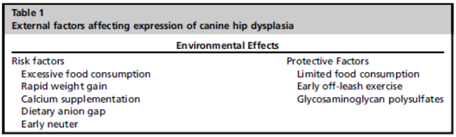
Swedish Experiences From 60 Years of Screening and Breeding Programs for Hip Dysplasia--Research, Success, and Challenges. Åke Hedhammar. Front.Vet. Sci. May 2020;7:228. Quote: A screening program for hip dysplasia (HD) was introduced in Sweden during the 1950s for German shepherd dogs, before for a few breeds and now any breed. Degree of canine HD was originally graded 1-4 (slight, mild, moderate, and severe) and used in Swedish screening program up to year 2000 and was thereafter replaced by letters A-E with A and B for no signs/near normal, C for mild, D for moderate, and E for severe HD. Final scoring is based on "the worst" side. In Sweden, 70% of all dogs are registered by the Swedish Kennel Club, and in relevant breeds, almost all breeding stock and 30-50% of all dogs are screened for HD. By an extensive database of all dogs registered since 1976 and mandatory identification by microchip, all results can be linked to dogs well-defined by identity and ancestral background. An implementation of structured screening and genetic health programs resulted in markedly decreased prevalence of HD already during the 1980s. The programs are based on open registries and on positive as well as negative results for identified individuals linked to their ancestral background. The successful decrease in moderate and severe HDs is illustrated for seven common breeds. However, there is also the challenge of a further decrease when already almost all breeding is performed with unaffected breeding stock. Handling that and the increased relative prevalence of less severe grades of HD (grade C) calls for breed-specific breeding strategies, taking into account the prevalence and clinical significance in each breed. ... By applied selection, the prevalence of moderate to severe HD has been reduced to one third in all of the most commonly predisposed breeds. ... Today in Sweden the prevalence of moderate and severe HD (D and E) in most breeds is lower than 10% (German shepherd dogs 7%, Labrador retrievers 4%, Golden retrievers 5%, Bernese mountain dogs 6%, Rottweilers 6%). It is only in a few giant breeds with a few registered dogs and no restrictions on breeding stock that the prevalence of grades D and E is over 10%. The successful reduction of HD is based on extensive screening mandatory regulated by the Swedish Kennel Club with the results in an open registry including positive as well as negative results and the wide use of this information in the selection of the breeding stock. Since the 1980s, majority of the breeding in most dog breeds are done with both parents screened and free of HD (Grade 0/A or B). The strength of the results is that the data represent the majority of the dogs of affected breeds in Sweden and that such a high proportion of these are screened for HD. In most predisposed breeds, not only breeding stock but also the majority of other dogs within the breed are screened, adding valuable information for the estimation of breeding values. ... Further decrease might rather be achieved by using estimated breeding values and genomic selection instead of more extensive and costly screening procedures. For the public perception of HD, the value of a clear distinction between grades D and E as a good predictor of the clinical entity vs. grade C as a tool to refine the selection criteria for breeding stock is indicated.
Retrospective Study of 240 Dogs Receiving Gabapentin for Chronic Pain Relief. Lily V Davis, Peter W Hellyer, Robin A Downing, Lori R Kogan. J. Vet.Med. & Research. August 2020;7(4):1194-1197. Quote: Our goal was to assess gabapentin dosage and tolerability in dogs taking it for chronic pain. ... All patients in this study were on gabapentin due to an inability to fully control their pain with NSAIDs or nutraceuticals alone. ... The majority (217 patients; 90.42%) had pain localized to their back. The other anatomic locations of pain included stifle (33; 13.8%), elbow/shoulder (21; 8.8%) and hip (11; 4.6%). Some patients' pain was localized to multiple areas and is counted in all categories. The most common diagnosis of pain was osteoarthritis (203; 84.6%), followed by generalized, nonspecific back pain (22;9.2%), intervertebral disk disease (18; 7.5%), and degenerative myelopathy (11; 4.6%). Some patients experienced two different kinds of pain and are embodied in both categories. One hundred and thirty-six (56.67%) patients did not have definitive anatomic cause for their pain, such as cruciate tears or intervertebral disc disease, as seen in Table 2. ... We retrospectively analyzed the medical records of 240 dogs taking gabapentin for chronic pain and systematically assessed: patient signalment, definitive diagnosis, location and description of pain, VAS scores immediately preceding and following the patient's maximum gabapentin dose, maximum gabapentin dosage, presence or absence of side effects related to gabapentin use, use of NSAID/immunomodulator drugs and nutraceuticals, presence or absence of levothyroxine supplementation, surgical procedures, and physical medicine. The range of tolerated gabapentin doses was 6.9 - 500 mg/kg/day [3.1- 227.3 mg/lb], PO, q12 hr (every 12 hrs) and only 10% of patients experienced the most common side effect of sedation. ... The results from this case series suggest that gabapentin is well-tolerated at much higher doses than what is typically prescribed. Side effects were uncommon, with no clear pattern based on dose, or dog size or age. Therefore, like many other analgesic medications, the efficacy of gabapentin appears patient-specific and should be dosed to effect until side effects are noted or analgesia is achieved.
CONNECT WITH US