
Cerebellar Infarcts -- Strokes --
in the Cavalier King Charles Spaniel
Cavalier King Charles spaniels appear predisposed to develop cerebellar infarcts, due to ischemic strokes. At least three published studies have reached that conclusion. See these articles: January 2005, September 2005, and October 2017.
 What It Is
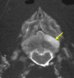
Ischemic stroke (or cerebellar ischemic stroke) is a sudden blockage of the blood flow to an area of the cerebellum of the brain caused either by a blood clot.* The blockage deprives the delivery of oxygen to that portion of the brain, creating a dead tissue lesion, typically wedge-shaped (see MRI image at right), called a cerebellar infarct. This usually results in the loss of some neurological function which had been controlled by the affected area of the brain.
* A similar stroke, caused by a rupture of one of the cerebellar arteries rather than its blockage, is labeled a cerebellar hemorrhagic stroke.
In an August 2015 study, a cavalier also has been found to have suffered a myocardial stroke -- in the heart rather than in the brain.
RETURN TO TOP
Symptoms
The onset of symptoms of a cerebellar infarct usually is quite sudden and dramatic. The cavalier may be behaving normally one minute, and then in an instant, become unsteady, un-coordinated, nauseous, walk uncontrollably in small circles*, or even faint and fall over. The dog may tilt its head, have facial paralysis, lose its vision or control of its bowels, or change its temperament.
In a September 2007 article, a cavalier diagnosed with a cerebella infarct displayed weakness in all four legs and a right-sided head tilt, and rapid, involuntary, vertical motion of the eyeball (nystagmus).
Some symptoms may progress over the first twelve hours, presumably due to brain swelling, while others may disappear quickly. Most symptoms should begin to lessen within a day or two.
* See Dr. Clare Rusbridge's YouTube video on the specific causes of circling.
RETURN TO TOP
Diagnosis
Cerebellar infarcts should be suspected in any case in which the onset of symptoms is sudden and otherwise unexplained. The dog should be taken to a veterinary clinic immediately; examination by a neurologist would be preferable. The veterinarian should conduct a complete neurological examination, in addition to blood pressure measurement, normal blood work and urinalysis, x-rays, and possibly an ultrasound and endocrine and thyroid profiles.
The symptoms may be confused with those of paradoxical vestibular syndrome, particularly the head tilt, which is the most consistent sign of unilateral vestibular dysfunction, and ataxia – loss of muscle coordination – and nystagmus, which is an involuntary rhythmic oscillation of the eyeball.
 The only accurate way to diagnose an infarction is using magnetic resonance
imagine (MRI), and to a limited extent, computed tomography (CT), to obtain
scans of brain images. Early enough CT imaging should enable the veterinarian to
determine whether the infarct was caused by a burst blood vessel (hemorrhagic),
but MRI is the best device for identifying a shut down blood vessel (ischemic),
which will better determine the treatment protocol.
The only accurate way to diagnose an infarction is using magnetic resonance
imagine (MRI), and to a limited extent, computed tomography (CT), to obtain
scans of brain images. Early enough CT imaging should enable the veterinarian to
determine whether the infarct was caused by a burst blood vessel (hemorrhagic),
but MRI is the best device for identifying a shut down blood vessel (ischemic),
which will better determine the treatment protocol.
(The image at right is an MRI scan of a CKCS skull, showing a wedge-shaped cerebellar infarct at lower right of the brain. Image from The Downs Veterinary Practice.)
There is a dispute among researchers as to the cause of the cavaliers' predisposition to this form of stroke. Dr. Curtis Dewey has attributed them to the “frequent presence of caudal occipital malformation syndrome (COMS) in these dogs.” COMS is more commonly referred to as Chiari-like malformation (CM).
However, in a 2005 study of several breeds of dogs with brain infarcts, the researchers found that CKCSs were "overrepresented" (18% of the entire group of dogs studied) and opined that the reason may be mitral valve disease (MVD) or blood platelets or CM. They stated:
"The reasons for overrepresentation of CKCS are unclear but may be due to the prevalence of heart disease, alterations in coagulation and platelet morphology, connective tissue pathology, or anatomic variation (ie, Chiari malformation) recognized in this breed. CKCS have a high prevalence of mitral valve regurgitation, as well as platelet abnormalities, 40 which might predispose them to cardioembolic infarction. Cavalier King Charles Spaniels also have a high prevalence of Chiari type malformation, which may alter vertebral-basilar blood flow and predispose them to strokes. This malformation was identified in 3 of the 6 CKCS of this study. A pathophysiologic link among the above conditions frequently seen in CKCS and the occurrence of ischemic stroke is speculative and remains to be further studied."
Dr. Dewey has responded to that hypothesis by stating:
"It has been postulated that this predisposition may be related to this breed's propensity to develop heart disease, inherited platelet abnormalities, or to local aberrations in regional arterial (e.b., basilar artery) blood flow due to caudal occipital malformation (COMS), which is common in the breed. ... In the author's experience, the combination of COMS and cerebellar infarct is common in the CKCS, whereas combinations involving cardiac disease and platelet abnormalities are not.”
RETURN TO TOP
Treatment
If the veterinarian is able to determine the underlying cause of the infarction, it should be treated promptly. Initially, mannitol, a diuretic, may be administered intravenously to overcome any brain swelling. Mannitol is the alcohol form of the sugar, mannose, and it is an osmotic diuretic and a mild renal vasodilator. It is used to treat excessive intra-cranial pressure and to expand blood vessel openings in the brain.
If systemic hypertension is found, enalapril maleate (Enacard, Vasotec), or another ACE-inhibitor may be prescribed. ACE-inhibitors block the angiotensin converting enzyme, which is necessary to produce a substance that causes blood vessels to tighten. So, ACE-inhibitors serve to relax the blood vessels, thereby lowering the blood pressure and increasing the supply of blood and oxygen to the brain.
Amlodipine (Norvasc) may also be administered. Amlodipine is a calcium channel blocker which relaxes blood vessels so that the blood can flow more easily.
Nicergoline (Fitergol, Sermion) is an alpha-blocking cerebral vasodilator which is a vasodilator that improves blood flow to the brain and stimulates the use of oxygen and glucose. It also blocks serotonin and dopamine receptors. It is used to treat migraine headaches that are of vascular origin and other problems of a vascular nature, such as dizziness and auditory problems.
Meclizine HCl (Antivert, Bonine, Bonine Max, Dramamine Less Drowsy) is an antiemetic which treats nausea, vomiting, and cizziness.
Palmitoylethanolamide (PEA) is a
N-acylethanolamine molecule in a family of long-chain fatty acid
 amides
called ALIAmides. PEA has been found to play a
protective role in neurological disorders caused by ischaemic stroke in
humans.
PEA levels have been observed to increase in tissue surrounding the
primary ischaemic lesion, in a patient with hemispheric stroke. it
was observed that when PEA was administered after an acute stroke, it
reduced infarct size in cortical and total infarct areas compared with
controls.
amides
called ALIAmides. PEA has been found to play a
protective role in neurological disorders caused by ischaemic stroke in
humans.
PEA levels have been observed to increase in tissue surrounding the
primary ischaemic lesion, in a patient with hemispheric stroke. it
was observed that when PEA was administered after an acute stroke, it
reduced infarct size in cortical and total infarct areas compared with
controls.
PEA is produced by the animal's body as needed in response to certain types of injuries. PEA is a product of normal fatty acid synthesis from palmitic acid. It is found in many common foods, particularly palm oil, soy beans, egg yolks, and peanuts. The commercial version is most commonly manufactured from palm oil*.
Not all PEA is alike. There are three types of PEA.
• Basic PEA, called "naive PEA", is insoluble in water and therefore the oral intake of it (rather than being injected directly into the abdomen) has very poor bioavailability, meaning that it does not get absorbed well in the dog's gut.
• Micronized PEA (m-PEA) is a patented technique that reduces the diameter of PEA particles, making them absorbable in the intestine, which has been found to be more effective than ordinary naive PEA in activating PEA levels in blood plasma in dogs. See this August 2014 article.
• Ultra-micronized PEA (um-PEA), also patented, reduces the PEA particle size further, to enable it to cross the blood-brain barrier, likewise has been found to be much more effective than naive PEA. See this August 2014 article.
 If a PEA product is not advertised as being micronized or
ultra-micronized, then
Dr. Clare Rusbridge advises that
"You
probably are wasting your money."
A variety of brands of
micronized and ultra-micronized PEA are offered on-line.
If a PEA product is not advertised as being micronized or
ultra-micronized, then
Dr. Clare Rusbridge advises that
"You
probably are wasting your money."
A variety of brands of
micronized and ultra-micronized PEA are offered on-line.
Recent research has produced evidence that ALIAmides can relieve dogs with hypersensitive skin disorders. In an August 2015 article, Italian researchers conducted an 8-week study of the effectiveness of oral ultra-micronized palmitoylethanolamide (um-PEA) in 160 dogs with moderate atopic dermatitis. Each dog received a daily dose of um-PEA at the rate of 10 mg/kg for 56 days. They report finding that um-PEA appeared to be effective and safe in reducing pruritus and skin lesions, and in improving the quality of life in dogs with moderate atopic dermatitis and moderate pruritus. This study was sponsored by patent holders of the use of PEA for inflammation and pain.
As for dosages, the studies using micronized PEA, the range was from 10 to 15 mg/kg/day, and the range for ultra-micronized was 24 mg/kg (for osteoarthritis in humans).
Read more about PEA on our PEA webpage.
* Palm oil: The palm oil cultivation industry has been destroying rainforests in Sumatra and Borneo in Indonesia and Malaysia, the only habitats of orangutans. If you are going to obtain PEA, we suggest that you do so only from vendors whose PEA has been manufactured with palm oil from sustainable sources and not the deforestation of rainforests. This link connects to a "PalmOil Scan Mobile App" which will enable you to determine if the PEA vendors you select obtain their palm oil from sustainable sources.
The prognosis is guarded if the symptoms are very severe and prolonged. However, dogs with less severe signs have shown rapid and dramatic recoveries. In a September 2007 article, a cavalier diagnosed with a cerebellar infarct displayed weakness in all four legs and a right-sided head tilt, and rapid, involuntary, vertical motion of the eyeball (nystagmus). It was treated with enalapril and meclizine, and within 3 weeks, the dog's "neurological condition had improved dramatically." Three months after discharge from the hospital, the dog was ambulatory in all 4 limbs.
RETURN TO TOP
Research News
December 2024:
Cavaliers ranked second in study of 125 dogs diagnosed with
ischemic stroke and additional related disorders.
 In
a
December 2024 article, UK researchers (Cecilia-Gabriella Danciu
[right], Rita Gonçalves, Carrete Jordina Caldero, Christoforos
Posporis, Javier Espinosa, Steven de Decker, Hanne Gredal, Sophie
Elizabeth Wyatt studied 125 dogs diagnosed with ischemic stroke and what
they described as a comorbidity -- another disorder possibly related to
the stroke, especially post-stroke epilepsy. Among the 125 dogs in the
study, cavalier King Charles spaniels (13/125 -- 10.4%) ranked second
only to greyhounds (14/125 -- 11.2%) among affected breeds. Among all of
the dogs, the most common comorbidities were
hypertension (20%) and
proteinuria (8%). Among cavaliers, the most common comorbidity was
mitral valve disease (MVD). The
authors discussed previous speculation about relationships between
strokes and other disorders in cavaliers, including MVD,
Chiari-like malformation ("which might
alter intracranial blood flow and predispose the breed to cardioembolic
infarction"), and macrothrombocytopenia
(over-sized blood platelets -- "In itself, this breed variation is not
associated with clinical signs, but it has been associated with
hypercoagulability measured with TEG [thromboelastography]."). However,
they report finding no connection between strokes in the cavalier
subjects of their study and any other disorder. They also reported that
post-stroke epilepsy was "uncommon" among 125 subjects of their study.
In
a
December 2024 article, UK researchers (Cecilia-Gabriella Danciu
[right], Rita Gonçalves, Carrete Jordina Caldero, Christoforos
Posporis, Javier Espinosa, Steven de Decker, Hanne Gredal, Sophie
Elizabeth Wyatt studied 125 dogs diagnosed with ischemic stroke and what
they described as a comorbidity -- another disorder possibly related to
the stroke, especially post-stroke epilepsy. Among the 125 dogs in the
study, cavalier King Charles spaniels (13/125 -- 10.4%) ranked second
only to greyhounds (14/125 -- 11.2%) among affected breeds. Among all of
the dogs, the most common comorbidities were
hypertension (20%) and
proteinuria (8%). Among cavaliers, the most common comorbidity was
mitral valve disease (MVD). The
authors discussed previous speculation about relationships between
strokes and other disorders in cavaliers, including MVD,
Chiari-like malformation ("which might
alter intracranial blood flow and predispose the breed to cardioembolic
infarction"), and macrothrombocytopenia
(over-sized blood platelets -- "In itself, this breed variation is not
associated with clinical signs, but it has been associated with
hypercoagulability measured with TEG [thromboelastography]."). However,
they report finding no connection between strokes in the cavalier
subjects of their study and any other disorder. They also reported that
post-stroke epilepsy was "uncommon" among 125 subjects of their study.
June 2023:
Cavalier experiences cardiopulmonary arrest and global
hypoxic-ischemic brain injury during routine castration.
 In
a
June 2023 article, UK clinicians Abbe Harper Crawford (right),
Elsa Beltran, Cecilia-Gabriella Danciu, Dylan Yaffy at the Royal
Veterinary College report a study of 6 dogs and 2 cats having suffered
global hypoxic-ischemic brain injuries (GHIBI), due to cardiopulmonary
arrest or anesthetic complications during surgical procedures. One of
the dogs was a cavalier King Charles spaniel which experienced the GHIBI
incident during a routine castration. The arrest resulted in a lack of
bloodflow (ischemia) to the brain, denying it needed glucose, oxygen,
and adenosine triphosphate (ATP) depletion causing the GHIBI. The CKCS
displayed generalized seizures, was treated with levetiracetam, and
recovered fully with no neurological deficits detected after 3 months.
They concluded that:
In
a
June 2023 article, UK clinicians Abbe Harper Crawford (right),
Elsa Beltran, Cecilia-Gabriella Danciu, Dylan Yaffy at the Royal
Veterinary College report a study of 6 dogs and 2 cats having suffered
global hypoxic-ischemic brain injuries (GHIBI), due to cardiopulmonary
arrest or anesthetic complications during surgical procedures. One of
the dogs was a cavalier King Charles spaniel which experienced the GHIBI
incident during a routine castration. The arrest resulted in a lack of
bloodflow (ischemia) to the brain, denying it needed glucose, oxygen,
and adenosine triphosphate (ATP) depletion causing the GHIBI. The CKCS
displayed generalized seizures, was treated with levetiracetam, and
recovered fully with no neurological deficits detected after 3 months.
They concluded that:
"Despite severe neurological deficits at onset, long-term positive outcome is possible. The duration of the hypoxic-ischemic insult and the rate of subsequent neurological improvement might provide indications of the likelihood of functional recovery in dogs and cats with GHIBI, with longer insults (particularly where resuscitation is delayed by out-of-hospital arrest) and slow neurological improvement after ROSC [return of spontaneous circulation] suggestive of a worse long-term outcome, as well as diffuse brainstem involvement."
June 2022:
Cavalier-poodle puppy suffers brain injuries due to oxygen loss
after cardiac arrest.
 In
a
June 2022 case report by Australian veterinary specialists (Jiah
Goh, Louis Mark Eramanis [right], M. Milne, M. Boller), an
18-week-old male cavalier King Charles spaniel & poodle mixed breed
suffered what was described as naturally-occuring cardiopulmonary arrest
(CPA) followed by neurological abnormalities. The dog was recovering
from having undergone an abdominal ultrasound scan under general
anesthesia at the time of the cardiac arrest. The attending
veterinarians performed cardiopulmonary resuscitation (CPR) to revive
the dog. Following the dog's resuscitation, he displayed decerebrate
rigidity (abnormal stiffness of the legs due to damage to the brain
stem) and stuporous mentation (difficulty arousing and impaired
awareness), along with episodes of barking and apnea (brief periods of
not breathing). Magnetic resonance imaging (MRI) confirmed suspected
"global brain ischemia" (GBI). Despite hospitalization and extensive
efforts over the seven days following the CPA, the puppy showed no
neurological improvement and continued to fail in other respects, and he
was euthanized. The post-mortem examination diagnosed severe,
wide-spread damage to the neurons in the brain, as well as in the heart,
due to lack of oxygen. The reported value of this study was that the
diagnosis from MRI scans was consistent with the brain tissue
examinations, suggesting that MRIs may be used to determine the extent
of global brain ischemia in dogs following cardiac arrest.
In
a
June 2022 case report by Australian veterinary specialists (Jiah
Goh, Louis Mark Eramanis [right], M. Milne, M. Boller), an
18-week-old male cavalier King Charles spaniel & poodle mixed breed
suffered what was described as naturally-occuring cardiopulmonary arrest
(CPA) followed by neurological abnormalities. The dog was recovering
from having undergone an abdominal ultrasound scan under general
anesthesia at the time of the cardiac arrest. The attending
veterinarians performed cardiopulmonary resuscitation (CPR) to revive
the dog. Following the dog's resuscitation, he displayed decerebrate
rigidity (abnormal stiffness of the legs due to damage to the brain
stem) and stuporous mentation (difficulty arousing and impaired
awareness), along with episodes of barking and apnea (brief periods of
not breathing). Magnetic resonance imaging (MRI) confirmed suspected
"global brain ischemia" (GBI). Despite hospitalization and extensive
efforts over the seven days following the CPA, the puppy showed no
neurological improvement and continued to fail in other respects, and he
was euthanized. The post-mortem examination diagnosed severe,
wide-spread damage to the neurons in the brain, as well as in the heart,
due to lack of oxygen. The reported value of this study was that the
diagnosis from MRI scans was consistent with the brain tissue
examinations, suggesting that MRIs may be used to determine the extent
of global brain ischemia in dogs following cardiac arrest.
June 2016:
Cavaliers are most common
and youngest victims of ischaemic stroke in study of 23
affected dogs.
 In a
June 2016 study of 23 dogs with neurological signs of rostral
cerebellar ischaemic stroke, the most -- 9 of them -- were cavalier King
Charles spaniels. The researchers (Barbara Thomsen [right], Laurent Garosi,
Geoff Skerritt, Clare Rusbridge, Tim Sparrow, Mette Berendt, Hanne
Gredal) found the cavalier was significantly younger at stroke onset
(mean age 7 years ± 9 months) compared to non-CKCSs (mean age 9 years
and 1 month ± 6 months). The most prominent neurological
symptoms were gait abnormalities, head tilt, nystagmus, decreased menace
response, postural reaction deficits, and proprioceptive (stimuli
connected with the position and movement of the body) deficits.
In a
June 2016 study of 23 dogs with neurological signs of rostral
cerebellar ischaemic stroke, the most -- 9 of them -- were cavalier King
Charles spaniels. The researchers (Barbara Thomsen [right], Laurent Garosi,
Geoff Skerritt, Clare Rusbridge, Tim Sparrow, Mette Berendt, Hanne
Gredal) found the cavalier was significantly younger at stroke onset
(mean age 7 years ± 9 months) compared to non-CKCSs (mean age 9 years
and 1 month ± 6 months). The most prominent neurological
symptoms were gait abnormalities, head tilt, nystagmus, decreased menace
response, postural reaction deficits, and proprioceptive (stimuli
connected with the position and movement of the body) deficits.
Ms. Thomsen (right) is a Ph.D. student at the Department of Veterinary Clinical and Animal Sciences, University Hospital for Companion Animals, University of Copenhagen. She conceived of the study and study design, participated in the coordination of the study, contributed to the acquisition and interpretation of data and drafted the manuscript.
August 2015:
Cerebellar infarction symptoms in cavaliers resemble acute vestibular
signs.
 In an
August 2015 abstract, UK neurologists (B. Thomsen, L. Garosi, G.
Skerritt, C. Rusbridge [right], T. Sparrow, M. Berendt, H. Gredal) report
finding cerebellar infarctions in 24 dogs, including 9 cavalier King
Charles spaniels, displaying ataxia (staggering or rolling gait),
hypermetria (voluntary muscular movements tend to overreach their
intended goals), head tilt, nystagmus (uncontrolled movements of the
eyes), reduced postural reactions (e.g., replacing a foot after being
lifted), and decreased menace response (eye blinking in response to an
on-coming object). They concluded that cerebellar ischemic strokes
should be considered an important differential diagnosis in dogs with
acute vestibular signs.
In an
August 2015 abstract, UK neurologists (B. Thomsen, L. Garosi, G.
Skerritt, C. Rusbridge [right], T. Sparrow, M. Berendt, H. Gredal) report
finding cerebellar infarctions in 24 dogs, including 9 cavalier King
Charles spaniels, displaying ataxia (staggering or rolling gait),
hypermetria (voluntary muscular movements tend to overreach their
intended goals), head tilt, nystagmus (uncontrolled movements of the
eyes), reduced postural reactions (e.g., replacing a foot after being
lifted), and decreased menace response (eye blinking in response to an
on-coming object). They concluded that cerebellar ischemic strokes
should be considered an important differential diagnosis in dogs with
acute vestibular signs.
 August 2015:
Cavalier in congestive heart failure suffers blood clots, a tear in the
left atrium wall, and a cardiac stroke. In an
August 2015 article by University of Pennsylvania veterinarians (Meg
M. Sleeper [right], Meredith E. Maczuzak, Susan J. Bender), they found that a
deceased cavalier King Charles spaniel which had been in congestive
heart failure due to mitral valve disease, also had suffered from a
partial tear of his left atrium wall, and blood clot blockage in his
left atrium and right foreleg, and a stroke (myocardial infarction).
August 2015:
Cavalier in congestive heart failure suffers blood clots, a tear in the
left atrium wall, and a cardiac stroke. In an
August 2015 article by University of Pennsylvania veterinarians (Meg
M. Sleeper [right], Meredith E. Maczuzak, Susan J. Bender), they found that a
deceased cavalier King Charles spaniel which had been in congestive
heart failure due to mitral valve disease, also had suffered from a
partial tear of his left atrium wall, and blood clot blockage in his
left atrium and right foreleg, and a stroke (myocardial infarction).
September 2007:
Cavalier recovers from symptoms due to cerebellar infarct.
 In
a September 2007
article about case studies of 4 dogs diagnosed with a cerebellar
wedge-shaped region, the clinicians ( John C. Irwin, Curtis W. Dewey
[right], Joseph D. Stefanacci) at LIVS on Long Island, NY, reported
about a 7-year-old cavalier that displayed weakness in all four legs and
a right-sided head tilt, and rapid, involuntary, vertical motion of the
eyeball (nystagmus). MRI confirmed that a cerebellar wedge-shaped region
was inactive (hypointense). He was treated with enalapril and meclizine,
and within 3 weeks, the dog's "neurological condition had improved
dramatically." Three months after discharge from the hospital, the dog
was ambulatory in all 4 limbs.
In
a September 2007
article about case studies of 4 dogs diagnosed with a cerebellar
wedge-shaped region, the clinicians ( John C. Irwin, Curtis W. Dewey
[right], Joseph D. Stefanacci) at LIVS on Long Island, NY, reported
about a 7-year-old cavalier that displayed weakness in all four legs and
a right-sided head tilt, and rapid, involuntary, vertical motion of the
eyeball (nystagmus). MRI confirmed that a cerebellar wedge-shaped region
was inactive (hypointense). He was treated with enalapril and meclizine,
and within 3 weeks, the dog's "neurological condition had improved
dramatically." Three months after discharge from the hospital, the dog
was ambulatory in all 4 limbs.
RETURN TO TOP
Related Links
Veterinary Resources
Enhanced recovery of diastolic function after global myocardial ischemia in the intact animal. Kevin Tveter, John St.Cyr, Joseph Schneider, Richard Bianco, John Foker. Pediatr. Res. 1988; 23:226A. Quote: "The relationship of myocardial ATP levels and postischemic dysfunction remains controversial. In an intact animal model of global ischemia (Isc) and recovery, we have found ribose (R) or adenine (A) and R accelerated the return of ATP levels (84% and 80% recovery by 24 hrs). The purpose of this study was to determine the effects of enhancing ATP recovery on postlsc function. Following 20 min of Isc on cardiopulmonary bypass, dogs received either R (80mM) (n=S), A (20mM) and R (80mM)(n=5) or saline (NS)(n=6) for 24 hrs. The end-systolic pressure-volume ratio CEmax• mmHg/ml), dP/dt (mmHg/sec) and diastolic circumferential stress (cr, dynes x 103/cm2)-strain (e:) relationships were determined from sonomicrometry and micromanometry data during transient vena caval occlusions. ... We conclude: (1) recovery of systolic function is essentially complete by 4 hrs. (2) Return of diastolic function is enhanced similarly to ATP recovery by R or A/R. (3) Because A did not further enhance ATP recovery, R appears to be the rate limiting ATP precursor."
Enhanced high energy phosphate recovery with ribose infusion after global myocardial ischemia in a canine model. John A. St. Cyr, Richard W. Bianco, Joseph R. Schneider, John R. Mahoney, Jr., Kevin Tveter, Stanley Einzig, John E. Foker. J. Surgical Res. February 1989;46:157-162. Quote: High energy phosphate levels are depressed following global ischemia and require several days to completely recover. Short-term methods to enhance ATP recovery have included infusion of ATP precursors, inhibition of enzymes that catabolize AMP, and membrane transport stabilization. Several precursors have been used to augment adenine nucleotide synthesis including adenosine, inosine, adenine, and ribose. Because of the short-term nature of previous experiments, recovery had been incomplete and the effects in the intact animal unknown. The purpose of this study was to determine the effects of ribose infusion in a long-term model of global ischemia and attempt to identify the precursor which limits myocardial ATP regeneration in the intact animal. Global myocardial ischemia (20 min, 37°C) was produced in dogs on cardiopulmonary bypass. With reperfusion either ribose (80 mM) in normal saline or normal saline alone was infused at 1 ml/min into the right atrium and the animals were followed for 24 hr. Ventricular biopsies were obtained through an indwelling ventricular cannula prior to ischemia, at the end of ischemia, and 4 and 24 hr postischemia and analyzed for adenine nucleotides and creatine phosphate levels. Radiolabeled microspheres were used to measure myocardial and renal blood flows and no significant difference was found between ribose-treated control groups. In both groups, myocardial ATP levels fell by at least 50% at the end of ischemia. No significant ATP recovery occurred after 24 hr in the control dogs, but in the ribose-treated animals, ATP levels rebounded to 85% of control by 24 hr. Total myocardial adenine nucleotide content and energy charge also recovered in the ribose group but not in the control animals. The ribose infusion, therefore, significantly enhanced the recovery of energy levels in the postischemic myocardium in the intact animals.
MRI appearance of cerebrovascular disease in seven spaniels. Mcconnell, J. F., Garosi, L., Dennis, R., Platt, S. R. & Abramson C. J. 2003 BSAVA Congress; pg 568.
Cerebellar Infarcts in Two Dogs Diagnosed With Magnetic Resonance Imaging. Jason M. Berg and Richard J. Joseph. J Am Anim Hosp Assoc.2003;39:203-207. Quote: "Two dogs presented with severe, peracute-onset, neurological signs. Neuroanatomical localization was cerebellovestibular. Magnetic resonance imaging (MRI) was performed and revealed focal, wedge-shaped lesions in the cerebellum. Diagnosis of cerebellar infarctions was made based on peracute-onset, clinical signs, MRI, and outcome as well as ancillary diagnostic information. Both dogs recovered completely. Cerebellar infarction should be included in the differential of any dog with peracute-onset, central cerebellovestibular signs regardless of severity of clinical signs. Outcome was excellent in these dogs."
Magnetic Resonance Imaging Findings of Presumed Cerebellar Cerebrovascular Accident in Twelve Dogs. J. F. McConnell, L. Garosi, S. R. Platt. Vety. Radiology & Ultrasound. Jan. 2005; 46(1):1-10. Quote: Magnetic resonance imaging findings of presumed cerebellar cerebrovascular accident in 12 dogs. J. F. Mcconnell, L. Garosi, S. R. Platt. Vet. Radiology & Ultrasound. January 2005;46(1):1-10. Quote: The magnetic resonance imaging (MRI) findings of presumed cerebrovascular accident in 12 dogs are described. ... The dogs ranged in age from 4.5 to 15 years with a median of 8.25 years and mean of 8.9 years. Seven dogs were male (four neutered) and five were neutered females. There were two mixed breed (one English Springer spaniel cross, one undetermined) and six pure breeds: four Cavalier King Charles spaniels (CKCS), two golden retrievers and one—English Cocker spaniel, Weimaraner, Border collie, and Greyhound. ... Fourteen lesions were seen, commonly (11 of 14) within the gray matter of the cerebellar hemispheres or vermis. Thirteen lesions were hyperintense on T2-weighted images (in 11 dogs) and one was hypointense. Eleven of 14 lesions were within the region supplied by the rostral cerebellar artery or one of its main branches and there was no, or minimal, mass effect. Contrast enhancement was only seen in six lesions and was mild in all. Gradient-echo images provided additional information in two dogs. The appearance of infarction in dogs with diffusion-weighted images (DWI) is similar to that in humans, and provided supportive evidence for the diagnosis of infarction in five dogs. The use of gradient-echo and DWI is recommended for the evaluation of suspected cerebrovascular accidents in dogs. Six of the 12 affected animals were spaniels or spaniel crosses, suggesting a possible breed predisposition.
Vascular Encephalopathies. C.W. Dewey. 50° Congresso Nazionale Multisala SCIVAC, 2005. Quote: “There appears to be a predisposition for spaniel breeds, especially cavalier King Charles spaniels, to develop cerebellar strokes. This is suspected to be due to frequent presence of caudal occipital malformation syndrome (COMS) in these dogs, with associated interference with normal basilar artery flow.”
Neurological diseases of the Cavalier King Charles spaniel. Rusbridge, C. J. Small Animal Practice. June 2005;46(6): 265-272. "CKCSs seem to have an increased tendency towards cerebrovascular disease (McConnell and others 2003), particularly infarction of the rostral cerebellar artery. Affected dogs present with signs of acuteonset, rapidly progressive central vestibular syndrome. Rostral cerebellar artery infarction in humans is associated with cardiogenic embolism and major artery occlusive disease such as carotid artery dissection (Yin and others 1994). The CKCS is predisposed to mitral valve disease Haggstrom and others 1992), increased platelet aggregation (Olsen and others 2001) and arterial disease (Buchanan and others 1997), all of which offer some explanation for a tendency towards cerebrovascular disease. In the UK, any CKCS presented with signs of intracranial haemorrhage or infarction should be screened for Angiostrongylus vasorum. This parasite can result in bleeding and coagulation disorders, and the CKCS appears to be predisposed to infection (Chapman and others 2004)."
Results of diagnostic investigations and long-term outcome of 33 dogs with brain infarction (2000-2004). Garosi, J.F. McConnell, S.R. Platt, G. Barone, J.C. Baron, A. de Lahunta, S.J. Schatzberg. J. Vet. Intern. Med. September 2005;19(3):725-731. Quote: Medical records of 33 dogs [including 6 cavalier King Charles spaniels] presented for acute onset, nonprogressive, intracranial dysfunction that had a magnetic resonance imaging diagnosis of brain infarction were reviewed. ... Cavalier King Charles Spaniels (CKCS) and Greyhounds, however, appeared overrepresented (18% and 15%, respectively) compared with CKCS and Greyhounds presented to the neurology service (3.5% and 0.87%, respectively) and the hospital population (3% and 0.66%, respectively) during the same study period. The reasons for overrepresentation of CKCS are unclear but may be due to the prevalence of heart disease, alterations in coagulation and platelet morphology, connective tissue pathology, or anatomic variation (ie, Chiari malformation) recognized in this breed. CKCS have a high prevalence of mitral valve regurgitation, as well as platelet abnormalities, 40 which might predispose them to cardioembolic infarction. Cavalier King Charles Spaniels also have a high prevalence of Chiari type malformation,41 which may alter vertebral-basilar blood flow and predispose them to strokes. This malformation was identified in 3 of the 6 CKCS of this study. A pathophysiologic link among the above conditions frequently seen in CKCS and the occurrence of ischemic stroke is speculative and remains to be further studied. ... Postmortem confirmation of brain infarction was available in 10 dogs. All dogs were evaluated by CBC, serum biochemistry, thyroid and adrenal testing, urinalysis, thoracic and abdominal imaging, and cerebrospinal fluid analysis. Results of coagulation profile and arterial blood pressure were available in 32/33 and 28/33 dogs, respectively. On the basis of the imaging findings, infarcts were classified depending on their type (territorial or lacunar) and location within the brain (telencephalic, 10/33; thalamic/midbrain, 8/33; cerebellar, 15/33). No marked associations among location or type of infarct and patient age and sex, occurrence of systemic hypertension, and the presence or absence of a concurrent medical condition were identified. Small breed dogs (< or =15 kg) were significantly more likely to have territorial cerebellar infarcts, whereas large breed dogs (>15 kg) were significantly more likely to have lacunar thalamic or midbrain infarcts. A concurrent medical condition was detected in 18/33 dogs with brain infarcts, with chronic kidney disease (8/33) and hyperadrenocorticism (6/ 33) being most commonly encountered. Of 33 dogs, 10 were euthanized because of the severity and lack of improvement of their neurologic status or the severity of their concurrent medical condition. No association was identified between type or location of infarct and patient outcome. Dogs with concurrent medical conditions had significantly shorter survival times than those with no identifiable medical condition and were significantly more likely to suffer from recurrent neurologic signs because of subsequent infarcts.
Clinical and Topographic Magnetic Resonance Characteristics of Suspected Brain Infarction in 40 Dogs. L. Garosi, J.F. McConnell, S.R. Platt, G. Barone, J.C. Baron, A. de Lahunta, and S.J. Schatzberg. J Vet Intern Med 2006;20:311–321. Quote: "Medical records of 40 dogs [including 8 cavalier King Charles spaniels] presented for evaluation of acute-onset, nonprogressive, intracranial dysfunction by means of magnetic resonance imaging (MRI) diagnosis of brain infarction were reviewed. Location of the brain infarcts was: 11 of 38, telencephalic; 8 of 38, thalamic/midbrain; 18 of 38, cerebellar; and 3 of 38, multifocal. Telencephalic infarcts developed within the territory of the middle cerebral (4/11), rostral cerebral (2/11), and striate (5/11) arteries. Thalamic/midbrain infarcts developed within the territory of perforating arteries of the caudal portion of the thalamus and rostral portion of the brainstem (8/8). All cerebellar infarcts (18/38) were within the territory of the rostral cerebellar artery or one of its branches. All infarcts appeared nonhemorrhagic, with marked contrast enhancement observed in only 3 of 38 dogs, all of which were imaged more than 7 days after the onset of signs of neurologic dysfunction. Diffusion-weighted imaging (DWI) sequences were available from 6 dogs, all imaged within 5 days of the onset of signs of neurologic dysfunction. Suspected infarcts were hyperintense on DWI sequences and were hypointense on the apparent diffusion coefficient map. Telencephalic infarcts caused abnormal mental status, contralateral postural reaction deficit, contralateral nasal hypalgesia, contralateral menace deficit, and ipsilateral circling. Thalamic/midbrain infarcts caused contralateral or ipsilateral postural reaction deficit, contralateral menace deficit, ipsilateral head tilt or turn, nystagmus, ventrolateral strabismus, and anisocoria. Cerebellar infarcts caused ipsilateral asymmetric cerebellar quality ataxia, head tilt, intermittent opisthotonus, nystagmus, and ipsilateral menace deficit with apparent normal vision."
Suspected cerebellar infarcts in 4 dogs. John C. Irwin, Curtis W.
Dewey, Joseph D. Stefanacci. J Vet Emer & Crit Care; Sept 2007; doi:
10.1111/j.1476-4431.2007.00220.x . Objective:
Reports of cerebellar infarction in veterinary literature are rare.
Documentation and descriptions of cerebellar infarction in human literature have
been well described. Brain lesions suspected to be infarcts have been recognized
with magnetic resonance imaging (MRI). ... Four dogs presented ... with the
history of acute onset cerebellovestibular dysfunction [including a
cavalier King Charles spaniel]. All animals were imaged using a 0.5 T
super-conducting Magna GE MRI unit. Suspected cerebellar infarction was
diagnosed in all dogs based on history, physical examination, and MRI findings.
Further diagnostics were performed based on the individual case and owner
compliance. Functional recovery was favorable in 3 dogs. One dog was euthanized
shortly following the onset of neurological dysfunction. ... Case no. 1 A
7-year-old, neutered male, Cavalier King Charles Spaniel
was referred to LIVS with a history of acute onset of
non-ambulatory tetraparesis and a right-sided head tilt 17
 hours
before admission. The dog appeared healthy before the onset of clinical
signs with no known toxin ingestion or trauma history. ... Upon
neurological examination, abnormalities included a right-sided head
tilt, positional vertical nystagmus, absent menace response bilaterally,
and non-ambulatory tetraparesis with conscious proprioceptive deficits
in all 4 limbs. Voluntary motor and proprioceptive deficits were worse
on the left side. A left-sided cerebellomedullary angle lesion resulting
in a paradoxical vestibular syndrome was suspected. ... The dog was
anesthetized and an MRI of the brain was performed. A large, left-sided,
rostral cerebellar wedge-shaped region was hypointense on T1-weighted
images and hyperintense on T2-weighted images. ... Figure 2 [right]
shows the hyperintense lesion on a T2-weighted image in axial
plane. ... The dog was discharged 3 days post-admission with enalapril
maleate (0.4 mg/kg q 12 h) and meclizine HCl (25mg q 24 h). At follow-up
3 weeks later, the neurological examination had improved dramatically.
Although the dog was still non-ambulatory with conscious proprioceptive
deficits, improved voluntary motor was evident bilaterally. The
nystagmus had resolved and a mild right-sided head tilt persisted. An
echocardiogram performed at that time was unremarkable. Three months
after discharge from the hospital, the dog was represented to the
internal medicine service in a diabetic ketoacidotic state. The dog was
ambulatory in all 4 limbs with conscious proprioceptive deficits of the
pelvic limbs and the head tilt had lessened in severity. A phone
conversation 1 year and 4 months after the initial diagnosis of
cerebellar infarct revealed the dog was ambulating well with the ability
to walk, run, jump, play, and climb stairs. The head tilt had improved
dramatically. Medications at that time included enalapril maleate (0.4
mg/kg q 12 h) and human insulin isophane suspension (11U SQ q 12 h). ...
Case 1 was diagnosed with hypertension and diabetes. Diabetes mellitus
is commonly associated with hypertension in humans. The mechanism for
hypertension in diabetes mellitus is believed to be an immunemediated
microangiopathy affecting the basement membrane of vessels.
Microangiopathy is not believed to be a lesion of clinical significance
in animals. Perhaps, the hypertension in Case 1 was primary.
hours
before admission. The dog appeared healthy before the onset of clinical
signs with no known toxin ingestion or trauma history. ... Upon
neurological examination, abnormalities included a right-sided head
tilt, positional vertical nystagmus, absent menace response bilaterally,
and non-ambulatory tetraparesis with conscious proprioceptive deficits
in all 4 limbs. Voluntary motor and proprioceptive deficits were worse
on the left side. A left-sided cerebellomedullary angle lesion resulting
in a paradoxical vestibular syndrome was suspected. ... The dog was
anesthetized and an MRI of the brain was performed. A large, left-sided,
rostral cerebellar wedge-shaped region was hypointense on T1-weighted
images and hyperintense on T2-weighted images. ... Figure 2 [right]
shows the hyperintense lesion on a T2-weighted image in axial
plane. ... The dog was discharged 3 days post-admission with enalapril
maleate (0.4 mg/kg q 12 h) and meclizine HCl (25mg q 24 h). At follow-up
3 weeks later, the neurological examination had improved dramatically.
Although the dog was still non-ambulatory with conscious proprioceptive
deficits, improved voluntary motor was evident bilaterally. The
nystagmus had resolved and a mild right-sided head tilt persisted. An
echocardiogram performed at that time was unremarkable. Three months
after discharge from the hospital, the dog was represented to the
internal medicine service in a diabetic ketoacidotic state. The dog was
ambulatory in all 4 limbs with conscious proprioceptive deficits of the
pelvic limbs and the head tilt had lessened in severity. A phone
conversation 1 year and 4 months after the initial diagnosis of
cerebellar infarct revealed the dog was ambulating well with the ability
to walk, run, jump, play, and climb stairs. The head tilt had improved
dramatically. Medications at that time included enalapril maleate (0.4
mg/kg q 12 h) and human insulin isophane suspension (11U SQ q 12 h). ...
Case 1 was diagnosed with hypertension and diabetes. Diabetes mellitus
is commonly associated with hypertension in humans. The mechanism for
hypertension in diabetes mellitus is believed to be an immunemediated
microangiopathy affecting the basement membrane of vessels.
Microangiopathy is not believed to be a lesion of clinical significance
in animals. Perhaps, the hypertension in Case 1 was primary.
... Cerebellar infarction appears to have characteristic MRI features. Affected tissue within the cerebellum is typically wedge-shaped with low signal intensity in T1-weighted images, high signal intensity in T2-weighted images, subtle rim enhancement without central contrast enhancement in T1 post-gadolinium, and selectively hyperintense in fluid-attenuated inversion recovery images. Anatomic regions serviced by rostral cerebellar arteries are affected. Vascular risk factors as compared with reports in human literature are also discussed.”
A Practical Guide to Canine and Feline Neurology. Curtis W. Dewey. John Wiley & Sons; 2008; 4-6, 194-195. Quote: ""Breed-associated neurologic abnormalities of dogs and cats. ... Cavalier King Charles Spaniels ... Cerebellar infarct". pp. 4-6. “Cerebellar infarcts appear to be most common in small breed dogs, most notably the Cavalier King Charles Spaniel (CKCS). It has been postulated that this predisposition may be related to this breed's propensity to develop heart disease, inherited platelet abnormalities, or to local aberrations in regional arterial (e.b., basilar artery) blood flow due to caudal occipital malformation (COMS), which is common in the breed. ... In the author's experience, the combination of COMS and cerebellar infarct is common in the CKCS, whereas combinations involving cardiac disease and platelet abnormalities are not.”
Ischaemic and haemorrhagic stroke in the dog. A Wessmann, K Chandler, L Garosi. Vety.J. June 2009; 180(3):290-303. Quote: "Cerebrovascular disease results from any pathological process of the blood vessels supplying the brain. Stroke, characterised by its abrupt onset, is the third leading cause of death in humans. This rare condition in dogs is increasingly being recognised with the advent of advanced diagnostic imaging. ... Cavalier King Charles Spaniels and greyhounds were overrepresented in one study of brain infarcts (Garosi et al., 2005a). Small breed dogs were more likely to have cerebellar territorial infarcts and large breed dogs were more likely to have lacunar thalamic-midbrain infarcts ... Magnetic resonance imaging (MRI) is the first choice diagnostic tool for stroke, particularly using diffusion-weighted images and magnetic resonance angiography for ischaemic stroke and gradient echo sequences for haemorrhagic stroke. An underlying cause is not always identified in either humans or dogs. Underlying conditions that may be associated with canine stroke include hypothyroidism, neoplasia, sepsis, hypertension, parasites, vascular malformation and coagulopathy. Treatment is mainly supportive and recovery often occurs within a few weeks. The prognosis is usually good if no underlying disease is found."
Computed tomography diagnosis of eight dogs with brain infarction. AEH Paul, Z Lenard, CS Mansfield. Australian Vet J. Oct. 2010; 88(10): 374-380. Quote: “Objective: Medical records of eight dogs [two Cavalier King Charles Spaniels (CKCS)] presenting with acute onset of neurological signs and a diagnosis of brain infarction as determined by computed tomography (CT) imaging were reviewed. Results: Ischaemic infarction in the territory of the rostral cerebellar artery was identified in three spaniel-breed dogs. All cerebellar infarcts were non-haemorrhagic. Telencephalic infarcts were identified in five dogs, in the territories of the middle cerebral artery (2/5) and rostral cerebral artery (3/5). One of these dogs had an ischaemic infarction, but all other infarctions appeared haemorrhagic. All dogs were geriatric (≥8 years old), with concurrent medical conditions identified in six dogs. One dog was euthanased after diagnosis because of the severity of its neurological signs and one dog was euthanased as a result of associated renal disease 2 months after diagnosis. Six dogs were alive at least 1 year after diagnosis. Conclusions CT is useful in the diagnosis of cerebrovascular accident in dogs, which can present as a spectrum of images with early changes in attenuation and subtle mass effects detected after infarction. CT is particularly sensitive for detecting haemorrhagic infarction, but under-represent ischaemic and lacunar infarctions when compared with MRI.”
Feline Cerebrovascular Disease: Clinical and Histopathologic Findings in 16 Cats. Ulrike Michal Altay, Geoff C. Skerritt, Monika Hilbe, Felix Ehrensperger, and Frank Steffen. J.Amer.Anim.Hosp.Assn. March 2011;47(2):89-97. Quote: "... Cerebellar infarcts in Cavalier King Charles spaniels have been reported with increasing frequency ... ."
Clinical and topographic magnetic resonance imaging characteristics of suspected thalamic infarcts in 16 dogs. Rita Gonçalves, Inés Carrera, Laurent Garosi, Peter M. Smith, J. Fraser McConnell, and Jacques Penderis Vet J, April 2011, 188(1)39-43. Quote: “Sixteen dogs with acute-onset, non-progressive signs of brain dysfunction and magnetic resonance imaging (MRI) characteristics compatible with thalamic infarction are described. ... There were 3 Cavalier King Charles spaniels (CKCS) ... Topographically the MRI lesions could be grouped in three thalamic regions, namely, paramedian (8/16), extensive dorsal (5/16) and ventrolateral (3/16). Paramedian lesions resulted in signs typical of vestibular dysfunction. Extensive dorsal lesions were associated with vestibular ataxia, circling and contralateral menace response deficits. Ventrolateral lesions resulted in circling and contralateral proprioceptive deficits. In several dogs, regions other than the thalamus were also affected: four extended into the midbrain; six extended to the internal capsule, and two dogs had a second lesion in the cerebellum. Three clinical syndromes were identified in association with thalamic infarction. These signs varied somewhat, most likely because lesions were not confined to specific nuclear boundaries and involved different combinations of thalamic nuclei."
Concurrent Medical Conditions and Long-term Outcome in Dogs with Nontraumatic Intracranial Hemorrhage. Mark Lowrie, Luisa De Risio, Ruth Dennis, Francisco Llabres-D´iaz, Laurent Garosi. Vety Radiology & Ultrasound. Apr 2012; 53(4):381-388.
Strokes: signs, causes and management. Sebastien Behr, Raquel Trevail, Roberto José-López, James Elford. Vet. Times. April 2013. Quote: "What exactly is a stroke? It is defined as an acute onset of focal neurological signs secondary to a cerebrovascular accident. A cerebrovascular accident is a disruption of the blood supply to an area of the brain. Cerebrovascular accident can be divided in two main groups: • ischaemic: when a blockage of an artery occurs; and • haemorrhagic: when bleeding in or around the brain occurs. Ischaemic cerebrovascular accidents are more common than haemorrhagic cerebrovascular accidents both in humans and small animals. Ischaemic cerebrovascular accident can either affect large blood vessels, causing a territorial infarct or can affect small intraparenchymal capillary blood vessels, causing lacunar infarcts. Lacunar infarcts can sometimes be silent and only be incidental MRI findings in some cases. Ischaemic strokes can sometimes be brief and temporary and in such cases are called transient ischaemic attacks (TIA) – the clinical signs would then only last for a few minutes or up to 24 hours maximum. ... The most commonly affected canine breeds for ischaemic cerebrovascular accident are the cavalier King Charles spaniel, greyhound and Labrador retriever."
Spontaneous ischaemic stroke in dogs: clinical topographic similarities to humans. H. Gredal1, G. C. Skerritt, P. Gideon, P. Arlien-Soeborg, M. Berendt. Acta Neurologica Scandinavica. Sept. 2013;128(3):e11–e16. Quote: "Background: Translation of experimental stroke research into the clinical setting is often unsuccessful. Novel approaches are therefore desirable. As humans, pet dogs suffer from spontaneous ischaemic stroke and may hence offer new ways of studying genuine stroke injury mechanisms. Aims: The objective of this study was to compare clinical symptoms and infarct topography of naturally occurring ischaemic stroke in pet dogs with human ischaemic stroke. Methods: Medical records and magnetic resonance imaging (MRI) of 27 dogs with spontaneous ischaemic stroke [including cavalier King Charles spaniels] were retrospectively investigated with respect to clinical symptoms and infarct topography. Symptomatology and MRI characteristics were compared with humans. Results: Seventy per cent were diagnosed with middle cerebral artery (MCA) occlusions. Motor dysfunction or sensory-motor dysfunction was reported in 78%, including specific signs of contra-lateral motor dysfunction in 11 of 27 (40%). Seizures were reported in 15 of 27 cases (56%). Conclusions: Spontaneously occurring ischaemic stroke in dogs share characteristics with human ischaemic stroke in terms of clinical symptoms and infarct topography. Investigating pet dogs with spontaneous ischaemic stroke may provide an alternative approach to the research of stroke injury mechanisms as they occur naturally, and should be further investigated."
Micronized/ultramicronized palmitoylethanolamide displays superior oral efficacy compared to nonmicronized palmitoylethanolamide in a rat model of inflammatory pain. Daniela Impellizzeri, Giuseppe Bruschetta, Marika Cordaro, Rosalia Crupi, Rosalba Siracusa, Emanuela Esposito, Salvatore Cuzzocrea. Neuroinflammation. August 2014; doi: 10.1186/s12974-014-0136-0. Quote: Background: The fatty acid amide palmitoylethanolamide (PEA) has been studied extensively for its anti-inflammatory and neuroprotective actions. The lipidic nature and large particle size of PEA in the native state may limit its solubility and bioavailability when given orally, however. Micronized formulations of a drug enhance its rate of dissolution and reduce variability of absorption when orally administered. The present study was thus designed to evaluate the oral anti-inflammatory efficacy of micronized/ultramicronized versus nonmicronized PEA formulations. Methods: Micronized/ultramicronized PEA was produced by the air-jet milling technique, and the various PEA preparations were subjected to physicochemical characterization to determine particle size distribution and purity. Each PEA formulation was then assessed for its anti-inflammatory effects when given orally in the carrageenan-induced rat paw model of inflammation, a well-established paradigm of edema formation and thermal hyperalgesia. Results: Intraplantar injection of carrageenan into the right hind paw led to a marked accumulation of infiltrating inflammatory cells and increased myeloperoxidase activity. Both parameters were significantly decreased by orally given micronized PEA (PEA-m; 10 mg/kg) or ultramicronized PEA (PEA-um; 10 mg/kg), but not nonmicronized PeaPure (10 mg/kg). Further, carrageenan-induced paw edema and thermal hyperalgesia were markedly and significantly reduced by oral treatment with micronized PEA-m and ultramicronized PEA-um at each time point compared to nonmicronized PeaPure. However, when given by the intraperitoneal route, all PEA formulations proved effective. Conclusions: These findings illustrate the superior anti-inflammatory action exerted by orally administered, micronized PEA-m and ultramicronized PEA-um, versus that of nonmicronized PeaPure, in the rat paw carrageenan model of inflammatory pain.
Do Dogs Have Strokes? Cerebrovascular Accident. Laurent Garosi. 11th ECC UK 2014 Lecture, Vet. Times. November 2014.
Myocardial infarct associated with a partial thickness left atrial tear in a dog with mitral insufficiency. Meg M. Sleeper, Meredith E. Maczuzak, Susan J. Bender. J. Vet. Cardiology. August 2015. Quote: "A 10-year-old male neutered cavalier King Charles Spaniel with a 1-year history of degenerative mitral valve disease presented for dyspnea and severe weakness. He was diagnosed with congestive heart failure, systolic dysfunction, presumptive myocardial infarction and a left atrial thrombus based on thoracic radiographs, electrocardiogram and echocardiographic findings. Clinical signs also suggested right foreleg embolism. The dog was euthanized due to the grave prognosis and a postmortem evaluation was performed. The postmortem examination confirmed myocardial infarction and was thought to be due to embolic showering from the thrombus attached to a partial thickness left atrial endocardial tear."
Neurological signs in 24 dogs with rostral cerebellar infarction. B. Thomsen, L. Garosi, G. Skerritt, C. Rusbridge, T. Sparrow, M. Berendt, H. Gredal. 27th ESVN-ECVN Symposium. J.Vet.Int.Med. August 2015; ESVN-ECVN Abstracts p.27. Quote: "Studies on canine ischemic stroke suggest an overrepresentation of cerebellar cases. Although not the leading localization in humans, a cerebellar stroke syndrome characterized by vertigo, ataxia, nystagmus, and headache is described. Purpose of the present study was to characterize neurological signs in dogs with cerebellar infarction. A retrospective multicenter study of dogs examined 2010–2014 was performed. Dogs with acute non-progressive neurological deficits and magnetic resonance imaging suggestive of cerebellar infarction were eligible for inclusion. Twenty-four dogs (16 females, 8 males) with a median age of 8 years (range 3–11 years) were included. Breeds comprised the Cavalier King Charles Spaniel (n = 9), Greyhound (n = 3), Labrador Retriever (n = 2), and ten other breeds. In all dogs, infarcts were located to the rostral cerebellum and the territory of the rostral cerebellar artery (left n = 13, right n = 10, midline n = 1). Common neurological deficits were ataxia (n = 24), hypermetria (n = 12), contralateral head tilt (n = 13), nystagmus (n = 8), reduced postural reactions (n = 8), and decreased menace response (ipsilateral n = 4, bilateral n = 2). Four dogs had additional non-cerebellar brain infarcts. Six dogs had a history suggestive of a possible transient ischemic attack. Cerebellar ischemic stroke should be considered an important differential diagnosis in dogs with acute vestibular signs. A syndrome of canine cerebellar ischemic stroke caused by infarction in the territory supplied by the rostral cerebellar artery with neurological deficits resembling its human counterpart does seem to exist."
Cerebral Infarction. Mark Troxel. Clinician's Brief. August 2015;23-27. Quote: Signalment: Cavalier King Charles spaniels: Possibly related to local alterations in intracranial pressure secondary to Chiari-like malformation.
Neurological signs in 23 dogs with suspected rostral cerebellar ischaemic stroke. Barbara Thomsen, Laurent Garosi, Geoff Skerritt, Clare Rusbridge, Tim Sparrow, Mette Berendt, Hanne Gredal. Acta Veterinaria Scandinavica. June 2016. Quote: Background: In dogs with ischaemic stroke, a very common site of infarction is the cerebellum. The aim of this study was to characterise neurological signs in relation to infarct topography in dogs with suspected cerebellar ischaemic stroke and to report short-term outcome confined to the hospitalisation period. A retrospective multicentre study of dogs with suspected cerebellar ischaemic stroke examined from 2010–2015 at five veterinary referral hospitals was performed. Findings from clinical, neurological, and paraclinical investigations including magnetic resonance imaging were assessed. Results: Twenty-three dogs, 13 females and 10 males with a median age of 8 years and 8 months, were included in the study. The Cavalier King Charles Spaniel (n = 9) was a commonly represented breed. All ischaemic strokes were located to the vascular territory of the rostral cerebellar artery including four extensive and 19 limited occlusions. The most prominent neurological deficits were gait abnormalities (ataxia with hypermetria n = 11, ataxia without hypermetria n = 4, non-ambulatory n = 6), head tilt (n = 13), nystagmus (n = 8), decreased menace response (n = 7), postural reaction deficits (n = 7), and proprioceptive deficits (n = 5). Neurological signs appeared irrespective of the infarct being classified as extensive or limited. All dogs survived and were discharged within 1–10 days of hospitalisation. Conclusions: Dogs affected by rostral cerebellar ischaemic stroke typically present with a collection of neurological deficits characterised by ataxia, head tilt, and nystagmus irrespective of the specific cerebellar infarct topography. In dogs with peracute to acute onset of these neurological deficits, cerebellar ischaemic stroke should be considered an important differential diagnosis, and neuroimaging investigations are indicated. ... The Cavalier King Charles Spaniel was significantly younger at stroke onset (mean age 7 years ± 9 months) compared to non-Cavalier King Charles Spaniels (mean age 9 years and 1 month ± 6 months) (P = 0.03). ... Furthermore, it has been speculated if the Chiari-like malformation, which is often found in Cavalier King Charles Spaniels, may cause flow disturbances and predispose affected dogs to rostral cerebellar infarction. ... Although dogs are often severely compromised at presentation, short-term prognosis is excellent and rapid clinical improvement may be observed within the first week following the ischaemic stroke.
Putative Cerebral Microbleeds in Dogs Undergoing Magnetic Resonance Imaging of the Head: A Retrospective Study of Demographics, Clinical Associations, and Relationship to Case Outcome. S. C. Kerwin, J. M. Levine, C. M. Budke, J. F. Griffin 4th, C. E. Boudreau. J. Vet. Intern. Med. May 2017. Quote: Background: Cerebral microbleeds (CMBs) are focal intraparenchymal signal voids on gradient-echo magnetic resonance imaging (MRI), corresponding to regions of chronic hemorrhage. In humans, they are associated with systemic disease and shorter survival times. Although similar findings have been identified in dogs, their epidemiology and clinical correlations have not been investigated. Objective: To determine epidemiological features, clinical associations, and associations with outcome for putative CMB-like foci (putative microbleeds [pMBs]) identified by T2*-weighted MRI in dogs. Animals: Five hundred and eighty-two dogs [including 22 cavalier King Charles spaniels] undergoing 3T brain MRI between 2011 and 2016. Methods: Retrospective case–control study. Demographic, diagnostic, and clinicopathological data were obtained from medical records and phone follow-up. Demographic variables were compared between dogs with and without evidence of pMBs. For dogs with such evidence, and a subset of matched controls, associations with clinical presentation, concurrent disease, and survival times were evaluated. Results: Dogs with pMBs were older (P < .001) and smaller (P = .004) than unaffected dogs. Compared to matched controls, they presented more frequently for vestibular signs (P = .030). Cortical atrophy occurred concurrently with pMBs in 26% (14/54) of dogs. Diagnosed renal disease was not significantly associated with pMBs, but proteinuria was more common in dogs with pMBs than in matched controls (odds ratio = 3.01, P = .005). Dogs with pMBs had a shorter median survival time than did matched controls (P = .011). Conclusions and Clinical Importance: Putative microbleeds occurred in 54 of 582 (9.3%) of dogs undergoing brain MRI, but may not be a normal consequence of aging. They were associated with shorter survival time and proteinuria in the study population.
An Update on Cerebrovascular Disease in Dogs and Cats. Christen Elizabeth Boudreau. Vet. Clin. Sm. Anim. October 2017. DOI:10.1016/j.cvsm.2017.08.009. Quote: Cerebrovascular disease (CVD) in dogs and cats can be either ischemic or hemorrhagic. Exclusive of trauma cases, ischemic CVD is more commonly recognized in both species. ... Certain dog breeds, including the Cavalier King Charles spaniel and greyhound, may be more predisposed to ischemic CVD. ... These conditions are often presumptively diagnosed. Exclusion of mimicking brain diseases and identification of predisposing factors are important in animals with suspect CVD. Advanced imaging, especially MRI, is the key diagnostic test for making a presumptive diagnosis of either ischemic or hemorrhagic CVD. Current standard-of-care treatments are largely supportive and aimed at minimizing secondary brain injury. Cases of ischemic CVD and nontraumatic intracranial hemorrhage may have a good prognosis with just supportive care, especially if no underlying cause is identified.
D-Dimer Concentrations and Thromboelastography in Five Dogs With Ischemic Stroke. Bodil Cathrine Koch, Luca Motta, Bo Wiinberg, Ulrik Westrup, Annemarie Thuri Kristensen, Geoff Skerritt, Mette Berendt, Hanne Gredal. ront. Vet. Sci. August 2019;6:255; doi: 10.3389/fvets.2019.00255. Quote: Ischemic stroke is a condition increasingly recognized in dogs; however, the number of publications on dogs with ischemic stroke is still limited and hemostatic parameters are infrequently reported. D-dimer levels have been shown to be elevated in people with acute ischemic stroke compared to a healthy control population and it has been proposed that a normal D-dimer can be used to exclude thromboembolism in dogs. In this case series, we report hemostatic parameters, including D-dimer and thromboelastography (TEG) along with clinical and imaging findings for five dogs [including one cavalier King Charles spaniel] diagnosed with ischemic stroke. ... Case 4: A six-year, two-month-old female cavalier King Charles spaniel presented with a history of a peracute onset of lethargy and circling to the right. The dog presented to the hospital seven days after the onset of clinical signs, and had already improved clinically, according to the owner. The general physical examination was normal. On neurological examination, the dog showed delayed proprioceptive positioning on the left thoracic and pelvic limbs and an inconsistent menace response on the left side. Circling was not detected on presentation. The remainder of the neurological examination was normal, and the findings were considered consistent with a right forebrain neurolocalization. ... An MRI scan of the brain was performed under general anesthesia and included T2W sagittal and transverse images and FLAIR transverse images. Sagittal and transverse T1W images were acquired before and after IV administration of gadolinium contrast (0.1 mmol/kg, gadolinium dimeglumine). In the right frontal and rostral aspects of the temporal lobe there was an intra-axial, focal, well-defined, and sharply demarcated lesion. The lesion was hyperintense on T2W and FLAIR sequences and hypointense on T1W images compared to normal gray matter. The changes were particularly evident within the cortical gray matter. The lesion was associated with mild mass effect and there was no contrast enhancement. These findings were suggestive of an ischemic stroke affecting the territory of the right middle cerebral artery. Changes consistent with Chiari-like malformation and a left-sided otitis media with effusion were also seen; both were considered incidental findings. Further investigations to evaluate for a predisposing cause for cerebrovascular disease was not undertaken based on the owner's decision. The dog improved with time, and was considered neurologically normal at re-examination 49 days after discharge, with no recurrence of neurological deficits. ... All dogs had a normal D-dimer concentration on presentation. A hypercoagulable state was identified in two dogs based on the results of the TEG, and was suspected in the remaining three cases based on a shortened TEG clot reaction time. Based on the findings in the present cases, a D-dimer within the normal reference range does not seem an appropriate negative predictor for canine ischemic stroke. The demonstration of a possible hypercoagulable state, as identified by the TEG, is an interesting finding which should be explored further to help reveal predisposing hypercoagulable conditions in dogs with ischemic stroke.
Preliminary investigation of serum cardiac troponin I in dogs with acute ischaemic stroke. R. Gonçalves, D. Sanchez-Masian, T. W. Maddox, J. Dukes-McEwan. J. Sm. Anim. Pract. December 2019;doi: 10.1111/jsap.13097 Objectives: To describe the incidence of elevated serum cardiac troponin I in dogs with acute ischaemic strokes, to evaluate its prognostic value in these patients and characterise a possible relationship between cardiac troponin I elevation in dogs with ischaemic strokes and underlying cardiac dysfunction. Materials and Methods: Prospective study of 18 dogs [including 1 cavalier King Charles spaniel] with acute ischaemic stroke diagnosed by MRI of the brain. Serum cardiac troponin I concentration, trans-thoracic echocardiography and six-lead electrocardiography were performed and findings were compared between dogs with good and poor outcome. Results: Serum cardiac troponin I was increased in 17 dogs (median 0.95 ng/mL; range 0.146 to 153). Focal hyperechoic regions of myocardium were visible in two dogs using trans-thoracic echocardiography and presumed to represent acute infarcts. A significant association was found between cardiac troponin I and creatinine concentrations. No difference in cardiac troponin I concentrations was detected between dogs that experienced good and poor outcomes. Clinically important cardiac dysfunction was identified in two dogs. Clinical Significance: Cardiac troponin I is commonly elevated in patients diagnosed with acute ischaemic stroke but, in this small study population, did not have prognostic value. Larger studies (recruiting a study population of 98 dogs for a power of 0.8 and a 0.05 alpha/critical value) would aid in further investigation of these preliminary results.
Brain magnetic resonance imaging and histopathology findings in a dog with global brain ischaemia following cardiopulmonary arrest. Jiah Goh, L. M. Eramanis, M. Milne, M. Boller. Australian Vet. J. June 2022; doi: 10.1111/avj.13178. Quote: Background: Global brain ischaemia following cardiopulmonary arrest is uncommonly reported in veterinary medicine yet neurologic injury after arrest is a known morbidity. Case report: An 18-week-old male entire Cavalier King Charles Spaniel-Poodle was referred following 3 days of neurologic abnormalities after cardiopulmonary arrest. After resuscitation, the animal had decerebrate rigidity, a stuporous mentation and intermittent episodes of vocalisation and apnoea. A brain magnetic resonance imaging (MRI) was undertaken 4 days after cardiopulmonary arrest, with standard sequences (T1-weighted, T2-weighted and fluid-attenuated inversion recovery) as well as diffusion-weighted imaging to better discern ischaemic injury and cytotoxic oedema for prognostic reasons. MRI findings were consistent with global brain ischaemia affecting the hippocampus, cerebellum and substantia nigra, the latter two not previously identified in canine cases of global brain ischaemia. The patient was euthanased on day eight post-cardiopulmonary arrest due to a lack of neurological improvement and developing sepsis as a complication. Ante-mortem identification of affected areas of the brain was confirmed on histological examination, with evidence of ischaemic injury seen in the cerebrum, hippocampus, cerebellum, basal nuclei and thalamus. Conclusion: This report describes ante-mortem MRI and postmortem findings in a dog with global brain ischaemia following cardiopulmonary arrest. A multimodal approach to neuroprognostication in patients recovering from cardiopulmonary arrest is recommended.
Clinical presentation, diagnosis, treatment, and outcome in 8 dogs and 2 cats with global hypoxic-ischemic brain injury (2010-2022). Abbe Harper Crawford, Elsa Beltran, Cecilia-Gabriella Danciu, Dylan Yaffy. J. Vet. Intern. Med. June 2023; doi: 10.1111/jvim.16790. Quote: Background: Global hypoxic-ischemic brain injury (GHIBI) results in variable degrees of neurological dysfunction. Limited data exists to guide prognostication on likelihood of functional recovery. Hypothesis: Prolonged duration of hypoxic-ischemic insult and absence of neurological improvement in the first 72 hours are negative prognostic indicators. Animals: Ten clinical cases with GHIBI. [One cavalier King Charles spaniel.] Methods: Retrospective case series describing 8 dogs and 2 cats with GHIBI, including clinical signs, treatment, and outcome. Results: Six dogs and 2 cats experienced cardiopulmonary arrest or anesthetic complication in a veterinary hospital and were promptly resuscitated ... during routine castration, Cavalier King Charles spaniel [CKCS]. ... Seven showed progressive neurological improvement within 72 hours of the hypoxic-ischemic insult. Four fully recovered and 3 had residual neurological deficits. ... Generalized seizures were observed in the CKCS. ... One dog presented comatose after resuscitation at the primary care practice. Magnetic resonance imaging confirmed diffuse cerebral cortical swelling and severe brainstem compression and the dog was euthanized. Two dogs suffered out-of-hospital cardiopulmonary arrest, secondary to a road traffic accident in 1 and laryngeal obstruction in the other. The first dog was euthanized after MRI that identified diffuse cerebral cortical swelling with severe brainstem compression. In the other dog, spontaneous circulation was recovered after 22 minutes of cardiopulmonary resuscitation. However, the dog remained blind, disorientated, and ambulatory tetraparetic with vestibular ataxia and was euthanized 58 days after presentation. ... Three dogs (CKCS, Labrador retriever and Bichon Frise) and 1 cat (BSH) recovered fully with no neurological deficits detected on re-examination 4-12 weeks post-GHIBI. ... At follow-up 4 years later, no additional seizures had been observed. The CKCS was continuing to receive levetiracetam 3 months after hospital discharge, but no additional seizures had been observed. ... Histopathological examination of the brain confirmed severe diffuse cerebral and cerebellar cortical necrosis. Conclusions and Clinical Importance: Duration of hypoxic-ischemic insult, diffuse brainstem involvement, MRI features, and rate of neurological recovery could provide indications of the likelihood of functional recovery after GHIBI.
Comorbidities, long-term outcome and poststroke epilepsy associated with ischemic stroke – A multicenter observational study of 125 dogs. Cecilia-Gabriella Danciu, Rita Gonçalves, Carrete Jordina Caldero, Christoforos Posporis, Javier Espinosa, Steven de Decker, Hanne Gredal, Sophie Elizabeth Wyatt. J. Vet. Intern. Med. December 2024; doi: 10.1111/jvim.17291. Quote: Background: Little is known regarding the comorbidities and prognostic factors associated with the long-term outcome of ischemic stroke in dogs. Although poststroke epilepsy is a well-recognized syndrome in people, it is unclear if this phenomenon also occurs in dogs. Hypothesis/objective: Document comorbidities, long-term outcome (survival and stroke recurrence), and occurrence of epileptic seizures associated with ischemic stroke. Animals: One hundred and twenty-five client-owned dogs. Methods: Multicenter observational study including dogs diagnosed with ischemic stroke between 2000 and 2021. Associations between comorbidities, stroke location and extent, poststroke epileptic seizures, and long-term outcome were investigated. Referring veterinarians and owners were contacted to obtain follow-up information. Results: Fifty-two dogs (41.6%) had a comorbidity. ... The most common presenting breeds were greyhound (14/125, 11.2%), Cross Breed (14/125, 11.2%), cavalier King Charles spaniel (13/125, 10.4%), shih tzu (7/125, 5.6%). ... The most common comorbidities were hypertension (20%) and proteinuria (8%). ... A substantial proportion of dogs in the current study were diagnosed with cardiac disease, with MMVD being the most common. Despite this, cardiac disease did not significantly reduce long-term survival. While we are unable to make any conclusions regarding other causes of cardiac disease because of the low numbers of affected dogs, we suggest that MMVD does not appear to be a prognostic factor in dogs diagnosed with ischemic stroke. Despite this, the link between ischemic stroke and MMVD as a possible risk factor is unknown. Cavalier King Charles spaniels were previously overrepresented as a breed diagnosed with ischemic stroke. This breed is also overrepresented with MMVD. In cavalier King Charles spaniels, alternative causes have been hypothesized to explain the high incidence of ischemic stroke including Chiari-like malformation which might alter intracranial blood flow and predispose the breed to cardioembolic infarction. Additionally, this breed also has macrothrombocytopenia, caused by a mutation in beta1-tubulin, which results in an abnormally low concentration of circulating platelets, but with a larger size.31 In itself, this breed variation is not associated with clinical signs, but it has been associated with hypercoagulability measured with TEG. Therefore, this might be a more important factor for the higher prevalence of ischemic stroke in this breed. Additionally, in another study, cardiac troponin I was investigated in dogs with ischemic stroke. Despite elevated cardiac troponin I, clinically relevant cardiac diseases were identified only in a small proportion of cases.34 Therefore, cardiac diseases were deemed unlikely to be an underlying cause for ischemic stroke in dogs. ... Eight dogs (6.4%) that did not survive to discharge had a territorial ischemic stroke. Overall median survival time for dogs with a comorbidity was 482 days (range, 1-3013) and 907 days (range, 1-3027) in dogs without comorbidities (Kaplan-Meier survival analysis P = .602). Twenty-four dogs (19.2%) had a suspected stroke recurrence and a total of 8/109 dogs (7.3%) developed poststroke epilepsy. No association was found between suspected stroke recurrence or development of poststroke epilepsy and survival (P = .812, P = .487). Conclusions and Clinical Importance: Despite no significant difference in survival of dogs diagnosed with ischemic stroke, with or without comorbidities, investigations for underlying causes are recommended to provide appropriate treatment. Poststroke epilepsy is uncommon.
CONNECT WITH US