
Retinal Dysplasia in
Cavalier King Charles Spaniels
-
 What
It Is
What
It Is - -- Total (TRD)
- -- Multifocal (MRD)
- -- SARDS
- Diagnosis
- Treatment
- Breeders' Responsibilities
- What You Can Do
- Research News
- Related Links
- Veterinary Resources
The most serious eye defects that afflict high percentages of cavalier King Charles spaniels are forms of retinal dysplasia (RD), according to the American College of Veterinary Ophthalmologists (ACVO).* Geographic multifocal retinal dysplasia (MRD) has been found to be fairly common among cavalier King Charles spaniels. See this 2022 book.
* See also, the Canine Inherited Disorders Database and Ophthalmic Disease in Veterinary Medicine and this 2013 report.
What It Is
Retinal dysplasia is a congenital malformation of the retina. It occurs when the two layers of the retina do not form together properly. There are two main types of RD affecting cavaliers -- total (TRD) and multifocal (MRD).
• Total (TRD)
In total retinal dysplasia (TRD), both eyes are involved, and a form of retinal detachment is visible in young puppies, which are blind and may show signs of nystagmus -- rapid and uncontrollable eye movements. Cavaliers with retinal detachments are completely blind.
• Multifocal (MRD)
In multifocal RD, "folds" appear in the inner retinal layer, called
retinal folds.
They vary in number, from one to several. In the "geographic"
form of multifocal retinal dysplasia (MRD), there are larger areas of
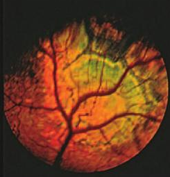 defective retinal
development. They appear as irregular or horseshoe-shaped areas. (See the eye of a CKCS with geographic retinal dysplasia, at
below left.) In the severe form of dysplasia, known as retinal detachment, the
retinal layers do not come together at all.
defective retinal
development. They appear as irregular or horseshoe-shaped areas. (See the eye of a CKCS with geographic retinal dysplasia, at
below left.) In the severe form of dysplasia, known as retinal detachment, the
retinal layers do not come together at all.
Retinal folds represent small blind spots which are probably not even noticed by the dog. However, geographic dysplasia may lead to large deficits in the visual field.
MRD rarely has any effect on the dog's vision.
The cause of most cases of retinal dysplasia in cavaliers is genetic. Of all purebred dogs, multifocal retinal dysplasia (MRD) and geographic retinal dysplasia are most commonly found in the cavalier King Charles spaniel, according to Dr. Sheila M. Crispin of the School of Veterinary Science, University of Bristol, UK.
• SARDS
Another cause of late-onset retinal dysplasia may be a condition known as sudden acquired retinal degeneration syndrome (SARDS), which may affect any breed of dog, usually middle to old aged and more females than males. The onset of visual loss is sudden, with complete vision loss in a day to four weeks. SARDS may be an autoimmune disease in which the dog's immune system attacks the body's normal cells.
In a November 2013 study of 100 dogs with SARDS, researchers found that, in addition to blindness, most of the dogs were reported to have increased thirst, urine output, and appetite along with weight gain.
RETURN TO TOP
Diagnosis
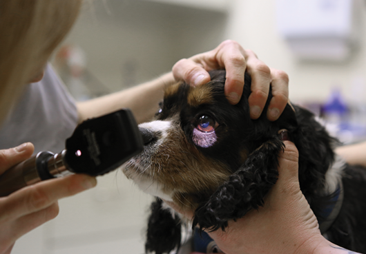
 All CKCSs should be examined by a board certified veterinary ophthalmologist
to determine whether the dogs have retinal dysplasia, and if so, the type and
degree of it. Board certified veterinary ophthalmologists are listed on the
website of the
American College of Veterinary Ophthalmologists
(ACVO).
All CKCSs should be examined by a board certified veterinary ophthalmologist
to determine whether the dogs have retinal dysplasia, and if so, the type and
degree of it. Board certified veterinary ophthalmologists are listed on the
website of the
American College of Veterinary Ophthalmologists
(ACVO).
Upon examination, the ophthalmologist can tell the degree of severity of the dysplasia. Most cases of retinal dysplasia do not progress after puppyhood, and the ophthalmologist may be able to predict the extent to which the dysplasia will interfere with the dog's field of vision. The cause of most cases of retinal dysplasia in cavaliers is genetic. Of all purebred dogs, multifocal retinal dysplasia (MRD) and geographic retinal dysplasia are most commonly found in the cavalier King Charles spaniel, according to Dr. Sheila M. Crispin of the School of Veterinary Science, University of Bristol, UK.
In cases of SARDS, no changes in the appearance of the affected eyes are apparent immediately, Degeneration of the retina may appear subsequently.
RETURN TO TOP
Treatment
There reportedly is no treatment for retinal dysplasia.
RETURN TO TOP
Breeders' Responsibilities
 The cause of most cases of retinal dysplasia in cavaliers is genetic. Of all purebred dogs,
multifocal retinal dysplasia (MRD) and geographic retinal dysplasia are most
commonly found in the cavalier King Charles spaniel.
The cause of most cases of retinal dysplasia in cavaliers is genetic. Of all purebred dogs,
multifocal retinal dysplasia (MRD) and geographic retinal dysplasia are most
commonly found in the cavalier King Charles spaniel.
The Genetics Committee of the ACVO classifies the mildest form of retinal dysplasia (retinal folds) as a "breeder option" for CKCSs. Therefore, the Canine Eye Registration Foundation (CERF) does not deny certification to cavalier King Charles spaniels which are affected with the disorder of retinal folds. However, both the Genetics Committee of the ACVO and the Canine Inherited Disorders Database recommend that CKCSs affected with geographic and retinal detachment forms of retinal dysplasia not be bred. Any littermates of breeding stock having retinal dysplasia should be taken into consideration. See The Blue Book.
The Cavalier King Charles Spaniel Club, USA recommends that, prior to breeding any cavalier, the dog have a normal rating or be within CERF "breeder options" from a screening by a board certified veterinary ophthalmologist.
The Canine Health Information Center (CHIC) is a centralized canine health database sponsored by the AKC/Canine Health Foundation (AKC/CHF) and OFA. The CHIC, working with participating parent clubs, provides a resource for breeders and owners of purebred dogs to research and maintain information on the health issues prevalent in specific breeds.
AKC's national breed clubs establish the breed specific testing protocols. Dogs complying with the breed specific testing requirements are issued CHIC numbers. The ACKCSC requires that, to qualify for CHIC certification, cavaliers must have a CERF eye examination, recommending that an initial CERF exam be performed at 8 to 12 weeks, with a follow up exam once the dog reaches 12 months, and annual exams thereafter until age 5 years, and every other year until age 9 years. However, all that is required to qualify for a CHIC certificate is that the breeding stock be examined by a veterinary ophthalmologist. It does not require that the results of the examination show no eye disorders.
Nevertheless, all cavalier breeding stock should be examined by board certified veterinary ophthalmologists at least annually and cleared by the veterinary specialists for all but the mildest form (retinal folds) of retinal dysplasia, the closer the examination to the breeding the better.
RETURN TO TOP
 What You Can Do:
Ocu-GLO Rx is a nutraceutical containing several natural antioxidants in a combination blend formulated specifically for canine eye health. Many veterinary ophthalmologists recommend this product to maintain healthy eyes. Even if your dog has not been diagnosed with a vision disorder, antioxidants contained in Ocu-GLO Rx are considered helpful in keeping dogs' eyes healthy.
RETURN TO TOP
Research News
January 2026:
MSU study finds inner retinal plaques in blood vessels of
cavalier diagnosed with retinal dysplasia.
 In an
April 2026 article, a team of specialists from Michigan State
University (Shaile Gehrke [right], Christine Harman, Amanda
Jacobson, Gail McRae, Andras Komaromy) compared two cases of dogs -- a cavalier King Charles spaniel
and a Labrador retriever -- which had been diagnosed with presumed
geographic retinal dysplasia. Using high resolution imaging --
confocal scanning laser ophthalmoscopy (cSLO), optical coherence
tomography (OCT), and OCT angiography (OCTA) -- they determined that,
instead of geographic retinal dysplasia, the cavalier had inner retinal
plaques in its retinal blood vessels. General anesthesia is required to
perform these tests. The investigators surmised the underlying cause in
this case of the CKCS was thickening of the retinal nerve fiber layer.
In an
April 2026 article, a team of specialists from Michigan State
University (Shaile Gehrke [right], Christine Harman, Amanda
Jacobson, Gail McRae, Andras Komaromy) compared two cases of dogs -- a cavalier King Charles spaniel
and a Labrador retriever -- which had been diagnosed with presumed
geographic retinal dysplasia. Using high resolution imaging --
confocal scanning laser ophthalmoscopy (cSLO), optical coherence
tomography (OCT), and OCT angiography (OCTA) -- they determined that,
instead of geographic retinal dysplasia, the cavalier had inner retinal
plaques in its retinal blood vessels. General anesthesia is required to
perform these tests. The investigators surmised the underlying cause in
this case of the CKCS was thickening of the retinal nerve fiber layer.
EDITOR'S NOTE: Inner retinal plaques are particles of cholesterol lodged in the blood vessels of the retina. The causes of these plaques can vary from genetic predisposition, autoimmune conditions, infections leading to inflammation, environmental conditions, and even dietary factors. Since the cavalier has been found to be predisposed to forms of retinal dysplasia, it appears likely that the cause of retinal plaques in the breed also is genetic. More specifically, in this MSU study, the investigators suspect that the underlying cause was thickening of the retinal nerve fiber layer. The main point of this MSU study is that if only ophthalmoscopy is used to diagnose the condition, what may appear to be geographic retinal dysplasia may in fact be determined by higher resolution imaging to be inner retinal plaques.
September 2015:
Italian researchers find retinal dysplasia and microphthalmia in a
family of cavaliers.
 In a
September 2015 publication of an
oral abstract presented before the European College of Veterinary
Ophthalmologists in May 2015, a team of Italian ophthalmologists (Luca
Mertel [right], MG Baldini, E Moretti, SP Marelli, A Picchi, M
Polli) report
on the parents and littermates of a family of cavalier King Charles
spaniels. The ruby dam "was affected by multiple
distichia and bilateral
peripheral tapetal multifocal retinal dysplasia." The black-&-tan sire
"had unilateral iris to iris persistent pupillary membranes." Pup #1
(ruby female) "had bilateral multifocal and geographical retinal
dysplasia". Pup #2 (black-&-tan male) "showed bilateral
microphthalmia".. Pup #3 (black-&-tan male) "had bilateral iris to iris
persistent pupillary membranes and unilateral right multifocal and
geographical horseshoe-shaped retinal dysplasia." They concluded:
In a
September 2015 publication of an
oral abstract presented before the European College of Veterinary
Ophthalmologists in May 2015, a team of Italian ophthalmologists (Luca
Mertel [right], MG Baldini, E Moretti, SP Marelli, A Picchi, M
Polli) report
on the parents and littermates of a family of cavalier King Charles
spaniels. The ruby dam "was affected by multiple
distichia and bilateral
peripheral tapetal multifocal retinal dysplasia." The black-&-tan sire
"had unilateral iris to iris persistent pupillary membranes." Pup #1
(ruby female) "had bilateral multifocal and geographical retinal
dysplasia". Pup #2 (black-&-tan male) "showed bilateral
microphthalmia".. Pup #3 (black-&-tan male) "had bilateral iris to iris
persistent pupillary membranes and unilateral right multifocal and
geographical horseshoe-shaped retinal dysplasia." They concluded:
"Microphthalmia with lens dysmorphogenesis and normal looking fundi seems to be a feature in the CKCS. Littermates may be affected with varies forms of retinal dysplasia, as in the Akita and the Chow Chow."
October 2013:
Dr. Peter Bedford reports multifocal retinal dysplasia is inherited in CKCSs.
In the
![]() Autumn 2013 issue of
EJCAP Online for the Federation of European Companion
Animal Veterinary Associations (FECAVA), UK ophthalmologist Dr. Peter G.C. Bedford
summarizes the research in hereditary eye disorders. He states that multifocal
retinal dysplasia (MRD) is inherited in the cavalier King Charles spaniel as a
recessive trait. He also states that in the CKCS, "the folds may be concentrated
within a single circular locus in the tapetal fundus. Unilateral involvement is
more commonly seen and here the disease is referred to as geographic retinal
dysplasia (GRD)."
Autumn 2013 issue of
EJCAP Online for the Federation of European Companion
Animal Veterinary Associations (FECAVA), UK ophthalmologist Dr. Peter G.C. Bedford
summarizes the research in hereditary eye disorders. He states that multifocal
retinal dysplasia (MRD) is inherited in the cavalier King Charles spaniel as a
recessive trait. He also states that in the CKCS, "the folds may be concentrated
within a single circular locus in the tapetal fundus. Unilateral involvement is
more commonly seen and here the disease is referred to as geographic retinal
dysplasia (GRD)."
2005: The Animal
Health Trust (AHT) is conducting research to try to establish the
pattern of
 inheritance of CKCSs with multifocal retinal dysplasia (MRD).
Dr. Keith Barnett (right), European
Specialist in Veterinary Ophthalmology, is leading a team of AHT colleagues who
are researching the DNA of such cavaliers. According to Dr. Barnett, cavaliers
could benefit from the MRD research in Golden Retrievers, where it is hoped that
the gene marker responsible for the condition will soon be identified, and that
cavaliers may possess a similar marker. Dr. Barnett believes his team has
identified the chromosome responsible for MRD, but they still are searching for
the gene.
inheritance of CKCSs with multifocal retinal dysplasia (MRD).
Dr. Keith Barnett (right), European
Specialist in Veterinary Ophthalmology, is leading a team of AHT colleagues who
are researching the DNA of such cavaliers. According to Dr. Barnett, cavaliers
could benefit from the MRD research in Golden Retrievers, where it is hoped that
the gene marker responsible for the condition will soon be identified, and that
cavaliers may possess a similar marker. Dr. Barnett believes his team has
identified the chromosome responsible for MRD, but they still are searching for
the gene.
Dr. Barnett requests that breeders who have CKCSs affected with MRD send blood samples from the affected dogs and their unaffected siblings, parents, and close relatives to identify the responsible gene. Contact Dr. Barnett at the AHT if you wish to participate in the research project. He may be reached at Animal Health Trust, Lanwades Park, Kentford, Newmarket, Suffolk CB8 7UU, United Kingdom; telephone: (+44) (0)8700 502424; email: Keith.Barnett@aht.org.uk Blood samples of 3 to 5 ml should be provided in ETDA anti-coagulant tubes. Alternatively, for very young or old donors, cheek swabs may be used. Samples should be marked for the attention of Dr. K. Barnett and sent to: Sarah Gray, The Animal Health Trust, Lanwades Park, Newmarket, Suffolk CB8 7UU UK. Please indicate clearly whether the samples are MRD pass or failures and the name of the Ophthalmologist who conducted the diagnosis.
2005: Canine Multi-focal Retinopathy (CMR) is a inherited eye disease known thus far to affect Great Pyrenees, Mastiffs and Coton de Tulear. Dr. Bruce Grahn of the University of Saskatchewan, Canada, first described CMR in the Great Pyrenees. A DNA-based test that reportedly accurately diagnoses multi-focal retinopathy occurring in these breeds has been developed and is being administered by OptiGen. The test also reportedly detects carriers of this condition and clears dogs that are genetically normal. For more information, see this page of the OptiGen website: www.optigen.com/opt9_test_cmr.html
RETURN TO TOP
Related Links
RETURN TO TOP
Veterinary Resources
Control of Canine Genetic Diseases. Padgett. G.A., Howell Book House 1998, pp. 198-199, 242.
Ocular Disorders Presumed to be Inherited in Dogs. A.C.V.O. 1999.
The geographic form of retinal dysplasia in dogs is not always a congenital abnormality. Holle, Stankovics, Sarna, Aguirre. Vet. Ophthalmology; 2:1 (61-66) Mar 1999.
What are your clinical diagnosis, lesion localization, and differential diagnoses? Cheryl L. Cullen and Bruce H. Grahn. Can Vet J; Sept. 2002;43(9):729-730.
Guide to Congenital and Heritable Disorders in Dogs. Dodds WJ, Hall S, Inks K, A.V.A.R., Jan 2004, Section II(270).
Breed Predispositions to Disease in Dogs & Cats. Alex Gough, Alison Thomas. 2004; Blackwell Publ. 44-45.
Notes on Veterinary Ophthalmology. Crispin S.M. Blackwell Publ. 2005.
Ophthalmic Disease in Veterinary Medicine. Martin C.L. Manson Publ. 2005.
Retinal dysplasia. Canine Inherited Disorders Database.
Ophthalmic Disease in Veterinary Medicine. Charles L. Martin. Manson Publ. 2009; page 475, table 15.1. Quote: "Presumed Inherited Ocular Diseases: Table 15.1: Breed predisposition to eye disease in dogs: Cavalier King Charles Spaniel: ... Retinal dysplasia: geographic / detachment, retinal folds".
Breed Predispositions to Disease in Dogs & Cats (2d Ed.). Alex Gough, Alison Thomas. 2010; Blackwell Publ. 54.
Ocular Disorders Presumed to be inherited in purebred dogs. Genetics Committee of the American College of Veterinary Ophthalmologists. Blue Book 6th Ed. 2013. pp. 241-247. Quote: "Cavalier King Charles Spaniel: Disorder: . Retinal dysplasia -- folds. Inheritance: Not defined. K. Retinal dysplasia -- geographic/detached. Inheritance: Not defined."
Hereditary Ocular Disease in the dog. Peter G C Bedford. EJCAP, Genetic/Hereditary Disease and Breeding. Oct. 2013;233(3):23-41. Quote: "Congenital inherited disease: Retinal Dysplasia: The term retinal dysplasia (RD) is commonly used to describe those inherited retinal conditions which are seen clinically as either neuroretinal folds or neuroretinal nonattachment. ... The simplest manifestation of R.D. is seen as neuroretinal folds, the affected tissue being separated from the underlying retinal pigment epithelium (RPE). Within a fold there should be abnormal proliferation of photoreceptor elements to justify the term dysplasia and differentiate this disease from the simple neuroretinal folds classically seen in the Shetland Sheepdog which disappear during early post natal development. In the puppy with RD the whole of the fundus may be involved, but in the affected adult the folds are normally restricted to the tapetal fundus. With the passage of time some folds may be rendered ophthalmoscopically inapparent, their presence being eventually replaced in later life by patches of retinal degeneration. The disappearance of folds from both the puppy and adult fundus can lead to difficulties in the explanation of a diagnosis on occasion and the development of a DNA test would be most helpful. This form of R.D. is referred to as multifocal retinal dysplasia (MRD) and is inherited in the Cavalier King Charles Spaniel (CKCS), the Hungarian Puli, the Golden Retriever (GR), the Labrador Retriever (LR) and the Rottweiler as a recessive trait. In some of the affected in the CKCS, the GR and the LR breeds the folds may be concentrated within a single circular locus in the tapetal fundus. Unilateral involvement is more commonly seen and here the disease is referred to as geographic retinal dysplasia (GRD).
Long-term outcome of sudden acquired retinal degeneration syndrome in dogs. Jane A. Stuckey, Jacqueline W. Pearce, Elizabeth A. Giuliano, Leah A. Cohn, Ellison Bentley, Amy J. Rankin, Margi A. Gilmour, Christine C. Lim, Rachel A. Allbaugh, Cecil P. Moore, Richard W. Madsen. JAVMA; Nov. 2013;243(10)1425-1431. Quote: "Objective: To investigate long-term outcomes and owner-perceived quality of life associated with sudden acquired retinal degeneration syndrome (SARDS) in dogs. Design: Survey study. Animals: 100 dogs with SARDS examined at 5 academic veterinary institutions from 2005 to 2010. Procedures: The diagnosis was based on documented acute vision loss, normal results of ophthalmic examinations, and evaluation of extinguished bright-flash electroretinograms. Primary owners of affected dogs completed a questionnaire addressing outcome measures including vision, systemic signs, and perceived quality of life for their dogs. Results: Age at diagnosis was significantly correlated with positive outcome measures; dogs in which SARDS was diagnosed at a younger age were more likely to have alleged partial vision and higher owner-perceived quality of life. Polyphagia was the only associated systemic sign found to increase in severity over time. Medical treatment was attempted in 22% of dogs; visual improvement was not detected in any. Thirty-seven percent of respondents reported an improved relationship with their dog after diagnosis, and 95% indicated they would discourage euthanasia of dogs with SARDS. Conclusions and Clinical Relevance: Blindness and concurrent systemic signs associated with SARDS appeared to persist indefinitely, but only polyphagia increased in severity over time. Most owners believed their pets had good quality of life and would discourage euthanasia of dogs with SARDS."
The genetics of eye disorders in the dog. Cathryn S. Mellersh. Canine Genetics & Epidemiology. April 2014. Quote: "Inherited forms of eye disease are arguably the best described and best characterized of all inherited diseases in the dog, at both the clinical and molecular level and at the time of writing 29 different mutations have been documented in the scientific literature that are associated with an inherited ocular disorder in the dog. The dog has already played an important role in the identification of genes that are important for ocular development and function as well as emerging therapies for inherited blindness in humans. Similarities in disease phenotype and eye structure and function between dog and man, together with the increasingly sophisticated genetic tools that are available for the dog, mean that the dog is likely to play an ever increasing role in both our understanding of the normal functioning of the eye and in our ability to treat inherited eye disorders. This review summarises the mutations that have been associated with inherited eye disorders in the dog."
Familial retinal dysplasia and microphthalmia with lens abnormalities in the cavalier King Charles spaniel (CKCS). L Mertel, MG Baldini, E Moretti, SP Marelli, A Picchi, M Polli. Vet. Opthalmology. September 2015;18(5):E5. Quote: "Purpose: To report congenital hereditary eye disorders including microphthalmia with cataract, posterior lenticonus and retinal dysplasia in a family of CKCS. The clinical findings of bilateral multifocal retinal dysplasia in a dam led to the ocular examination of the stud and the litter. Multiple congenital ocular anomalies of this family are described. Methods: All dogs were examined following the ECVO eye scheme and the horizontal corneal diameter was measured (mm) in the awake animals using a caliper. Results: The dam (34 months, ruby) was affected by multiple distichia and bilateral peripheral tapetal multifocal retinal dysplasia. The sire (27 months, black and tan) had unilateral iris to iris persistent pupillary membranes. Pup #1 (female, 2 months, ruby) had bilateral multifocal and geographical retinal dysplasia characterized by a horseshoe-shaped area in the right dorsolateral tapetal periphery and a circular dysplastic lesion in the left dorsomedial tapetal periphery. Pup #2 (male, 2 months, black and tan) showed bilateral micro-phthalmia (11 mm OU) with nuclear, cortical and posterior capsular cataract and right posterior lenticonus. Pup #3 (male, 2 months, black and tan) had bilateral iris to iris persistent pupillary membranes and unilateral right multifocal and geo-graphical horseshoe-shaped retinal dysplasia. Conclusion: Microphthalmia with lens dysmorpho-genesis and normal looking fundi seems to be a feature in the CKCS. Littermates may be affected with varies forms of retinal dysplasia, as in the Akita and the Chow Chow."
Essentials of Veterinary Ophthalmology (4th ed.). Gelatt, Kirk N.; Plummer, Caryn E. Wiley-Blackwell. 2022. Quote: Breeds of Dogs Affected with Retinal Dysplasia: ... Cavalier King Charles Spaniel - Type of RD: Geographic -- Other Abnormalities: Microphthalmia, cataracts, posterior lenticonus.
The Blue Book. Ocular Disorders Presumed to be inherited in purebred dogs. Genetics Committee of the American College of Veterinary Ophthalmologists. Blue Book 15th Ed. 2023. pp. 297-302.
Comparison of Presumed Geographic Retinal Dysplasia Between a Cavalier King Charles Spaniel and a Labrador Retriever. Shaile Gehrke, Christine Harman, Amanda Jacobson, Gail McRae, Andras Komaromy. Clinical Case Rpts. April 2026; doi: 10.1002/ccr3.72373. Quote: Geographic RD is recognized as an inherited condition in the Cavalier King Charles Spaniel (CKCS). The breeding recommendations by the Genetics Committee of the American College of Veterinary Ophthalmologists (ACVO) for RD-affected CKCS are similar to most other canine breeds: While dogs with geographic RD should not be bred, dogs with focal/multifocal RD or retinal folds can still be bred ('breeder option'). The Hereditary Eye Diseases (HED) Committee of the European College of Veterinary Ophthalmologists (ECVO) states that breeding is optional for dogs affected by focal/multifocal or geographic RD unless the breed club issues different advice. According to the Blue Book on Ocular Disorders Presumed to Be Inherited in Purebred Dogs, published by the ACVO Genetics Committee, of the 17,092 CKCS examined between 2019 and 2023, 1.6% had geographic RD, while 3.2% had retinal folds. Even though these prevalences are higher than in the Labrador Retriever with 0.4% geographic RD and 0.9% retinal folds, no detailed morphological and clinical reports on geographic RD in the CKCS have been published to the best of our knowledge. It is unknown if these lesions represent true RD or are similar to the inner retinal plaques that develop later than 10 weeks of age in Labrador Retrievers, Golden Retrievers, and German Shepherds. ... This report represents the first detailed description of presumed geographic retinal dysplasia in a CKCS and provides additional documentation of inner retinal plaques in a Labrador Retriever using cSLO, OCT, and OCTA. The inner retinal plaques described in these two dogs, which cannot be differentiated from geographic RD without high-resolution imaging, closely resemble those previously reported in Labrador Retrievers, Golden Retrievers, and German Shepherds. Both the CKCS and Labrador Retriever in this study exhibited significant retinal nerve fiber layer thickening, forming a plaque-like horseshoe-shaped retinal lesion along the dorsal venule, and an additional smaller, similar lesion along a ventral venule in the CKCS. All of the retinal plaques contained multifocal suspected rosettes. In addition, the CKCS also had suspected rosettes in the outer retina with the lesions. Some of these rosettes had hyporeective lumina, while others had hyperreective lumina. ... To fill this knowledge gap, our retrospective study aims to describe high-resolution imaging of geographic RD in a CKCS using confocal scanning laser ophthalmoscopy (cSLO), optical coherence tomography (OCT), and OCT angiography (OCTA). We compared our findings with the images of a Labrador Retriever with unilateral inner retinal plaques, since this condition has been previously characterized. Case 1: Cavalier King Charles Spaniel (CKCS): A 15-month-old intact female CKCS (body weight = 7.1 kg) was referred for advanced imaging following the identification of a geographic RD-like lesion during a Companion Animal Eye Registry (CAER) examination. The dog exhibited no visual deficits and was otherwise healthy, with no evidence of systemic disease or medications. ... Neither dog received a detailed ophthalmic examination before four months of age. ... Vascular anomalies were not readily visible via funduscopy, but were prominent on OCTA in both our cases. ... Imaging OD of the CKCS revealed marked thickening of the retinal nerve fiber layer, forming a plaque-like lesion with multifocal either hypo- or hyperreflective lumina in the inner and outer retina, suspected to be rosettes. Similar findings were observed in the non-tapetal circular lesion OS, with thickening and rosettes. ... Suspected aneurysms and disruptions in the vascular plexus were observed, with the CKCS showing a higher density of vascular anomalies at the periphery of the lesions. ... A major limitation of any case report is the small number of cases, which limits the ability to draw conclusions for the larger canine population. For example, variations in plaque features among dogs likely reflect differences in lesion severity or individual variability rather than breed-specificity. Nevertheless, our case report suggests that the presumed geographic RD observed in some CKCS may contain inner retinal plaques resulting from thickening of the retinal nerve fiber layer. ... In summary, we provide further evidence that in at least some dogs, retinal lesions appearing like geographic RD on ophthalmoscopy may contain inner retinal plaques; an observation that can only be made with high-resolution imaging.
CONNECT WITH US