
Microphthalmia in Cavalier King Charles Spaniels: Genetically Caused, Abnormally Small Eyeball
-
 What
It Is
What
It Is - Symptoms
- Related Conditions
- Diagnosis
- Treatment
- Breeders' Responsibilities
- What You Can Do
- Research News
- Related Links
- Veterinary Resources
Microphthalmia (microphthalmos) is a congenital and often inherited defect which is particularly common in the cavalier King Charles spaniel, according to the American College of Veterinary Ophthalmologists (ACVO).*
* See also, Ophthalmic Disease in Veterinary Medicine.
What It Is
Microphthalmia describes a condition in which one or both of the dog's eyes is abnormally small, resulting in restricted vision and possible blindness. It is attributed to the underdevelopment of the eyes during the fetal stage, presumably due to a genetic abnormality. It is suspected of being an inherited condition in the cavalier breed.
Microphthalmia may be associated with other developmental problems such as hydrocephalus and white matter abnormalities.
RETURN TO TOP
 Symptoms
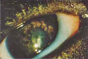
Microphthalmia varies in size of the eye and in degree of severity. The affected eye otherwise may function normally, or may have other defects, particluarly a cataract and including retinal folds and cherry eye.
The photo at right is of the eye of a CKCS puppy with microphthalmia and the additional defects of iris hypoplasia and an anterior cataract.
RETURN TO TOP
Related Conditions
Microphthalmia may be combined with a coloboma. A coloboma is a fetal development defect in which normal tissue in or surrounding the eye is missing at birth. A tissue gap or hole exists due to incomplete closure of the embryonic fissure during fetal development. Normally there is a small gap at the bottom of a developing eye that fills in and closes at a particular stage of development prior to whelphing.
 This
condition may lead to the formation of an cyst within the eye orbit. The
cyst can expand and extend into surrounding tissues, including causing
bone deformation. In this
September 2024 article, a cavalier King Charles spaniel named
Freddie (right) was diagnosed by MRI with both microphthalmia
and, in his left eye, a retinal-choroidal coloboma. The MRI imaging
showed that the cyst extended into the retrobulbar space and
nasopharynx, also causing some deformation of the presphenoid bone.
Freddie's left eye had no lens or iris. In
this case, the eye was removed, and examination of it confirmed the
absence of ocular structures which caused the congenital gap.
This
condition may lead to the formation of an cyst within the eye orbit. The
cyst can expand and extend into surrounding tissues, including causing
bone deformation. In this
September 2024 article, a cavalier King Charles spaniel named
Freddie (right) was diagnosed by MRI with both microphthalmia
and, in his left eye, a retinal-choroidal coloboma. The MRI imaging
showed that the cyst extended into the retrobulbar space and
nasopharynx, also causing some deformation of the presphenoid bone.
Freddie's left eye had no lens or iris. In
this case, the eye was removed, and examination of it confirmed the
absence of ocular structures which caused the congenital gap.
RETURN TO TOP
Diagnosis
The ophthalmologist may perform an ultrasound scan of the eye to map it, including the space behind the eyeball as well as the retina and lens, looking for other abnormalities. The dog's vision also will be tested.
In cases of suspected related conditions, magnetic resonance imaging (MRI) may be necessary to assess the extent of the associated abnormalities.
RETURN TO TOP
Treatment
There is no treatment for microphthalmia. Removal of the eyeball may be necessary if it causes permanent irritation.
All CKCSs should be examined at least annually by a board certified veterinary ophthalmologist. They are listed on the American College of Veterinary Ophthalmologists website.
RETURN TO TOP
Breeders' Responsibilities
 The Genetics Committee of the ACVO recommends that CKCSs affected with
microphthalmia not be bred. See
The Blue Book. The Canine Inherited Disorders Database also
recommends that cavaliers suffering from microphthalmia not be bred, nor should
the dog's parents and any of its siblings.
The Genetics Committee of the ACVO recommends that CKCSs affected with
microphthalmia not be bred. See
The Blue Book. The Canine Inherited Disorders Database also
recommends that cavaliers suffering from microphthalmia not be bred, nor should
the dog's parents and any of its siblings.
The Cavalier King Charles Spaniel Club, USA recommends that, prior to breeding any cavalier, the dog have a normal rating from a screening by a board certified veterinary ophthalmologist.
All CKCS breeding stock should be examined by board certified veterinary ophthalmologists and cleared by the veterinarians for microphthalmia.
RETURN TO TOP
 What You Can Do:
Ocu-GLO Rx is a nutraceutical containing several natural antioxidants in a combination blend formulated specifically for canine eye health. Many veterinary ophthalmologists recommend this product to maintain healthy eyes. Even if your dog has not been diagnosed with a vision disorder, antioxidants contained in Ocu-GLO Rx are considered helpful in keeping dogs' eyes healthy.
RETURN TO TOP
Research News
September 2024:
Cavalier is diagnosed by MRI with microphthalmia and coloboma
cyst in his eye.
In
this
September 2024 article, clinicians Becky Mosley and Clare
Rusbridge report the case study of a cavalier King Charles spaniel named
Freddie (right) that was diagnosed by MRI with both
microphthalmia and, in his left eye, a retinal-choroidal coloboma. A
coloboma is a fetal development defect in which normal tissue in or
surrounding the eye is missing at birth. A tissue gap or hole exists due
to incomplete closure of the embryonic fissure during fetal development.
Normally there is a small gap at the bottom of a developing eye that
fills in and closes at a particular stage of development prior to
whelphing. The MRI imaging showed that the cyst extended into the
retrobulbar space and nasopharynx, also causing some deformation of the
surrounding bone. In this case, the eye was removed, and examination of
it confirmed the absence of ocular structures which caused the
congenital gap.
November 2016:
Cavalier eye sizes are large for small dogs but fit in the
medium dog group.
 In a
November 2016 article, UK ophthalmologists (Carolin L. H. Chiwitt
[right],
Stephen J. Baines, Paul Mahoney, Andrew Tanner, Christine L. Heinrich,
Michael Rhodes, Heidi J. Featherstone) found that the size of the eyes
of the cavalier King Charles spaniel, a small dog, was larger than other
small dogs and fit within the eye size of medium sized dogs. The
researchers measured the eye sizes of 100 dogs in ten breeds, incuding
10 cavalier King Charles spaniels.
In a
November 2016 article, UK ophthalmologists (Carolin L. H. Chiwitt
[right],
Stephen J. Baines, Paul Mahoney, Andrew Tanner, Christine L. Heinrich,
Michael Rhodes, Heidi J. Featherstone) found that the size of the eyes
of the cavalier King Charles spaniel, a small dog, was larger than other
small dogs and fit within the eye size of medium sized dogs. The
researchers measured the eye sizes of 100 dogs in ten breeds, incuding
10 cavalier King Charles spaniels.
The researchers used both B-mode ocular ultrasound and computed tomography (CT) to do so in a procedure called "ocular biometry", which is the measurement of the dimensions of the eye, its components, and their interrelationships. They divided the 100 dogs into three size groups -- small, medium, and large based upon the UK Kennel Club assignments -- and placed the CKCS in the small dog group. They measured eye length, width, and height using the both of the devices -- ultrasound and CT.
They found that the CKCS had a relatively large eye for the small breed group, and there was no significant difference between the CKCS eye and those in the medium breed group. They also found that their measurements using both B-mode ultrasound and CT can be applied for the diagnosis of microphthalmos and buphthalmos.
September 2015:
Italian researchers find retinal dysplasia and microphthalmia in a
family of cavaliers.
 In a
September 2015 publication of an
oral abstract presented before the European College of Veterinary
Ophthalmologists in May 2015, a team of Italian ophthalmologists (Luca
Mertel [right], MG Baldini, E Moretti, SP Marelli, A Picchi, M
Polli) report
on the parents and littermates of a family of cavalier King Charles
spaniels. The ruby dam "was affected by multiple
distichia and bilateral
peripheral tapetal multifocal retinal dysplasia." The black-&-tan sire
"had unilateral iris to iris persistent pupillary membranes." Pup #1
(ruby female) "had bilateral multifocal and geographical retinal
dysplasia". Pup #2 (black-&-tan male) "showed bilateral
microphthalmia".. Pup #3 (black-&-tan male) "had bilateral iris to iris
persistent pupillary membranes and unilateral right multifocal and
geographical horseshoe-shaped retinal dysplasia." They concluded:
In a
September 2015 publication of an
oral abstract presented before the European College of Veterinary
Ophthalmologists in May 2015, a team of Italian ophthalmologists (Luca
Mertel [right], MG Baldini, E Moretti, SP Marelli, A Picchi, M
Polli) report
on the parents and littermates of a family of cavalier King Charles
spaniels. The ruby dam "was affected by multiple
distichia and bilateral
peripheral tapetal multifocal retinal dysplasia." The black-&-tan sire
"had unilateral iris to iris persistent pupillary membranes." Pup #1
(ruby female) "had bilateral multifocal and geographical retinal
dysplasia". Pup #2 (black-&-tan male) "showed bilateral
microphthalmia".. Pup #3 (black-&-tan male) "had bilateral iris to iris
persistent pupillary membranes and unilateral right multifocal and
geographical horseshoe-shaped retinal dysplasia." They concluded:
"Microphthalmia with lens dysmorphogenesis and normal looking fundi seems to be a feature in the CKCS. Littermates may be affected with varies forms of retinal dysplasia, as in the Akita and the Chow Chow."
October 2013:
![]() Dr. Peter Bedford reports microphthalmos and congenital cataracts are linked in CKCSs. In the Autumn
2013 issue of EJCAP Online for the Federation of
European Companion Animal Veterinary Associations (FECAVA), UK ophthalmologist Dr. Peter G.C. Bedford
summarizes the research in hereditary eye disorders. He states that congenital
nuclear cataract may accompany microphthalmos,
persistence of remnant pupillary membrane (PPM) and retinal dysplasia in the
Cavalier King Charles Spaniel.
Dr. Peter Bedford reports microphthalmos and congenital cataracts are linked in CKCSs. In the Autumn
2013 issue of EJCAP Online for the Federation of
European Companion Animal Veterinary Associations (FECAVA), UK ophthalmologist Dr. Peter G.C. Bedford
summarizes the research in hereditary eye disorders. He states that congenital
nuclear cataract may accompany microphthalmos,
persistence of remnant pupillary membrane (PPM) and retinal dysplasia in the
Cavalier King Charles Spaniel.
RETURN TO TOP
Related Links
RETURN TO TOP
Veterinary Resources
Imperforate and micro-lachrymal puncta in the dog. K. C. Barnett. J. Sm. Anim. Pract. August 1979;20(8):481-490. Quote: Lachrymation, the excessive secretion of tears, and epiphora, the inadequate drainage of tears, are common presenting clinical signs. It has long been supposed that the majority of cases of epiphora occur because part, or parts, of the naso-lachrymal duct system is congenitally absent or blocked and, if the former, that treatment is not possible. Imperforate and micro-lachrymal puncta are reported in detail in the dog. The main breeds affected are the Golden Retriever and Cocker Spaniel. Diagnosis and surgical treatment are discussed. The condition is considered to be more common than is realized. ... Cases in this series occured in the yellow Labrador (three), Samoyed (two) and single cases in the American Cocker Spaniel, English Springer Spaniel, Cavalier King Charles Spaniel, ... Case 26: Cavalier King Charles -- tri --- M -- 6 mos -- microphthalmos of upper and lower right lids.
Posterior lenticonus, cataracts and microphthalmia: congenital ocular defects in the Cavalier King Charles Spaniel. Narfstrom K, Dubielzig R. J. Sm. Anim. Pract. 1984; 25: 669-677. Quote: Eleven cases of congenital ocular defects were found in the screening of 144 Cavalier King Charles Spaniels in Sweden. Mainly posterior lenticonus, cataracts and microphthalmia were observed in the affected dogs, most of which were interrelated. Pathology was obtained from one of the cases demonstrating bilateral posterior lens capsule rupture with an unusual cellular reaction of the exposed lens material. ... The lenticular abnormalities found in the Cavalier King Charles Spaniel breed of dog mainly affect the nucleus, posterior cortex and the posterior capsule. These defects, together with an intact posterior lens capsule, indicate an early developmental insult in the formation of the foetal lens. Numerous theories have been proposed to explain posterior lenticonus formation. Most of the histologic evidence supports a congenital defect involving an abnormal embryonic remnant in the epithelial cell proliferation before the lens capsule forms or a hyperplasia of the lens accompanied by a thinning of the capsule over the defect. In the Cavalier dog the latter explanation seems to be the most likely. Persistence of hyaloid vessels or remnants of these is further evidence of an early developmental aberration. Microphthalmia is a common finding in conjunction with other severe ocular malformations which were also found in the present study.
Hereditary cataract in the Miniature Schnauzer. K. C. Barnett. J.Small Animal Prac.; Nov. 1985;26(11):635-644. Quote: "Posterior lenticonus, cataract and microphthalmos, as reported here and in the USA (Gelatt et al., 1983b) has also been reported in the Cavalier King Charles Spaniel in Sweden (Narfstrom & Dubielzig, 1984)."
Control of Canine Genetic Diseases. Padgett, G.A., Howell Book House 1998, pp. 198-199, 241.
Ocular Disorders Presumed to be Inherited in Purebred Dogs. Genetics Committee, A.C.V.O. 1999.
Guide to Congenital and Heritable Disorders in Dogs. Dodds WJ, Hall S, Inks K, A.V.A.R., Jan 2004, Section II(199).
Breed Predispositions to Disease in Dogs & Cats. Alex Gough, Alison Thomas. 2004; Blackwell Publ. 44-45.
Ophthalmic Disease in Veterinary Medicine. Martin C.L. Manson Publ. 2005.
Canine Inherited Disorders Database: http://ic.upei.ca/cidd/disorder/microphthalmia-ocular-dysgenesis
Ophthalmic Disease in Veterinary Medicine. Charles L. Martin. Manson Publ. 2009; page 475, table 15.1. Quote: "Presumed Inherited Ocular Diseases: Table 15.1: Breed predisposition to eye disease in dogs: Cavalier King Charles Spaniel: ... Microphthalmia with multiple ocular anomalies".
Ocular conditions affecting the brachycephalic breeds. Peter G.C. Bedford. 2010. RVC. Quote: "There are two types of disease which affect the eye of the brachycephalic breeds and both are directly or indirectly related to genetic predisposition. First and by far the commonest are those conditions which are due to be conformation of skull and are related to the exophthalmos which is the common feature of these breeds. Second there are those conditions which have been unwittingly bred into some brachycephalic breeds in the pursuit of desired breed characteristics. In this lecture I will present an overview of all the diseases that the small animal practitioner is likely to encounter in the brachycephalic breeds of pedigree dog. The fourteen breeds I have included for discussion are the Affenpinscher, Boston Terrier, Boxer, Bulldog, Cavalier King Charles and the King Charles Spaniels, (mesaticephalic) French Bulldog, Griffon Bruxellois, Japanese Chin, Lhasa Apso, Pekingese, Pug, Shih Tzu and Tibetan Spaniel. ... Corneal Lipid Dystrophy: The term applies to the characteristic cholesterol and triglyceride deposits in the superficial corneal stroma seen most commonly in the Cavalier King Charles Spaniel. It is clinically benign and seldom affects vision to any noticeable degree. ... Hereditary Cataract: Hereditary cataract is seen in the Boston Terrier and the Cavalier King Charles Spaniel. ... Microphthalmos (MoD): Again the American literature suggests that microphthalmos (MoD) may be inherited in the Cavalier King Charles Spaniel."
Genetic Connection: A Guide to Health Problems in Purebred Dogs, Second Edition. Lowell Ackerman. July 2011; AAHA Press; pg 169. Quote: "In the ... Cavalier King Charles spaniel ... microphthalmia is associated with multiple ocular anomalies."
Ocular Disorders Presumed to be inherited in purebred dogs. Genetics Committee of the American College of Veterinary Ophthalmologists. Blue Book 6th Ed. 2013. pp. 241-247. Quote: "Cavalier King Charles Spaniel: Disorder: A. Microphthalmia with multiple ocular defects. Inheritance: Not defined."
Hereditary Ocular Disease in the dog. Peter G C Bedford. EJCAP Online, Genetic/Hereditary Disease and Breeding. Autumn 2013;233(3):23-41. Quote: "Congenital inherited disease: ... Congenital Cataract: Cataract is defined as any opacity of the lens and/or its capsule. Congenital inherited cataract always involves the central embryonic and foetal nuclear portion of the lens, whilst those lens fibres which make up the cortex usually remain transparent. Thus congenital nuclear cataract is often described as stationary, with the effect on the dog's sight being dictated by the extent of the opacity. Such patients may be managed long term by using long acting mydriatic drugs, but when cortical involvement occurs lens removal may prove necessary. The condition occurs as a recessive trait in the Miniature Schnauzer and it may become established as an inherited entity in the Old English Sheepdog, the Golden Retriever and the West Highland White Terrier unless breeders respond adequately to early findings in these breeds. Congenital nuclear cataract may also accompany microphthalmos, PPM and retinal dysplasia in breeds like the Bloodhound, the Cavalier King Charles Spaniel, the Cocker Spaniel, the Dobermann, the Golden Retriever, the Old English Sheepdog, the Rottweiler, the Rough Collie, the Standard Poodle and the West Highland White Terrier as part of a multi-ocular defect (MOD).
Familial retinal dysplasia and microphthalmia with lens abnormalities in the cavalier King Charles spaniel (CKCS). L Mertel, MG Baldini, E Moretti, SP Marelli, A Picchi, M Polli. Vet. Opthalmology. September 2015;18(5):E5. Quote: "Purpose: To report congenital hereditary eye disorders including microphthalmia with cataract, posterior lenticonus and retinal dysplasia in a family of CKCS. The clinical findings of bilateral multifocal retinal dysplasia in a dam led to the ocular examination of the stud and the litter. Multiple congenital ocular anomalies of this family are described. Methods: All dogs were examined following the ECVO eye scheme and the horizontal corneal diameter was measured (mm) in the awake animals using a caliper. Results: The dam (34 months, ruby) was affected by multiple distichia and bilateral peripheral tapetal multifocal retinal dysplasia. The sire (27 months, black and tan) had unilateral iris to iris persistent pupillary membranes. Pup #1 (female, 2 months, ruby) had bilateral multifocal and geographical retinal dysplasia characterized by a horseshoe-shaped area in the right dorsolateral tapetal periphery and a circular dysplastic lesion in the left dorsomedial tapetal periphery. Pup #2 (male, 2 months, black and tan) showed bilateral micro-phthalmia (11 mm OU) with nuclear, cortical and posterior capsular cataract and right posterior lenticonus. Pup #3 (male, 2 months, black and tan) had bilateral iris to iris persistent pupillary membranes and unilateral right multifocal and geo-graphical horseshoe-shaped retinal dysplasia. Conclusion: Microphthalmia with lens dysmorpho-genesis and normal looking fundi seems to be a feature in the CKCS. Littermates may be affected with varies forms of retinal dysplasia, as in the Akita and the Chow Chow."
Ocular biometry by computed tomography in different dog breeds. Carolin L. H. Chiwitt, Stephen J. Baines, Paul Mahoney, Andrew Tanner, Christine L. Heinrich, Michael Rhodes, Heidi J. Featherstone. Vet. Ophth. November 2016. Quote: Objective: To (i) correlate B-mode ocular ultrasound (US) and computed tomography (CT) (prospective pilot study), (ii) establish a reliable method to measure the normal canine eye using CT, (iii) establish a reference guide for some dog breeds, (iv) compare eye size between different breeds and breed groups, and (v) investigate the correlation between eye dimensions and body weight, gender, and skull type (retrospective study). Procedure: B-mode US and CT were performed on ten sheep cadaveric eyes. CT biometry involved 100 adult pure-bred dogs with nonocular and nonorbital disease, representing eleven breeds [including ten cavalier King Charles spaniels]. Eye length, width, and height were each measured in two of three planes (horizontal, sagittal, and equatorial). Results: B-mode US and CT measurements of sheep cadaveric eyes correlated well (0.70-0.71). The shape of the canine eye was found to be akin to an oblate spheroid (a flattened sphere). A reference guide was established for eleven breeds. Eyes of large breed dogs (GSD, Labrador Retriever, Boxer) were significantly larger than those of medium ((Border Collie, SBT, Cocker Spaniel, ESS) and small breed dogs (CKCS, Border Terrier, JRT, WHWT) (P < 0.01), and eyes of medium breed dogs were significantly larger than those of small breed dogs (P < 0.01). ... There was no significant difference between the CKCS eye (a small breed) and the medium breed group. ... Eye size correlated with body weight (0.74-0.82) but not gender or skull type. Conclusions: Computed tomography is a suitable method for biometry of the canine eye, and a reference guide was established for eleven breeds. Eye size correlated with breed size and body weight. ... The CKCS had a relatively large eye for the small breed group. ... Because correlation between B-mode US and CT was shown, the obtained values can be applied in the clinical setting, for example, for the diagnosis of microphthalmos and buphthalmos.
The Blue Book. Ocular Disorders Presumed to be inherited in purebred dogs. Genetics Committee of the American College of Veterinary Ophthalmologists. Blue Book 15th Ed. 2023. pp. 297-302.
Microphthalmia with Retinal-Choroidal Colobomas (Orbital Cysts) in the
Cavalier King Charles Spaniel. Becky Mosley,
Clare Rusbridge. Unpublished case study. September 2024. Quote: What is
it? Microphthalmia with retinal-choroidal colobomas is a congenital eye
condition characterized by an underdeveloped eye (microphthalmia) and
defects (hole) in the retina and choroid. The "hole" occurs due to
incomplete closure of the embryonic fissure during fetal development.
This
condition often leads to the formation of orbital cysts, which can
extend into surrounding structures like the retrobulbar space or even
into the nasopharynx. In the case described, the cysts were observed to
slowly expand and cause bone deformation, but they did not show
expansion into the cranium How is it Diagnosed? Diagnosis typically
involves clinical examination and advanced imaging, such as MRI, to
assess the eye structure and any associated abnormalities. In the case
of Freddie (right), an MRI revealed a large retinal-choroidal
coloboma associated with the microphthalmic eye. The imaging showed
extension of the cyst into the retrobulbar space and nasopharynx, with
some bone deformation. Microphthalmia may be associated with other
developmental problems such as hydrocephalus and corpus callosal (white
matter) abnormalities. Histopathology of the removed eye confirmed the
absence of typical ocular structures. Is it Inherited? Microphthalmia is
recognized in the Cavalier King Charles Spaniel breed
and is suspected to be an inherited condition. While specific genetic
patterns are not detailed in the case report, the reference to human
cases links this condition to genetic abnormalities on chromosome 15
(15q12-q15). How is it Treated? Management of this condition depends on
its impact on the dog. Since many dogs with microphthalmia and colobomas
may not exhibit significant problems beyond blindness, monitoring is
often recommended. If there is extensive globe expansion by the cyst
then the eye may need to be removed and It is advised to keep watch for
any new signs, such as pain or respiratory changes. What is the Impact
on the Dog? The impact on the dog can vary. Many affected dogs are
primarily blind but otherwise have a good quality of life. The cysts
themselves may not significantly impact the dog's daily activities
unless they lead to pain or complications.
CONNECT WITH US