
Primary Secretory Otitis Media (PSOM)
in the Cavalier King Charles Spaniel
-
 IN
SHORT
IN
SHORT - IN DEPTH
- What It Is
- Relation to Other Disorders
- Symptoms
- Diagnosis
- Treatment
- What You Can Do
- Research News
- Related Links
- Veterinary Resources
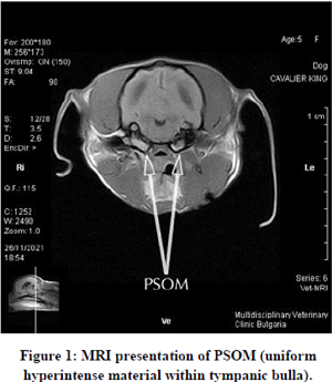
Primary secretory otitis media (PSOM) -- also known as "glue ear" or "middle ear effusion" (MEE) or "otitis media with effusion" (OME) -- is frequently diagnosed in cavalier King Charles spaniels. It is an inflammation of the dog's middle ear which consists of a highly viscous mucus plug which fills the middle ear cavity and may cause the tympanic membrane to bulge. The mucus has also been referred to as "hyperintense material".
PSOM has been reported almost exclusively in cavaliers. It has been reported to first occur in CKCSs as young as 11 months and as old as 12 years. Because the pain and other sensations in the head and neck areas, resulting from PSOM, are similar to some symptoms caused by syringomyelia (SM), some examining veterinarians may have mis-diagnosed SM in cavaliers which actually have PSOM and not SM. Other breeds in which PSOM has been diagnosed are boxers, dachshund, and shih tzu.
RETURN TO TOP
 IN
SHORT:
IN
SHORT:
The cause of PSOM is unknown. It is suspected to be due to a dysfunction of the middle ear or the Eustachian (auditory) tube: either (a) the increased production of mucus in the middle ear, or (b) decreased drainage of the middle ear through the auditory tube, or (c) both.
Not all PSOM-affected dogs display clinical symptoms, but of those which do, the principal ones are moderate to severe pain in the head or neck, holding the neck in a guarded position, and tilting the head. Other signs may include scratching at the ears, itchy ears, head tilt, excessive yawning, crying out in pain, ataxia, drooping ear or lip, inability to blink an eye, rapid eyeball movement, facial paralysis or nerve palsy, Vestibular disease, some loss of hearing, seizures, and fatigue. These symptoms, in many cases, are very similar to those of syringomyelia and, to some extent, to those of progressive hereditary deafness. Therefore, the examining veterinarian should take care to consider these other possible causes of the dog's symptomatic behaviors.
PSOM may be detected by veterinary neurology or dermatology specialists from either magnetic resonance imaging (MRI) or a computed tomography (CT) scan. Both require that the dog be under general anesthesia. It also may be observed using an operating microscope with good lighting and at a suitable magnification.
 Treatment traditionally has consisted of performing a myringotomy,
making a small cut in the eardrum (tympanic membrane), followed by
flushing the middle ear to force out the mucus plug. (See photo at
right.) Topical and/or
systemic corticosteroids and antibiotics then are administered. The
procedure may have to be repeated, in some cases several times,
depending upon how the dog responds. An alternative procedure is a
ventral bulla osteotomy,which involves making an incision on the under
side of the neck behind the jaw bone. The auditory bulla, a hollow bony
sheath that encloses parts of the middle ear, then is exposed and is
opened.
Treatment traditionally has consisted of performing a myringotomy,
making a small cut in the eardrum (tympanic membrane), followed by
flushing the middle ear to force out the mucus plug. (See photo at
right.) Topical and/or
systemic corticosteroids and antibiotics then are administered. The
procedure may have to be repeated, in some cases several times,
depending upon how the dog responds. An alternative procedure is a
ventral bulla osteotomy,which involves making an incision on the under
side of the neck behind the jaw bone. The auditory bulla, a hollow bony
sheath that encloses parts of the middle ear, then is exposed and is
opened.
Some specialist veterinarians have been prescribing N-Acetyl-L-Cysteine (NAC), a mucolytic -- mucus thinning agent or expectorant -- for cavaliers with PSOM, following surgeries.
RETURN TO TOP
IN DEPTH:
What it is
The cause of PSOM is unknown. Dr. Lynette Cole reports that it is speculated to be due to a dysfunction of the middle ear or the Eustachian (auditory) tube: either (a) the increased production of mucus in the middle ear, or (b) decreased drainage of the middle ear through the auditory tube, or (c) both.
In a 2010 study (and a 2013 report of the same study), UK veterinary researchers examined MRI scans of the skulls of 34 cavaliers, each of which had been scanned twice over periods from one month to 46 months. They concluded in their report that PSOM is a progressive condition in the CKCS and can progress from none to unilateral or bilateral; or from unilateral to bilateral on sequential scans, and that PSOM is an acquired condition in the CKCS and will not resolve spontaneously once it has developed. However, a few breeders report that second MRI scans of their PSOM-affected cavaliers show that the PSOM has disappeared without treatment.
• middle ear
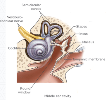 There
are two middle ears, the right one and the left one, each of which
consists of the ear drum, an air-filled
tympanic cavity, and three small bones (ossicles)
labelled the malleus, incus, and stapes, along with
their muscles and ligaments, and the tympanic membrane (eardrum).
(See the diagram at right.)
There
are two middle ears, the right one and the left one, each of which
consists of the ear drum, an air-filled
tympanic cavity, and three small bones (ossicles)
labelled the malleus, incus, and stapes, along with
their muscles and ligaments, and the tympanic membrane (eardrum).
(See the diagram at right.)
In an April 2024 article, UK researchers examined the structure and scaling of the middle ears of 17 dog breeds, including a cavalier King Charles spaniel. They found that the cavalier "stood out the most" with larger volumes of the ossicular bones (and also larger labyrinth volume) than any other breed. They suggested that these differences may be related to PSOM in the breed.
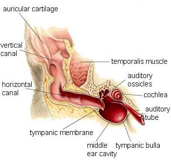
• auditory tube
The
auditory tube (Eustachian tube) connects the middle ear to the back of the nose. The tube
serves to ventilate and maintain equal air
 pressure both inside and outside of the
middle ear (tympanic cavity), to allow the eardrums (tympanic membranes) to vibrate
properly. The tube also allows fluid from mucus membranes in the middle
ear to drain through the nose. If the Eustachian tube is not working
properly, the air in the middle ear is absorbed, but it cannot be
replaced, causing air pressure inside the middle ear to be lower than
the air pressure outside, in the ear canal, creating a partial vacuum.
This difference in air pressure causes the mucus fluid to collect
inside the middle ear. The fluid then begins to become thicker and build
up, becoming an ever-enlarging mucus plug.
pressure both inside and outside of the
middle ear (tympanic cavity), to allow the eardrums (tympanic membranes) to vibrate
properly. The tube also allows fluid from mucus membranes in the middle
ear to drain through the nose. If the Eustachian tube is not working
properly, the air in the middle ear is absorbed, but it cannot be
replaced, causing air pressure inside the middle ear to be lower than
the air pressure outside, in the ear canal, creating a partial vacuum.
This difference in air pressure causes the mucus fluid to collect
inside the middle ear. The fluid then begins to become thicker and build
up, becoming an ever-enlarging mucus plug.
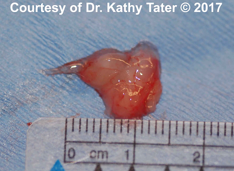
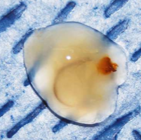
In a ten year study conducted in Sweden and reported in 2003, 61 cases of primary secretory otitis media were diagnosed in 43 cavaliers. In that study, conducted by Wiwian Stern-Bertholtz, Lennart Sjöström, and Nils Wallin-Håkanson, they explain the condition technically as follows:
"The Eustachian tube is kept closed by the surface tension caused by contact between air and mucus. A particular agent, identified as a combination of different phospholipids, decreases the surface tension inthe Eustachian tube of dogs, thus reducing the pressure needed to open the tube. When the tube is closed, the pressure in the middle ear is reported to become negative in relation to the pressure in the tube, which is equivalent to atmospheric pressure. This negative pressure, caused by lack of aeration, draws out the sterile transudate from the glandular tissues in the middle ear to the surface of the mucous membrane. The negative pressure remains and the process of accumulation of mucus carries on as long as the tympanic membrane is intact and the Eustachian tube is closed. Failure to open the Eustachian tube and thereby release the secretory products is believed to be the cause of secretory otitis media. An obstruction of the osseous part of the Eustachian tube is reported to be the most common cause. In PSOM, the overfilling of the middle ear with mucus and the subsequent bulging of the tympanic membrane, and the pain and neurological signs that are common, indicate that the pressure within the middle ear is high rather than low, at least in the final part of the disease process." Photo above shows mucus plugs removed from a cavalier. Courtesy, Downs Veterinary Practice, Bristol, UK.
In a September 2021 presentation, German and UK veterinary researchers sought to determine the role of the Eustachian tube (ET) in PSOM in cavaliers and two other brachycephalic breeds, French bulldogs and pugs. They used computed tomography (CT) images of the ears of 72 dogs, including 47 PSOM-affected ears of the brachycephalic breeds and 97 unaffected ears of the control breeds. They report that the prime function of the ET is ventilation and pressure equalization of the tympanic bullae, and that, when that fails, PSOM arises. They found that a shorter, and significant flatter ET with a significant different angulation in the brachycephalic dogs. They also found a marked reduction of tympanic bulla volume, and thicker muscles in the nasopharynx, and cartilage weakness in brachycephalic dogs suggests that the ventilation and pressure equalization system of the ET in brachycephalic dogs is a very dysfunctional, weak system. They concluded that their study demonstrates that ET width and angulation might contribute to development of PSOM.
RETURN TO TOP
• tympanic cavity
The tympanic cavity (also called the middle ear cavity) is a rounded, hollow space behind the eardrum, (tympanic membrane) which is encased in the tympanic bulla, a thin, bubble-like bony vessel. It is within this cavity that the PSOM mucus is located. (See the "middle ear cavity" in the diagram above.) In cavaliers, this cavity has been found to be much smaller and flatter than the large, rounded cavity in other breeds.
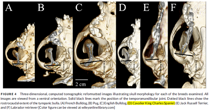
In a March 2006 article, the radiologists authors noted that, while the canine "tympanic cavity is usually quite large and rounded", in the cavalier, "it is smaller and flatter." Similarly, in a July 2017 article, a team of UK researchers used computed tomography (CT) to study the tympanic bullae of four brachycephalic breeds (pug, French bulldog, English bulldog, and cavalier) and compared them to two control breeds (Labrador retriever and Jack Russell terrier). They found that the CKCS had significantly flatter tympanic bullae than the other brachy breeds. (See Figure 4, below, of the July 2017 article's comparisons of the tympanic cavities of the cavalier and the five other breeds.) They also found middle ear effusion material in 68% of the cavaliers. They speculated that the flatter shape of the CKCS tympanic bulla may explain the reason for such a high incidence of PSOM in the breed. The authors stated:
"Cavalier King Charles Spaniels have been described to have a unique disease resulting in the formation of a buildup of highly viscous mucus within the middle ear (primary secretory otitis media or otitis media with effusion) of dogs without clinical evidence of otitis externa. ... The finding of an anatomical variation in the shape of the tympanic bulla of Cavalier King Charles Spaniels may offer a potential explanation for the pathogenesis of this disease in addition to the previously suggested changes in the orientation and function of the auditory tube. However, given the lack of histopathology and contrast-enhanced CT scan evaluations in the current study, this hypothesis is speculative. Further investigation to identify potential pathway alteration of the auditory tube in the Cavalier King Charles Spaniel would be an interesting addition for further clarification of this disease process."
RETURN TO TOP
• cavalier's unique morphology
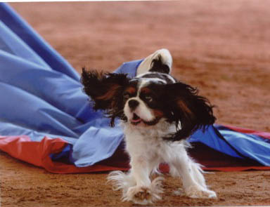
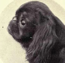 A uniqueness of the CKCS's
head shape -- its morphology -- is that
the breed was created in the 1920s from a more extreme snub-nosed
English toy spaniel -- the King Charles spaniel -- by breeding to
elongate the muzzle, rather than to shorten it. It is as if the
cavalier's muzzle has been treated like an accordion -- first compressed
to create the predecessor King Charles spaniel, and then stretched.
(See a profile of a 1910 English toy spaniel, at right.) Therefore,
the cavalier's unusual head development -- lengthening the muzzles of
shorter-faced ancestors -- may explain the uniqueness of PSOM in this
breed. See our blog article on this topic:
"The accordion-muzzled cavalier King Charles spaniel".
A uniqueness of the CKCS's
head shape -- its morphology -- is that
the breed was created in the 1920s from a more extreme snub-nosed
English toy spaniel -- the King Charles spaniel -- by breeding to
elongate the muzzle, rather than to shorten it. It is as if the
cavalier's muzzle has been treated like an accordion -- first compressed
to create the predecessor King Charles spaniel, and then stretched.
(See a profile of a 1910 English toy spaniel, at right.) Therefore,
the cavalier's unusual head development -- lengthening the muzzles of
shorter-faced ancestors -- may explain the uniqueness of PSOM in this
breed. See our blog article on this topic:
"The accordion-muzzled cavalier King Charles spaniel".
In a July 2010 article, UK investigators studied the MRI scans of 68 cavaliers, 54% of them also being diagnosed with PSOM. They also observed that the brachycephalic characteristics of an abnormally thick palate and reduced naso-pharyngeal aperature were "significantly associated" with PSOM. They concluded that:
"These results suggest that auditory tube dysfunction and OME may represent a previously overlooked consequence of brachycephalic conformation in dogs."
RETURN TO TOP
Relation to Other Disorders
In a 2010 report, UK researchers found an association between PSOM and brachycephalic conformation in cavaliers. They stated: "In CKCS, greater thickness of the soft palate and reduced nasopharyngeal aperture are significantly associated with OME [otitis media with effusion, meaning PSOM]." However, they did not explain why PSOM is so nearly limited to the cavalier, while so many other breeds are brachycephalic. They also concluded that bilateral PSOM was associated with CKCS with more extreme nasopharyngeal conformation, than unaffected CKCS.
In an October 2010 presentation before a meeting of the UK's Association of Veterinary Soft Tissue Surgeons, Robert N. White, a board certified veterinary soft tissue surgeon practicing at Willows Veterinary Centre and Referral Service in Solihull, West Midlands, observed that the cavalier does not appear to be a classically brachycephalic breed, despite the extent of BAOS in the breed, and that the extent of both PSOM and syringomyelia (SM) in the breed suggests that the CKCS may suffer from a combination syndrome of the three disorders, all associated with Chiari-like malformation (CM).
Infection as a cause of PSOM in the cavalier has been discounted by the researchers. In a May 2016 report of the examination of the ears of 41 cavaliers, the researchers concluded that bacterial infections are unlikely to be the cause of PSOM in the breed.
In a September 2022 article, German researchers examined 170 brachycephalic dogs for which computed tomography (CT) scans were performed prior to planned surgery for conditions due to brachycephalic airway obstruction syndrome (BAOS). Of those 170 dogs, 55, all either French bulldogs (35/66 -- 53%) or pugs (20/79 -- 25%) had CT indications of middle ear effusion (MEE). Tympanocentesis (draining the MEE material by first puncturing the eardrum with a needle) was performed to obtain samples of the MEE material for examination. Bacteria was found in 45% of the cases. In 73 of the ears, cells were examined using cytology testing, and inflammatory cells were found in all of them. The researchers confirmed that the MEE found in these French bulldogs and pugs is different from the non-inflammatory, cell-free effusions found in cavalier King Charles spaniels.
RETURN TO TOP
Symptoms
 Not
all cavaliers which have been found to have PSOM will display any
symptoms of it. As a result of magnetic resonance imaging (MRI) scans of
cavaliers with no signs of PSOM, studies have found that from 28% to 54%
of those CKCSs with no symptoms of PSOM nevertheless had PSOM in one or both middle ear cavities.
Not
all cavaliers which have been found to have PSOM will display any
symptoms of it. As a result of magnetic resonance imaging (MRI) scans of
cavaliers with no signs of PSOM, studies have found that from 28% to 54%
of those CKCSs with no symptoms of PSOM nevertheless had PSOM in one or both middle ear cavities.
Of those dogs with symptoms, the principal ones are:
• moderate to severe pain in the head or neck
• holding the neck in a guarded position
• tilting the head.
Other signs may include:
• scratching at the ears
• itchy ears
• head tilt
• head rubbing
• excessive yawning
• crying out in pain
• ataxia -- lack of muscle coordination
• drooping ear or lip
• inability to blink an eye
• rapid eyeball movement
• facial paralysis or nerve palsy
• drooling
• vestibular disease
• some loss of hearing
• seizures
• fatigue.
These symptoms, in many cases, are very similar to those of several other disorders, including syringomyelia and, to some extent, to those of progressive hereditary deafness. Therefore, the examining veterinarian should take care to consider these other possible causes of the dog's symptomatic behaviors.
In a 2009 UK study of 23 cavaliers with PSOM, the researchers (who choose to refer to the disorder as middle ear effusion) tested the dogs' hearing with the Brainstem Auditory Evoked Reponses (BAER) test and found that, even though the dogs' owners considered their dogs' hearing capabilities to be normal, the BAER tests demonstrated a conductive hearing loss in ears affected by middle ear effusion (PSOM). See same study in 2011 Veterinary Journal.
In an April 2015 report involving 27 cavaliers affected with PSOM, the researcher found that:
"In 74% (20/27) of the cases the dogs were deaf, 15% (4/27) of the dogs showed ataxia, 7% (2/27) showed a head tilt and 7% (2/27) showed a facial paralysis of the affected side. In 59% (16/27) of the cases the dogs were scratching, 52% (14/27) of the dogs were rubbing and 56% (15/27) were shaking their heads. In 19% (5/27) of the cases the dogs seemed to experience pain localized to the head or ears according to the owners. Gradations of these symptoms were divided in mild, moderate, severe and extreme. ...
"In the 27 dogs in this study, 14 of the 27 dogs were rubbing their head. Head rubbing (against the floor
or other surfaces) has, with the exception of dermatitis and allergies (Bruet, Bourdeau et al. 2012), only been associated with syringomyelia and CM and an unknown syndrome of behavioral signs of discomfort in the CKCS in former literature (Rusbridge 2005, Rusbridge, Carruthers et al. 2007). Only 7 of these 14 dogs were also diagnosed with CM/SM at the moment of the occurring clinical signs. After the myringotomy procedure, all clinical signs, including the head rubbing, were resolved in all 14 dogs for a period varying from four weeks to years. It seems that in these cases, the head rubbing was caused by the overfilled bulla(e) tympanica(e) and therefore this clinical sign can also be associated with PSOM."
Teeth chattering has been reported as having a possible relationship with PSOM in a May 2024 article, in which 4 cavaliers (and 2 cavalier-Bichons) were among a total of 11 dogs displaying teeth chattering. At least 2 of the CKCSs also had PSOM and were treated with myringotomies and middle ear flushes. The teeth chattering in both dogs ended after the PSOM treatments, but in one of them, the chattering and head shaking recurred 10 days later.
(NOTE: Veterinarians will tell us that dogs do not have "symptoms" but instead have "signs". But, for us laymen, the word "signs" can be confusing because of its different meanings. So, for us, "symptoms" it is.)
RETURN TO TOP
Diagnosis
- Otoscope
- Video-Otoscope
- Tympanometry
- Computed Tomography (CT)
- Magnetic Resonance Imaging (MRI)
- Brain-stem Auditory Evoked Response (BAER)
- Other Instuments
•
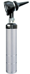 otoscope
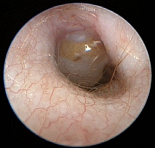
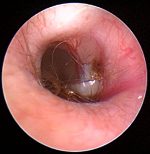
Diagnosis may be conducted using a variety of modalities. In cavaliers in particular, if the case is severe enough that the pars flaccida, the top portion of the dog's tympanic membrane (eardrum), is bulging, the condition may be visible on x-rays and even diagnosed manually with an otoscope (right). However, in cases in which the dog is in pain, or there are large amounts of wax or the dog does not tolerate the examination, PSOM cannot be excluded or confirmed solely by otoscopy.
RETURN TO TOP
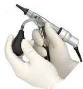
• video-otoscope
A more sophisticated form of otoscopy is video-otoscopy. It uses a
high-powered, fiber-optic camera which enables in-depth views of the two
sections of the canal -- vertical and horizontal -- and the eardrum.
General anesthesia is required for this procedure. It is combined with a
port that enables flushing and suction of the
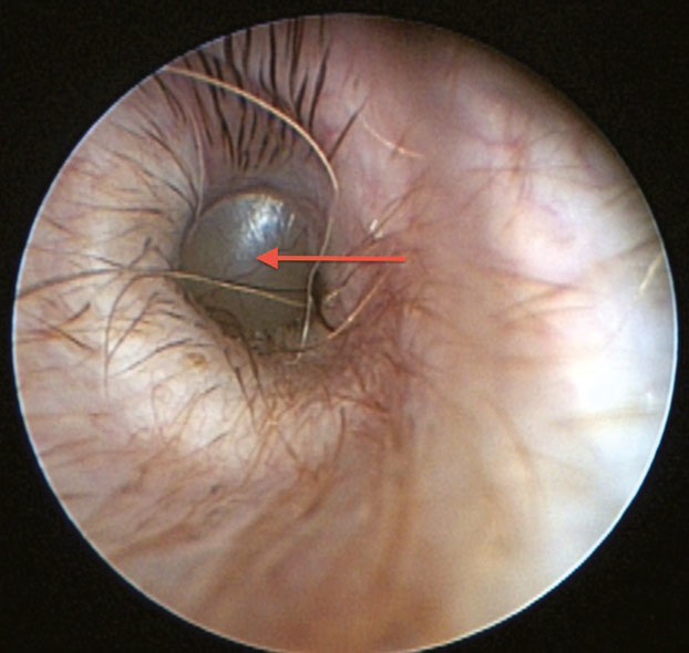 canal,
to remove wax, mucous, and debris. This is necessary to thoroughly clean
the canal for best visualization of the eardrum.
canal,
to remove wax, mucous, and debris. This is necessary to thoroughly clean
the canal for best visualization of the eardrum.
In this video-otoscopic image at the left, of the left ear canal and tympanic membrane of a cavalier, the red arrow points to a large, bulging pars flaccida. (Photo from Dr. Lynette Cole's December 2015 report.) However, cavaliers may have PSOM even though their pars flaccida is flat rather than bulging.
Video-otoscopy also is used in preparation of and during the surgical procedure, myringotomy, described below.
RETURN TO TOP
• tympanometry
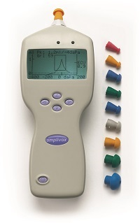 Tympanometry (impedance audiometry) is a noninvasive
method of examining the function of the middle ear while varying the
atmospheric pressure in the external ear canal and inferring the amount
of sound energy that is transmitted through the tympanum by measuring
the reflected sound energy. In a
February 2015 study, Dr. George Strain reported that the sensitivity
and specificity of tympanometry for the diagnosis of PSOM in cavaliers
were 84 and 47%, respectively. He recommended that clinical studies of
conscious dogs with PSOM need to be performed to validate the clinical
usefulness of these recordings. (See tympanometer at right.)
Tympanometry (impedance audiometry) is a noninvasive
method of examining the function of the middle ear while varying the
atmospheric pressure in the external ear canal and inferring the amount
of sound energy that is transmitted through the tympanum by measuring
the reflected sound energy. In a
February 2015 study, Dr. George Strain reported that the sensitivity
and specificity of tympanometry for the diagnosis of PSOM in cavaliers
were 84 and 47%, respectively. He recommended that clinical studies of
conscious dogs with PSOM need to be performed to validate the clinical
usefulness of these recordings. (See tympanometer at right.)
RETURN TO TOP
• computed tomography (CT)
Dr. Cole reported finding that tympanometry detected the PSOM in only
47% of ears with a flat pars flaccida.
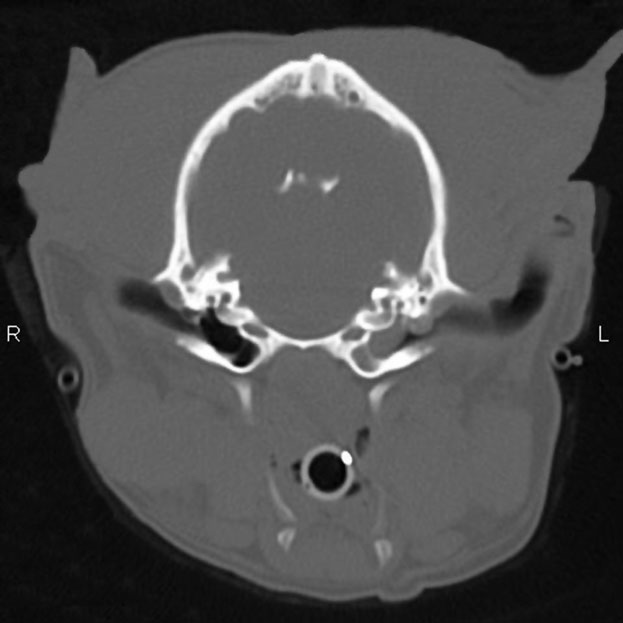 She concluded that, in cavaliers
with a flat pars flaccida, only computed tomography (CT) scans can reliably detect PSOM in
the CKCS. CT scans require that the dog be under general anesthesia. The CT scan at the left, of a cavalier King Charles spaniel with
left-sided [L] unilateral PSOM, shows the soft tissue density completely
filling the bulla on the left side [L] and the airfilled bulla on the
right side. (From Dr. Cole's December 2015 report.)
She concluded that, in cavaliers
with a flat pars flaccida, only computed tomography (CT) scans can reliably detect PSOM in
the CKCS. CT scans require that the dog be under general anesthesia. The CT scan at the left, of a cavalier King Charles spaniel with
left-sided [L] unilateral PSOM, shows the soft tissue density completely
filling the bulla on the left side [L] and the airfilled bulla on the
right side. (From Dr. Cole's December 2015 report.)
In a December 2015 report, Dr. Cole examined 60 cavalier King Charles spaniels which had clinical signs suggesting PSOM. To diagnose the disorder, they used otoscopy, tympanometry, pneumotoscopy and tympanic bulla ultrasonography, in addition to using computed tomography (CT), which they stated was "the gold standard for the diagnosis of PSOM in the CKCS."
RETURN TO TOP
 •
magnetic resonance imaging (MRI)
•
magnetic resonance imaging (MRI)
Diagnostic imaging by magnetic resonance imaging (MRI) scan is another option, however it is much more expensive than otocsopy and CT scans and also requires general anesethesia. CT usually is the preferred method because it is more sensitive and specific for changes to the middle ear's bony structures. In the MRI image at the right (courtesy, Downs Veterinary Practice, Bristol, UK.), the two bowl-shaped bullae are shown to contain accumulated mucus.
RETURN TO TOP
• brain-stem auditory evoked response (BAER)
Brain-stem auditory evoked response test
(BAER) is non-invasive and provides an objective assessment of
auditory function in canines. The BAER test
objectively examines a dog's hearing by bypassing the need to rely
subjectively on the patient's response. While BAER can detect hearing
loss at specific decibel levels, it cannot distinguish between hearing
loss due to conductivity, such as due to blockage from a PSOM
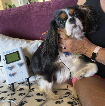 mucus
plug, and loss due to sensorineural causes such as congenital
hereditary deafness or age-induced hearing loss (presbycusis).
mucus
plug, and loss due to sensorineural causes such as congenital
hereditary deafness or age-induced hearing loss (presbycusis).
BAER measures the timing of electrical waves from the brain stem in response to a click, as a sound stimulus, in the ear. Within milliseconds of each click being made in a hearing dog's ear, a series of standard electrical waves appear on the BAER instrument's screen. The first wave comes from a nerve which transmits sound information to the brain. Then three or four other waves come from the areas of the brain stem which generate the hearing signal to the front of the brain and then to the cerebrum where the signal is interpreted as a sound. If the dog cannot hear the clicks, the waves will not appear on the screen. There are two types of BAER tests, air-conducted and bone-conducted.
BAER testing therefore will be used to confirm the suspected PSOM symptom of an hearing deficiency and its extent. Click here for a list of BAER test sites. Also, check our health clinics webpage for upcoming clinics offering BAER tests. Look for the symbol U
RETURN TO TOP
• other instuments
 Other possible alternative instruments for diagnosis of PSOM
include pneumatoscopy (an otoscope with a pneumatoscope
attachment -- see image at right -- which can show the mobility
of the eardrum in response to air pressure changes) and tympanic bulla
ultrasonography (ultrasound with a probe placed near the
bulla).
Other possible alternative instruments for diagnosis of PSOM
include pneumatoscopy (an otoscope with a pneumatoscope
attachment -- see image at right -- which can show the mobility
of the eardrum in response to air pressure changes) and tympanic bulla
ultrasonography (ultrasound with a probe placed near the
bulla).
However, in the December 2015 report described above, the researchers found that pneumotoscopy detected the PSOM in only 79% of ears with a flat pars flaccida, and tympanic bulla ultrasonography detected the PSOM in only 47% of ears with a flat pars flaccida. A November 2016 article compared ultrasound imaging with video otoscopy (an otoscope that also has a video camera that transmits images to a video screen) of otitits media in 32 dogs (one CKCS). That author concluded that video otoscopy was more reliable than ultrasound for the diagnosis of canine otitis media, but that ultrasound is a less invasive screening tool in nonsedated dogs.
Veterinary dermatologists in the United States may be located on the American College of Veterinary Dermatology website.
RETURN TO TOP
Treatment
- medicines
- myringotomy
- tympanostomy
- ventral bulla osteotomy
- ear canal ablation
- ventilation tube (grommet) insertion
- post-surgery medications
PSOM has been determined to be progressive and unlikely to spontaneously cure itself or stop its progression. It cannot be cured even by treatment, and so treatment is limited to easing or eliminating its symptoms. In many cases, the surgical procedures described below can result in regaining lost hearing due to PSOM. However, the surgeries are not likely to stop its progression, and therefore repeated surgical procedures may be necessary. Also, these surgeries may result in their own side effects, which can be similar to those of PSOM, including head tilt, drooping ear or lip, and/or facial paralysis.
• medicines
Theoretically, repeated use of drugs which thin mucous viscosity (mucolytic agents) can aid drainage of the PSOM mucous. Such drugs include N-Acetyl-L-Cysteine (NAC), (N-acetylcysteine) and bromhexine (Bisolvon). However, No studies have been published as yet to demonstrate the effectiveness of these medications.
• myringotomy
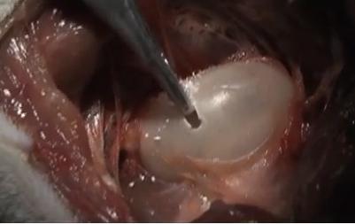
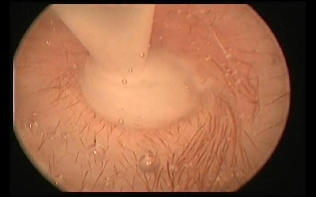 Treatment
primarily has consisted of performing a myringotomy
(see photo at right), making a small cut in the eardrum (tympanic
membrane), followed by flushing the middle ear to force out the mucus
plug. The photograph at right is of a myringotomy in progress. The ring
in the middle of the photo is the eardrum. The tube tip at the top is
the device used to flush the inner ear and force out the mucus. You may
watch a
Treatment
primarily has consisted of performing a myringotomy
(see photo at right), making a small cut in the eardrum (tympanic
membrane), followed by flushing the middle ear to force out the mucus
plug. The photograph at right is of a myringotomy in progress. The ring
in the middle of the photo is the eardrum. The tube tip at the top is
the device used to flush the inner ear and force out the mucus. You may
watch a
 close up video of a myringotomy actually being
performed on a cavalier named Baylee on
YouTube here.
close up video of a myringotomy actually being
performed on a cavalier named Baylee on
YouTube here.
Following the myringotomy, the specialist typically will repeat the CT scan, to see if all of the mucus has been removed, and then a BAER test to determine if hearing has been restored. Topical and/or systemic corticosteroids and antibiotics then are administered. The procedure may have to be repeated periodically, in some cases several times, depending upon how the dog responds.
In an April 2015 report involving 31 myringotomies and 5 tympanostomies (see below) on 27 cavaliers affected with PSOM, the researcher found that:
"[A]fter a single myringotomy procedure the mean recurrence time is 19.9 months with a median of 13 months and a recurrence rate of 61%. After tympanostomy the time to recurrence was shorter then after myringotomy (p = 0.022), which is contrary to the theory which describes that the continual tympanic cavity ventilation by using ventilation tubes may provide a longer symptom-free period. No signs of progression from unilateral to bilateral PSOM were seen."
RETURN TO TOP
• tympanostomy
 In a
March 2008 study conducted
by Australian researchers, they inserted tympanostomy tubes
(TT)
(right) within the
myringotomy incision in order to provide continual tympanic cavity
ventilation and drainage. They found that in the cases of the three
CKCSs which they operated on, all three dogs were asymptomatic at the
time of follow-up, 8, 6 and 4 months later, and they concluded that the
use of tympanostomy tubes may be an acceptable alternative to repeated
myringotomy. However, Dr. Cole reports that "no long-term prospective
studies have been published on the outcome after extrusion of the
tympanostomy tubes as far as the length of time the bulla remains
effusion free. In addition, no studies have reported on the efficacy of
a more “permanent” or long-term tympanostomy tube for treatment of
PSOM." In a
February 2013 report, a team of UK researchers also have questioned
the effectiveness of repeated tympanostomies.
In a
March 2008 study conducted
by Australian researchers, they inserted tympanostomy tubes
(TT)
(right) within the
myringotomy incision in order to provide continual tympanic cavity
ventilation and drainage. They found that in the cases of the three
CKCSs which they operated on, all three dogs were asymptomatic at the
time of follow-up, 8, 6 and 4 months later, and they concluded that the
use of tympanostomy tubes may be an acceptable alternative to repeated
myringotomy. However, Dr. Cole reports that "no long-term prospective
studies have been published on the outcome after extrusion of the
tympanostomy tubes as far as the length of time the bulla remains
effusion free. In addition, no studies have reported on the efficacy of
a more “permanent” or long-term tympanostomy tube for treatment of
PSOM." In a
February 2013 report, a team of UK researchers also have questioned
the effectiveness of repeated tympanostomies.
In an April 2015 report involving 31 myringotomies (see above) and 5 tympanostomies on 27 cavaliers affected with PSOM, the researcher found that:
"[A]fter a single myringotomy procedure the mean recurrence time is 19.9 months with a median of 13 months and a recurrence rate of 61%. After tympanostomy the time to recurrence was shorter then after myringotomy (p = 0.022), which is contrary to the theory which describes that the continual tympanic cavity ventilation by using ventilation tubes may provide a longer symptom-free period. No signs of progression from unilateral to bilateral PSOM were seen."
In an August 2015 study of 12 cavalier King Charles spaniels with PSOM, a team of UK clinicians report on the results of 22 video-otoscopy-guided tympanostomy tube placements from 2012 to 2014 at The Royal Veterinary College. The tympanostomy tubes were successfully placed in the tympanic membrane in the cavaliers, under video-otoscopic guidance using a rigid endoscope and grasping forceps. Outcomes were reported by telephonic answers to questionnaires of 11 of the CKCSs. The results:
• 3 dogs achieving normal hearing.
• 6 dogs demonstrated partial improvement of hearing.
• 10 dogs were reported with improved quality of life.
• Pruritus (severe itching) of the ears resolved in 3 of 9 dogs.
• Clinical signs recurred in 4 dogs because of tube dislodgement.
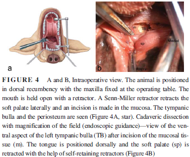 In an
August 2017 article, a team of French veterinary surgeons experimented with cadaver dogs to determine if
accessing the tympanic bulla through the mouth would be a practical
approach to installing a tympanostomy tube (TT) to allow for drainage
preventing recurring PSOM in cavalier King Charles spaniels. (See
the study's Figure 4 at right.) None of the
dogs were CKCSs in this study. They found that, in all cases, the
tympanic bulla osteotomy was performed without requiring access through
the ear canal and without damaging the inner ear or any neurovascular
structures. They noted that this oral approach to the tympanic bulla was
easier in mesaticephalic and dolichocephalic dogs than in brachycephalic
breeds. They concluded that, in spite of its small diameter, TT provides
constant ventilation and drainage of the middle ear cavity, resulting in
improved hearing and quality of life. They went on:
In an
August 2017 article, a team of French veterinary surgeons experimented with cadaver dogs to determine if
accessing the tympanic bulla through the mouth would be a practical
approach to installing a tympanostomy tube (TT) to allow for drainage
preventing recurring PSOM in cavalier King Charles spaniels. (See
the study's Figure 4 at right.) None of the
dogs were CKCSs in this study. They found that, in all cases, the
tympanic bulla osteotomy was performed without requiring access through
the ear canal and without damaging the inner ear or any neurovascular
structures. They noted that this oral approach to the tympanic bulla was
easier in mesaticephalic and dolichocephalic dogs than in brachycephalic
breeds. They concluded that, in spite of its small diameter, TT provides
constant ventilation and drainage of the middle ear cavity, resulting in
improved hearing and quality of life. They went on:
"We speculate that the transoral approach of the tympanic bulla could be used in cases of refractory PSOM treated with TT, or in cases where repeated TT placement and associated anesthetic episodes, are contraindicated due to the general condition of the patient. Removal of the entire ventral portion of the TB creates a larger opening than the TT and should therefore result in superior drainage."
RETURN TO TOP
• ventral bulla osteotomy
 An
alternative procedure is a ventral bulla osteotomy
(see photo above at right), which involves making an incision on the
under side of the neck behind the jaw bone. The auditory bulla, a hollow
bony sheath that encloses parts of the middle ear, then is exposed and
is opened. In the photograph above at right, the exposed bulla is being
opened.
An
alternative procedure is a ventral bulla osteotomy
(see photo above at right), which involves making an incision on the
under side of the neck behind the jaw bone. The auditory bulla, a hollow
bony sheath that encloses parts of the middle ear, then is exposed and
is opened. In the photograph above at right, the exposed bulla is being
opened.
• ear canal ablation
In rare cases, where the PSOM continues to re-occur after the surgical treatments described above, some veterinary surgeons reportedly have removed the entire ear canal. This procedure is called "total ear canal ablation" (TECA) and is a much more common remedy in end-stage cases of chronic, irreversible bacterial infections of the ear canal. It is performed in connection with a ventral bulla osteotomy. TECA surgery removes the entire ear canal, leaving the ear flap but sewing shut the opening beneath it. It can be a very tedious, risky surgery, since many nerves headed to the brain pass through the same area. In some cases, dogs reportedly still have some hearing ability following these surgeries. The dogs can sense sounds from vibrations reaching to the cochlear apparatus through the sinuses and skull.
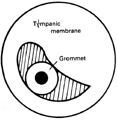
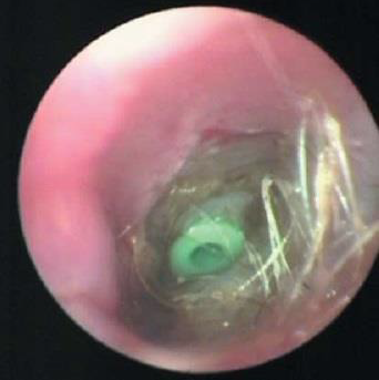
• ventilation tube (grommet) insertion
In a 1989 case report, UK surgeons inserted a ventilation tube (grommet) in the eardrum of a cavalier, following a myringotomy and flushing of the mucus plug. The grommet is intended to ventilate the middle ear cavity and equalize the middle ear pressure with the atmospheric pressure, to reduce pressure in the middle ear cavity and hopefully prevent subsequent mucus production. The grommet is intended to extrude after the eardrum membrane heals. However, in this case, the veterinarians observed that after the grommet was extruded, they mucus material unexpectedly continued to be produced. (See the diagram from this study, at right.)
RETURN TO TOP
• post-surgery medications
 Following
any of these surgical procedures, specialists usually will prescribe
prednione to counter any possible infection due to the inflammation
resulting from the surgery, and antibiotics and/or antifungals if any
external infection has been detected.
Following
any of these surgical procedures, specialists usually will prescribe
prednione to counter any possible infection due to the inflammation
resulting from the surgery, and antibiotics and/or antifungals if any
external infection has been detected.
Some specialist veterinarians have been prescribing N-Acetyl-L-Cysteine (NAC), (N-acetylcysteine) and bromhexine (Bisolvon) for cavaliers with PSOM, following surgeries. (See, e.g., this May 2014 report.) However, no studies have been performed to determine the effectiveness of these mucolytic agents in managing PSOM. Thorne Research offers NAC.
RETURN TO TOP
What You Can Do
 There
is a possibility that any cavalier will become deaf or partially
deaf. You can prepare for this possibility by learning how to
communicate with your dog by alternative ways, especially hand
signals and purposeful body motions. If a dog knows how to otherwise
understand what you expect of him, before he becomes deaf, then
communicating with him after he loses his hearing will be much
easier.
There
is a possibility that any cavalier will become deaf or partially
deaf. You can prepare for this possibility by learning how to
communicate with your dog by alternative ways, especially hand
signals and purposeful body motions. If a dog knows how to otherwise
understand what you expect of him, before he becomes deaf, then
communicating with him after he loses his hearing will be much
easier.
There are books and websites which offer helpful advice in communicating with deaf dogs. An excellent such book is Living With a Deaf Dog: A Book of Advice, Facts and Experiences About Canine Deafness by Susan C. Becker. An excellent website is DeafDogs.org
While it will not remedy PSOM, it is a good practice to keep your dogs' ears clean. Here is a YouTube video that shows how it can be done. We recommend ear cleaning products like Vet's + Best Ear Relief Wash + Dry and using them every week to ten days.
RETURN TO TOP
Research News
April 2024:
Middle ear bones of cavaliers are vastly larger than other
breeds in a UK anatomy study.
 In an
April 2024 article, UK researchers Matthew J. Mason
(right) and
Madaleine A. Lewis examined the right middle ear regions of 17 dogs of
different breeds, including a cavalier King Charles spaniel, from
CT-scans and micro-CT reconstructions of preserved skulls. The sizes of
the breeds ranged from a tiny chihuahua to a Saint Bernard. They
compared all of the components of the middle ears and comparative size
differences. These included the three small bones (ossicles), the malleus, incus, and stapes,
and the labyrinth chamber volumes of the inner ears. They report
finding:
In an
April 2024 article, UK researchers Matthew J. Mason
(right) and
Madaleine A. Lewis examined the right middle ear regions of 17 dogs of
different breeds, including a cavalier King Charles spaniel, from
CT-scans and micro-CT reconstructions of preserved skulls. The sizes of
the breeds ranged from a tiny chihuahua to a Saint Bernard. They
compared all of the components of the middle ears and comparative size
differences. These included the three small bones (ossicles), the malleus, incus, and stapes,
and the labyrinth chamber volumes of the inner ears. They report
finding:
"From our regression analyses, the breed that stood out most was the Cavalier King Charles Spaniel, which had larger ossicular and labyrinth volumes than expected for its skull length. Its ossicles considered collectively were 60% larger in volume than expected, its labyrinth just over 35% larger. The ossicles did not, however, differ noticeably in morphology to those of the other dogs. Since only one specimen was investigated we cannot be certain that this is typical of the breed, but it is interesting to note that cases of primary secretory otitis media are disproportionately common in Cavalier King Charles Spaniels (Cole, 2012; Stern-Bertholtz et al., 2003), suggesting that there may be something unusual about their middle ears. Cole et al. (2015) commented that these dogs appeared to have ‘small and shallow’ bullae, which could potentially be linked to the condition if drainage of fluids from the Eustachian tube is impeded. Relative to skull length, our specimen had a middle ear cavity volume that fell slightly below the regression line (Figure 6a), but nothing stands out regarding its cavity morphology (Figure 4p). Other authors have found a link between primary secretory otitis media and abnormal soft-tissue morphology in the nasopharyngeal region (Hayes et al., 2010), which would not be visible in our scans."
March 2023:
Recommended protocol for diagnosing and treating PSOM in
cavaliers.
 In
a
March 2023 article, Bulgarian clinician Ivelina Vacheva (right)
reported on her care of 14 cavalier King Charles spaniels, during 2021,
all with symptoms of primary secretory otitis media (PSOM). On each of
them, she performed otoscopy (visual examination of ht ear canal and
eardrum with an otoscope); ear cytology (samples from the ear canal
using a swab); culture and sensitivity testing (using a sterile swab);
MRI (which she described as “a crucial part of PSOM diagnosis);
video-otoscopy (using a fiber-optic camera enabling in-depth
In
a
March 2023 article, Bulgarian clinician Ivelina Vacheva (right)
reported on her care of 14 cavalier King Charles spaniels, during 2021,
all with symptoms of primary secretory otitis media (PSOM). On each of
them, she performed otoscopy (visual examination of ht ear canal and
eardrum with an otoscope); ear cytology (samples from the ear canal
using a swab); culture and sensitivity testing (using a sterile swab);
MRI (which she described as “a crucial part of PSOM diagnosis);
video-otoscopy (using a fiber-optic camera enabling in-depth
 visualization
of the canal and eardrum). She stated that video-otoscopy requires that
the dog be under general anesthesia. During the video-otoscopy, she
performed a deep ear cleaning, which she describes as a very important
step before a myringotomy, to achieve a thoroughly clean canal by
numerous flushings and mechanical cleanings. Finally, she performed a
myringotomy and then a middle ear flushing. Following the treatment, she
prescribed ear drops with dexamethasone at different strengths for a
total of four weeks, and N-acetylcysteine for 4 weeks, and prednisolone
for 15 days. She stated that receiving feedback for the dog’s owner was
extremely important during the post-treatment period. Of the 14 dogs
with signs consistent with PSOM, 8 were diagnosed with PSOM, 3 of which
had PSOM in both ears.
visualization
of the canal and eardrum). She stated that video-otoscopy requires that
the dog be under general anesthesia. During the video-otoscopy, she
performed a deep ear cleaning, which she describes as a very important
step before a myringotomy, to achieve a thoroughly clean canal by
numerous flushings and mechanical cleanings. Finally, she performed a
myringotomy and then a middle ear flushing. Following the treatment, she
prescribed ear drops with dexamethasone at different strengths for a
total of four weeks, and N-acetylcysteine for 4 weeks, and prednisolone
for 15 days. She stated that receiving feedback for the dog’s owner was
extremely important during the post-treatment period. Of the 14 dogs
with signs consistent with PSOM, 8 were diagnosed with PSOM, 3 of which
had PSOM in both ears.
EDITOR'S NOTE: It appears Dr. Vacheva recommends performing the MRI before the video-otoscopy, and then while the video camera remains in the ear, to perform the surgery -- the myringotomy. We should consider the role that anesthesia plays in all three of these procedures. If an MRI really is necessary, then ideally it should be performed back-to-back with the video-otoscopy examination and followed immediately by the surgery. That way only one anesthesia session is involved in the combination of diagnosis and treatment.
September 2022:
Middle ear effusions in French bulldogs and pugs are found to
differ from PSOM in cavaliers.
 In
a
September 2022 article, German researchers (Riccarda Schuenemann
[right], Anne Kamradt, Katrin Truar, Gerhard Oechtering) examined
170 brachycephalic dogs for which computed tomography (CT) scans were
performed prior to planned surgery for conditions due to brachycephalic
airway obstruction syndrome (BAOS). Of those 170 dogs, 55, all either
French bulldogs (35/66 -- 53%) or pugs (20/79 -- 25%) had CT indications
of middle ear effusion (MEE). Tympanocentesis (draining the MEE material
by first puncturing the eardrum with a needle) was performed to obtain
samples of the MEE material for examination. Bacteria was found in 45%
of the cases. In 73 of the ears, cells were examined using cytology
testing, and inflammatory cells were found in all of them. The
researchers confirmed that the MEE found in these French bulldogs and
pugs is different from the non-inflammatory, cell-free effusions found
in cavalier King Charles spaniels.
In
a
September 2022 article, German researchers (Riccarda Schuenemann
[right], Anne Kamradt, Katrin Truar, Gerhard Oechtering) examined
170 brachycephalic dogs for which computed tomography (CT) scans were
performed prior to planned surgery for conditions due to brachycephalic
airway obstruction syndrome (BAOS). Of those 170 dogs, 55, all either
French bulldogs (35/66 -- 53%) or pugs (20/79 -- 25%) had CT indications
of middle ear effusion (MEE). Tympanocentesis (draining the MEE material
by first puncturing the eardrum with a needle) was performed to obtain
samples of the MEE material for examination. Bacteria was found in 45%
of the cases. In 73 of the ears, cells were examined using cytology
testing, and inflammatory cells were found in all of them. The
researchers confirmed that the MEE found in these French bulldogs and
pugs is different from the non-inflammatory, cell-free effusions found
in cavalier King Charles spaniels.
July 2022:
German student studies head shapes of 15 cavaliers to
determine the cause of PSOM in the breed.
 In a
June 2022 veterinary school thesis, doctoral candidate
Sarah-Fabienne Charlott Possiel at the University of Veterinary Medicine
Hannover in Germany examined the computed tomography (CT) imagery of 40
dogs of brachycephalic breeds, including 15 cavalier King Charles
spaniels (CKCS), 15 French bulldogs, and 10 pugs, to increase
understanding of the causes of primary secretory otitis media (PSOM) in
those breeds. Of the 15 cavaliers, 2 had no PSOM, 7 had PSOM in one ear,
and 6 had PSOM in both ears. She focued primarily upon the dogs'
auditory tubes,
tympanic bullae, and middle ears, measuring them and creating
three-dimensional (3-D)
reconstruction models of them. She reports finding that the cavaliers'
bulla was significantly thickened and flattened, and that they had a
significant narrowing of the bony part of the auditory tube, which also
pointed in a slightly different direction than those of other breeds. She concluded that:
In a
June 2022 veterinary school thesis, doctoral candidate
Sarah-Fabienne Charlott Possiel at the University of Veterinary Medicine
Hannover in Germany examined the computed tomography (CT) imagery of 40
dogs of brachycephalic breeds, including 15 cavalier King Charles
spaniels (CKCS), 15 French bulldogs, and 10 pugs, to increase
understanding of the causes of primary secretory otitis media (PSOM) in
those breeds. Of the 15 cavaliers, 2 had no PSOM, 7 had PSOM in one ear,
and 6 had PSOM in both ears. She focued primarily upon the dogs'
auditory tubes,
tympanic bullae, and middle ears, measuring them and creating
three-dimensional (3-D)
reconstruction models of them. She reports finding that the cavaliers'
bulla was significantly thickened and flattened, and that they had a
significant narrowing of the bony part of the auditory tube, which also
pointed in a slightly different direction than those of other breeds. She concluded that:
"[B]rachycephaly has resulted in multiple changes in tubal morphology, affecting the bony portion as well as the location of the entire auditory tube. These changes in the morphology of the auditory tube predispose to tube dysfunction with subsequent effusion formation in brachycephalic dogs. In addition, the smaller bulla volume and thickened bulla wall of these dogs also appear to play a major role in tubal dysfunction."
September 2021:
UK and German study of PSOM-affected dogs finds their Eustachian
tube width and angulation may be a very dysfunctional, weak system.
 In
a
September 2021 presentation at the 2021 ESVN-ECVN Symposium, German
and UK veterinary researchers (F. Possiel, S. De Decker, H.A. Volk
[right], A.V. Volk) sought to determine the role of the Eustachian
tube (ET) in primary secretory otitis media (PSOM) in cavalier King
Charles spaniels and other brachycephalic breeds, French bulldogs and
pugs. They used computed tomography (CT) images of the ears of 72 dogs,
including 47 PSOM-affected ears of the brachycephalic breeds and 97
unaffected ears of the control breeds. They report that the prime
function of the ET is ventilation and pressure equalization of the
tympanic bullae, and that, when that fails, PSOM arises. They found that
a shorter, and significant flatter ET with a significant different
angulation in the brachycephalic dogs. They also found a marked
reduction of tympanic bulla volume, and thicker muscles in the
nasopharynx, and cartilage weakness in brachycephalic dogs suggests that
the ventilation and pressure equalization system of the ET in
brachycephalic dogs is a very dysfunctional, weak system. They concluded
that their study demonstrates that ET width and angulation might
contribute to development of PSOM.
In
a
September 2021 presentation at the 2021 ESVN-ECVN Symposium, German
and UK veterinary researchers (F. Possiel, S. De Decker, H.A. Volk
[right], A.V. Volk) sought to determine the role of the Eustachian
tube (ET) in primary secretory otitis media (PSOM) in cavalier King
Charles spaniels and other brachycephalic breeds, French bulldogs and
pugs. They used computed tomography (CT) images of the ears of 72 dogs,
including 47 PSOM-affected ears of the brachycephalic breeds and 97
unaffected ears of the control breeds. They report that the prime
function of the ET is ventilation and pressure equalization of the
tympanic bullae, and that, when that fails, PSOM arises. They found that
a shorter, and significant flatter ET with a significant different
angulation in the brachycephalic dogs. They also found a marked
reduction of tympanic bulla volume, and thicker muscles in the
nasopharynx, and cartilage weakness in brachycephalic dogs suggests that
the ventilation and pressure equalization system of the ET in
brachycephalic dogs is a very dysfunctional, weak system. They concluded
that their study demonstrates that ET width and angulation might
contribute to development of PSOM.
May 2021:
Dr. Cole describes how to do a myringotomy on a cavalier with
PSOM.
 In a
May 2021 article, Drs. Lynette Cole and Tim Nuttall
(right) explain when and
how to do a myringotomy on a cavalier with PSOM. They note that this
procedure is appropriate to give access to the middle ear for sampling,
flushing, and inserting a topical treatment. Their method is not limited
to treating cavaliers with PSOM, and they point out that even though
PSOM was first found in CKCSs and is present in up to 70% of them, it
can affect any brachycephalic breed. They provide a couple of
interesting CT scan images of cavaliers' eardrums, one bulging (at
left) and one flat (at right) but both affected with PSOM.
In a
May 2021 article, Drs. Lynette Cole and Tim Nuttall
(right) explain when and
how to do a myringotomy on a cavalier with PSOM. They note that this
procedure is appropriate to give access to the middle ear for sampling,
flushing, and inserting a topical treatment. Their method is not limited
to treating cavaliers with PSOM, and they point out that even though
PSOM was first found in CKCSs and is present in up to 70% of them, it
can affect any brachycephalic breed. They provide a couple of
interesting CT scan images of cavaliers' eardrums, one bulging (at
left) and one flat (at right) but both affected with PSOM.
May 2020:
UK study of 16 dogs with PSOM includes 8 cavaliers, finding
bacterial or inflammatory connections unlikely.
 In
a
May 2020 article by a team of UK veterinary researchers (Elspeth M.
Milne [right], Tim Nuttall, Katia Marioni-Henry, Chiara
Piccinelli, Tobias Schwarz, Ali Azar, Jennifer Harris, Juliet Duncan,
Michael Cheeseman), they examined 16 live brachycephalic dogs, including
8 (50%) cavalier King Charles spaniels, and 5 postmortems (none were
CKCSs) for middle ear effusions (MEE) -- their name for primary
secretory otitis media (PSOM) -- to study the cellular features of MEE
mucus. They also report additional factors regarding MEE-affected dogs.
The other breeds of live dogs were French Bulldog (5), boxer (4), and
English bulldog (1). The deceased dogs were boxers (3), French bulldog
(1), and English bulldog (1). Among the cavaliers, only one had MEE
only, meaning no other possibly related disorders. In the other seven
CKCSs, all had both MEE and Chiari-like malformation and syringomyelia
(CM/SM); 2 CKCSs were diagnosed with idiopathic epilepsy; 2 with
diabetes mellitus, and 1 with brachycephalic obstructive airway syndrome
(BOAS). They found that mucosa was thicker in MEE-affected dogs, and
that there was no bacterial growth in 79% of the examined effusions.
In
a
May 2020 article by a team of UK veterinary researchers (Elspeth M.
Milne [right], Tim Nuttall, Katia Marioni-Henry, Chiara
Piccinelli, Tobias Schwarz, Ali Azar, Jennifer Harris, Juliet Duncan,
Michael Cheeseman), they examined 16 live brachycephalic dogs, including
8 (50%) cavalier King Charles spaniels, and 5 postmortems (none were
CKCSs) for middle ear effusions (MEE) -- their name for primary
secretory otitis media (PSOM) -- to study the cellular features of MEE
mucus. They also report additional factors regarding MEE-affected dogs.
The other breeds of live dogs were French Bulldog (5), boxer (4), and
English bulldog (1). The deceased dogs were boxers (3), French bulldog
(1), and English bulldog (1). Among the cavaliers, only one had MEE
only, meaning no other possibly related disorders. In the other seven
CKCSs, all had both MEE and Chiari-like malformation and syringomyelia
(CM/SM); 2 CKCSs were diagnosed with idiopathic epilepsy; 2 with
diabetes mellitus, and 1 with brachycephalic obstructive airway syndrome
(BOAS). They found that mucosa was thicker in MEE-affected dogs, and
that there was no bacterial growth in 79% of the examined effusions.
March 2020: Review of MRIs of 518 Dutch cavaliers shows 20% had PSOM. In a March 2020 master's thesis, Utrecht Univ. student Maxime Laterveer reviewed 572 MRI brain scans of 518 Dutch cavalier King Charles spaniels screened in 2016 through 2018, for primary secretory otitis media (PSOM). She reports finding that the prevalence of PSOM in the left ear was 19.4% and in the right ear was 20.0%. She also found that PSOM did not improve upon screening for unaffected parents.
January 2019:
In a study of 68 PSOM-affected ears of cavaliers, 67% had no
bacterial growth.
 In a
January 2019 article, veterinary dermatologists and pathologists
Lynette K. Cole (right), Päivi J. Rajala-Schultz, Gwendolen Lorch, and Joshua B.
Daniels examined 69 ears of 41 cavalier King Charles spaniels diagnosed
with primary secretory otitis media (PSOM), searching for bacterial
growth which may have contributed to the PSOM. They report finding that
55 of 68 (81%) of the external ear canals (EEC) and 46 of 69 (67%) of
the middle ears (ME) had no bacterial growth; 38 (56%) had no growths in
either the EEC or the ME. Of the remaining EECs and MEs, the researchers
found eight different bacterial species, the most frequent being
coagulase-negative staphylococci, followed by Staphylococcus
pseudintermedius. They concluded:
In a
January 2019 article, veterinary dermatologists and pathologists
Lynette K. Cole (right), Päivi J. Rajala-Schultz, Gwendolen Lorch, and Joshua B.
Daniels examined 69 ears of 41 cavalier King Charles spaniels diagnosed
with primary secretory otitis media (PSOM), searching for bacterial
growth which may have contributed to the PSOM. They report finding that
55 of 68 (81%) of the external ear canals (EEC) and 46 of 69 (67%) of
the middle ears (ME) had no bacterial growth; 38 (56%) had no growths in
either the EEC or the ME. Of the remaining EECs and MEs, the researchers
found eight different bacterial species, the most frequent being
coagulase-negative staphylococci, followed by Staphylococcus
pseudintermedius. They concluded:
"In conclusion, bacterial otitis externa is an uncommon finding in CKCS with PSOM and the majority of ME with PSOM are culture-negative, with the cytological presence of bacteria in mucous being poorly correlated with conventional bacterial culture results. Additional studies evaluating the microbiome as well as additional exploration of the potential role of staphylococci in CKCS with PSOM are needed to further understand the pathogenesis of PSOM."
EDITOR'S NOTE: This whole thing is a little odd. In
June 2016, an
abstract of this study was presented to the Veterinary Dermatology
conference. None of the data varies between this current article and the
2016 abstract. However, in 2016, the authors definitively concluded
that:
"Based on the results of this study, bacterial infections are unlikely to be the cause of PSOM in the CKCS."
At that time, this was a very significant finding because there was
doubt about what role bacteria may have played in the onset of PSOM in
the breed. The 2016 abstract pretty much put such doubt to bed, and the
focus turned to structural causes related to a unique form of
brachycephaly in the CKCS. Now, the same researchers with the identical
data are suggesting that maybe more sophisticated studies, evaluating
the microbiome and exploring the potential role of staphylococci, be
performed.
This reversal of conclusions smacks of a possible case
of over-diagnosis. We suggest that funding be better spent examining a
control group of 60+ ears of non-PSOM-affected cavaliers to see how
common these bacteria are in many ears of this breed. We think the
finding will be that bacteria plays absolutely no role in the onset or
progression of PSOM, and that these researchers' 2016 conclusion was the
accurate one to draw from this data.
February 2018:
French researchers report study of two CKCSs with PSOM which displayed lip drooling
and facial paralysis.
 In a
February 2018 article, a team of French veterinary researchers
(Morgane Debuigne, Amandine Drut [right], N. Soetart, D.
Guillemaille, T. Brément, M. Fusellier, D. Fanuel, V. Bruet)
report finding primary secretory otitis media (PSOM) as the cause of two
cavalier King Charles spaniels drooling from their lips and with facial
paralysis. One of the two cavaliers also was diagnosed with Chiari-like
malformation and syringomyelia (CM/SM). The clinicians performed
myringotomies and flushed the tympanic bullae of both CKCSs and then treated
with corticosteroids. The dogs' clinical signs improved for several
months. They concluded:
In a
February 2018 article, a team of French veterinary researchers
(Morgane Debuigne, Amandine Drut [right], N. Soetart, D.
Guillemaille, T. Brément, M. Fusellier, D. Fanuel, V. Bruet)
report finding primary secretory otitis media (PSOM) as the cause of two
cavalier King Charles spaniels drooling from their lips and with facial
paralysis. One of the two cavaliers also was diagnosed with Chiari-like
malformation and syringomyelia (CM/SM). The clinicians performed
myringotomies and flushed the tympanic bullae of both CKCSs and then treated
with corticosteroids. The dogs' clinical signs improved for several
months. They concluded:
"These two cases highlight the importance of otitis media in the differential diagnosis of facial paralysis and focus on the originality of primary secretory otitis media in the Cavalier King Charles Spaniels."
October 2017:
In a French study of 205 dogs with middle ear disorders, only
cavaliers had PSOM.
 In
an
October 2017 article reporting a French study of 205 dogs identified
with signs of middle ear diseases, the researchers (Audrey Belmudes,
Charline Pressanti, Paul Y. Barthez, Eloy Castilla-Castaño, Lionel
Fabries, Marie C. Cadiergues [right]) used computed tomography
(CT) and found that of the 13 cavalier King Charles spaniels, 10 had
primary secretory otitis media. Nine of the CKCSs had PSOM in both ears
and the other cavalier had PSOM in only one ear. All of those cavaliers
were classified as being deaf. None of the other breeds had PSOM.
In
an
October 2017 article reporting a French study of 205 dogs identified
with signs of middle ear diseases, the researchers (Audrey Belmudes,
Charline Pressanti, Paul Y. Barthez, Eloy Castilla-Castaño, Lionel
Fabries, Marie C. Cadiergues [right]) used computed tomography
(CT) and found that of the 13 cavalier King Charles spaniels, 10 had
primary secretory otitis media. Nine of the CKCSs had PSOM in both ears
and the other cavalier had PSOM in only one ear. All of those cavaliers
were classified as being deaf. None of the other breeds had PSOM.
September 2017:
PSOM is found in up to 38% of CKCSs examined in a
Netherlands/Belgium study of 1,000+ dogs.
 In a
September 2017 article detailing a 12-year (2004 to 2015) study of
1,020 cavalier King Charles spaniels diagnosed using MRI scans in the
Netherlands and Belgium, researchers (Katrien Wijnrocx, Leonie W. L. Van
Bruggen, Wieteke Eggelmeijer, Erik Noorman, Arnold Jacques, Nadine Buys,
Steven Janssens, Paul J. J. Mandigers [right]) report finding the presence of
middle ear effusion (PSOM) in 19% to 21% of CKCSs younger than 3 years
(out of 809 dogs), and 32% to 38% of cavaliers older than 3 years (out
of 211 dogs). The same increase was noted for dogs that were MRI-scanned
multiple times. They stated:
In a
September 2017 article detailing a 12-year (2004 to 2015) study of
1,020 cavalier King Charles spaniels diagnosed using MRI scans in the
Netherlands and Belgium, researchers (Katrien Wijnrocx, Leonie W. L. Van
Bruggen, Wieteke Eggelmeijer, Erik Noorman, Arnold Jacques, Nadine Buys,
Steven Janssens, Paul J. J. Mandigers [right]) report finding the presence of
middle ear effusion (PSOM) in 19% to 21% of CKCSs younger than 3 years
(out of 809 dogs), and 32% to 38% of cavaliers older than 3 years (out
of 211 dogs). The same increase was noted for dogs that were MRI-scanned
multiple times. They stated:
"There was no correlation, in this study, for CM [Chiari-like malformation] and the presence of middle ear effusion, which seems to suggest that middle ear effusion is more associated with a brachycephalic conformation rather than the actual CM grading. This is remarkable as CM is clearly associated with a brachycephalic conformation. Remarkably, a small but statistically significant correlation was found for CDD [central canal dilation] and middle ear effusion. If this observation is correct it merits further research, as it has not been investigated before. Furthermore it must be noted that in a CKCS, it might be difficult to notice clear signs that could be caused by the middle ear effusion as most of them suffer from CM as well."
EDITOR'S NOTE:
The study's authors conclude "remarkably" that PSOM does not correlate
with Chiari-like malformation, even though both disorders are attributed
to a brachycephalic conformation. But what they do not do is explain why
our breed, the cavalier King Charles spaniel, which is no where near as
snub-nosed as several other brachycephalic breeds, has such a high
incidence of PSOM. We have speculated that this is due to the jumbled
result of creating the CKCS from the much shorter-muzzled English toy
spaniel (King Charles spaniel) beginning in the late 1920s. See our blog
article on this topic:
"The accordion-muzzled cavalier King Charles spaniel".
August 2017:
French surgeons test an oral approach to installing tympanostomy
tubes in cavaliers with PSOM.
In an
August 2017 article, a team of French veterinary surgeons (Maria
Manou, Pierre H. M. Moissonnier, Nicolas Jardel, Aymeric Tissier,
Rosario Vallefuoco) experimented with cadaver dogs to determine if
accessing the tympanic bulla through the mouth would be a practical
approach to installing a tympanostomy tube (TT) to allow for drainage
preventing recurring PSOM in cavalier King Charles spaniels. (See
the study's Figure 4 at right.) None of the
dogs were CKCSs in this study. They found that, in all cases, the
tympanic bulla osteotomy was performed without requiring access through
the ear canal and without damaging the inner ear or any neurovascular
structures. They noted that this oral approach to the tympanic bulla was
easier in mesaticephalic and dolichocephalic dogs than in brachycephalic
breeds. They concluded that, in spite of its small diameter, TT provides
constant ventilation and drainage of the middle ear cavity, resulting in
improved hearing and quality of life. They went on:
"We speculate that the transoral approach of the tympanic bulla could be used in cases of refractory PSOM treated with TT, or in cases where repeated TT placement and associated anesthetic episodes, are contraindicated due to the general condition of the patient. Removal of the entire ventral portion of the TB creates a larger opening than the TT and should therefore result in superior drainage."
July 2017:
Cavaliers are found to have significantly flatter tympanic
bullae than other brachycephalic breeds.
 In a
July 2017 article, a team of UK researchers (Ben Mielke, Richard
Lam, Gert ter Haar [right]) studied the tympanic bullae of four brachycephalic
breeds (pug, French bulldog, English bulldog, and cavalier King Charles
spaniel -- 25 dogs of each breed) and compared them to two control
breeds (Labrador retriever and Jack Russell terrier). They found that
the CKCS had significantly flatter tympanic bullae than the other brachy
breeds. (See the article's Figure 4, below.) They also found middle ear effusion material in 68% of the
cavaliers. They speculated that the flatter shape of the CKCS tympanic
bulla may explain the reason for such a high incidence of PSOM in the
breed. The authors stated:
In a
July 2017 article, a team of UK researchers (Ben Mielke, Richard
Lam, Gert ter Haar [right]) studied the tympanic bullae of four brachycephalic
breeds (pug, French bulldog, English bulldog, and cavalier King Charles
spaniel -- 25 dogs of each breed) and compared them to two control
breeds (Labrador retriever and Jack Russell terrier). They found that
the CKCS had significantly flatter tympanic bullae than the other brachy
breeds. (See the article's Figure 4, below.) They also found middle ear effusion material in 68% of the
cavaliers. They speculated that the flatter shape of the CKCS tympanic
bulla may explain the reason for such a high incidence of PSOM in the
breed. The authors stated:
"Cavalier King Charles Spaniels have been described to have a unique disease resulting in the formation of a buildup of highly viscous mucus within the middle ear (primary secretory otitis media or otitis media with effusion) of dogs without clinical evidence of otitis externa. ... The finding of an anatomical variation in the shape of the tympanic bulla of Cavalier King Charles Spaniels may offer a potential explanation for the pathogenesis of this disease in addition to the previously suggested changes in the orientation and function of the auditory tube. However, given the lack of histopathology and contrast-enhanced CT scan evaluations in the current study, this hypothesis is speculative. Further investigation to identify potential pathway alteration of the auditory tube in the Cavalier King Charles Spaniel would be an interesting addition for further clarification of this disease process."
October 2016: German study finds that video otoscopy beats ultrasound in diagnosing otitis media. In a November 2016 article, a team of German researchers (J. Classen, A. Bruehschwein, A. Meyer-Lindenberg, R.S. Mueller) compared ultrasound imaging with video otoscopy (an otoscope that also has a video camera that transmits images to a video screen) of otitits media in 32 dogs (one CKCS). The author concluded that video otoscopy was more reliable than ultrasound for the diagnosis of canine otitis media, but that ultrasound is a less invasive screening tool in nonsedated dogs.
May 2016: Study of 41 cavaliers shows that bacterial infection is an unlikely cause of PSOM in CKCSs. In a June 2016 abstract, Dr. Lynette Cole led a study (with P. Rajala-Schultz, G. Lorch) of the ears of 41 cavalier King Charles spaniels to determine if bacterial infection causes primary secretory otitis media (PSOM) in the breed. Of the 69 ears examined, no bacterial growth was found in the ear secretions in 81% of the external ear canals and 67% of the middle ears. Only 18% had bacterial in the external ear canals, and 33% had bacteria in their middle ears. The team cautiously concluded that "bacterial infections are unlikely to be the cause of PSOM in the CKCS." (See also this January 2019 report.)
January 2016:
US Neurologist Rossmeisl finds PSOM, CM/SM, brachycephalic airway
obstruction syndrome and laryngeal paralysis in a cavalier.
 In a
January 2016 article, Virginia Tech veterinary neurologist John
Rossmeisl reports on a 13-year-old, male castrated cavalier King Charles
spaniel which had symptoms of vestibular disease -- a left head tilt and
circling to the left, which progressed to left lateral recumbency,
positional strabismus (OS), and rotary nystagmus (OU) -- and also the
absence of postural reactions in all limbs, exaggerated spinal reflexes
in all limbs, and respiratory distress. He performed an MRI and
otoscopic examination and performed a myringotomy. (See specimen of
mucus at right.) He found no evidence
of inflammation or infection. He diagnosed: (a) primary secretory otitis
media (PSOM), causing the peripheral vestibular signs; (b)
CM/SM,
causing the central nervous system symptoms; (c)
brachycephalic airway
obstruction syndrome (BAOS) and laryngeal paralysis, causing the dog’s
inability to walk and required tracheal intubation and supplemental
oxygen. The owner chose euthanasia rather than surgery to relieve the
BAOS.
In a
January 2016 article, Virginia Tech veterinary neurologist John
Rossmeisl reports on a 13-year-old, male castrated cavalier King Charles
spaniel which had symptoms of vestibular disease -- a left head tilt and
circling to the left, which progressed to left lateral recumbency,
positional strabismus (OS), and rotary nystagmus (OU) -- and also the
absence of postural reactions in all limbs, exaggerated spinal reflexes
in all limbs, and respiratory distress. He performed an MRI and
otoscopic examination and performed a myringotomy. (See specimen of
mucus at right.) He found no evidence
of inflammation or infection. He diagnosed: (a) primary secretory otitis
media (PSOM), causing the peripheral vestibular signs; (b)
CM/SM,
causing the central nervous system symptoms; (c)
brachycephalic airway
obstruction syndrome (BAOS) and laryngeal paralysis, causing the dog’s
inability to walk and required tracheal intubation and supplemental
oxygen. The owner chose euthanasia rather than surgery to relieve the
BAOS.
October 2015:
George Strain reviews PSOM in the cavalier King Charles spaniel.
In a September 2015 article,
 Dr. George Strain
(right) of LSU veterinary school
summarizes the current knowledge about PSOM in cavaliers. He describes
it as a late onset conductive hearing loss almost exclusively in the
CKCS. He says that "no proven hereditary cause has been identified, but
the preponderance of occurrence in the one breed supports this as a
possible factor." He notes that "a correlation between PSOM and
pharyngeal conformation has been suggested [citing this 2010 article],
possibly from a thick soft palate and reduced nasopharyngeal aperture,
which might argue against a hereditary basis. He further reports:
Dr. George Strain
(right) of LSU veterinary school
summarizes the current knowledge about PSOM in cavaliers. He describes
it as a late onset conductive hearing loss almost exclusively in the
CKCS. He says that "no proven hereditary cause has been identified, but
the preponderance of occurrence in the one breed supports this as a
possible factor." He notes that "a correlation between PSOM and
pharyngeal conformation has been suggested [citing this 2010 article],
possibly from a thick soft palate and reduced nasopharyngeal aperture,
which might argue against a hereditary basis. He further reports:
"Signs include hearing loss, neck scratching, otic pruritus, head shaking, abnormal yawning, head tilt, facial paralysis, or vestibular disturbances . Testing has confirmed that the hearing loss is conductive. Implantation of tympanostomy tubes has been successful in a small number of cases, but treatment is typically by myringotomy followed by flushing of the mucus plug, repeated as needed. No studies have been reported of genetic association studies."
September 2015:
UK researchers report mixed results of tympanostomy tube treatment for
PSOM in cavaliers. In an
August 2015 study of 12 cavalier
King Charles spaniels with PSOM, a team of UK clinicians (V. Guerin, R.
Hampel, G. Ter Haar) report on the
results of 22 video-otoscopy-guided tympanostomy tube placements from
2012 to 2014 at The Royal Veterinary College. The tympanostomy tubes
were successfully placed in the tympanic membrane in the cavaliers,
under video-otoscopic guidance using a rigid endoscope and grasping
forceps. Outcomes were reported by telephonic answers to questionnaires
of 11 of the CKCSs. The results:
• 3 dogs achieving normal hearing.
• 6 dogs demonstrated partial improvement of hearing.
• 10 dogs were reported with improved quality of life.
• Pruritus (severe itching) of the ears resolved in 3 of 9 dogs.
• Clinical signs recurred in 4 dogs because of tube dislodgement.
August 2015:
US researchers determine that only computed tomography (CT) can
reliably detect
non-bulging PSOM in cavaliers.
 In
a
December 2015 study by a
team of Ohio veterinarians (Lynette K. Cole [right], Valerie F. Samii, Susan O.
Wagner, Päivi J. Rajala-Schultz), they examined 60 cavalier King Charles
spaniels which had clinical signs suggesting PSOM. To diagnose the
disorder, they used otoscopy, tympanometry, pneumotoscopy and tympanic
bulla ultrasonography, in addition to using computed tomography (CT),
which they stated was "the gold standard for the diagnosis of PSOM in
the CKCS."
In
a
December 2015 study by a
team of Ohio veterinarians (Lynette K. Cole [right], Valerie F. Samii, Susan O.
Wagner, Päivi J. Rajala-Schultz), they examined 60 cavalier King Charles
spaniels which had clinical signs suggesting PSOM. To diagnose the
disorder, they used otoscopy, tympanometry, pneumotoscopy and tympanic
bulla ultrasonography, in addition to using computed tomography (CT),
which they stated was "the gold standard for the diagnosis of PSOM in
the CKCS."
All of the methods diagnosed PSOM in ears with large bulging "pars flaccida" (the triangular, flaccid portion of the eardrum). However, cavaliers may have PSOM even though their pars flaccida is flat rather than bulging.
Specifically, they found:
(a) 43 (72%) had PSOM, of which 30 had PSOM in both ears and 13 in only one ear (73 affected ears);
(b) Only 21 of the 73 ears with PSOM had a large bulging pars flaccida;
(c) The remaining 52 affected ears had a flat pars flaccida;
(d) Tympanometry detected the PSOM in only 47% of ears with a flat pars flaccida;
(e) Pneumotoscopy detected the PSOM in only 79% of ears with a flat pars flaccida;
(f) Tympanic bulla ultrasonography detected the PSOM in only 47% of ears with a flat pars flaccida.
They concluded that, in cavaliers with a flat pars flaccida, none of the other diagnostic tests (otoscopy, tympanometry, pneumotoscopy and tympanic bulla ultrasonography) can be recommended in place of a CT scan for the diagnosis of PSOM.
April 2015:
Dutch research
finds myringotomies are more successful than tympanostomies in slowing
recurrences of PSOM. In
an
April 2015 master's thesis, N. Bolder at Utrecht University, Netherlands, reports
that in a study of 27 cavalier King Charles spaniels diagnosed with
primary secretory otitis media (PSOM), 31 myringotomies and 5
tympanostomy procedures were performed. The purpose of the study was to
"assess the time interval in which recurrence of clinical signs occurs
after treatment of PSOM with a myringotomy or tympanostomy procedure and
to examine if there was any progression of this disease over time or
affecting sides."
In
an
April 2015 master's thesis, N. Bolder at Utrecht University, Netherlands, reports
that in a study of 27 cavalier King Charles spaniels diagnosed with
primary secretory otitis media (PSOM), 31 myringotomies and 5
tympanostomy procedures were performed. The purpose of the study was to
"assess the time interval in which recurrence of clinical signs occurs
after treatment of PSOM with a myringotomy or tympanostomy procedure and
to examine if there was any progression of this disease over time or
affecting sides."
Mr. Bolder reported finding that:
"[A]fter a single myringotomy procedure the mean recurrence time is 19.9 months with a median of 13 months and a recurrence rate of 61%. After tympanostomy the time to recurrence was shorter then after myringotomy (p = 0.022), which is contrary to the theory which describes that the continual tympanic cavity ventilation by using ventilation tubes may provide a longer symptom-free period. No signs of progression from unilateral to bilateral PSOM were seen."
Additionally, the researcher reported these clinical signs associated with PSOM in the cavaliers:
"In 74% (20/27) of the cases the dogs were deaf, 15% (4/27) of the dogs showed ataxia, 7% (2/27) showed a head tilt and 7%% (2/27) showed a facial paralysis of the affected side. In 59% (16/27) of the cases the dogs were scratching, 52% (14/27) of the dogs were rubbing and 56% (15/27) were shaking their heads. In 19% (5/27) of the cases the dogs seemed to experience pain localized to the head or ears according to the owners (figure 10). Gradations of these symptoms were divided in mild, moderate, severe and extreme. ...
"In the 27 dogs in this study, 14 of the 27 dogs were rubbing their head. Head rubbing (against the floor or other surfaces) has, with the exception of dermatitis and allergies (Bruet, Bourdeau et al. 2012), only been associated with syringomyelia and CM and an unknown syndrome of behavioral signs of discomfort in the CKCS in former literature (Rusbridge 2005, Rusbridge, Carruthers et al. 2007). Only 7 of these 14 dogs were also diagnosed with CM/SM at the moment of the occurring clinical signs. After the myringotomy procedure, all clinical signs, including the head rubbing, were resolved in all 14 dogs for a period varying from four weeks to years. It seems that in these cases, the head rubbing was caused by the overfilled bulla(e) tympanica(e) and therefore this clinical sign can also be associated with PSOM."
February 2015:
Dr. George Strain finds tympanometry can be recorded in conscious dogs
to assist in the evaluation of the middle ear conditions.
In a
February 2015 study, Drs. George Strain
(right) and Asia J. Fernandes of
Louisiana State University veterinary school evaluated tympanometric
measurments of 16 dogs' ears. Tympanometry (impedance audiometry) is a
noninvasive method of examining the function of the middle ear while
varying the atmospheric pressure in the external ear canal and inferring
the amount of sound energy that is transmitted through the tympanum by
measuring the reflected sound energy. The researchers found that
tympanograms can be recorded in conscious dogs to assist in the
evaluation of the middle ear structures. They also reported that the
sensitivity and specificity of tympanometry for the diagnosis of PSOM in
cavaliers were 84 and 47%, respectively. They recommended that clinical
studies of conscious dogs with PSOM need to be performed to validate the
clinical usefulness of these recordings.
June 2014: Good News! Dr. Charles Bluestone retires! Perhaps now he and his federal funding will leave our cavaliers alone. See our previous discussions of this problematic busy-body. April 2013. February 2012. November 2011. December 2010.
May 2014: Texas A&M specialists report on PSOM case in a cavalier. In a May 2014 report, Texas A&M veterinary specialists Megan Stout Steele and Jonathan Levine describe the case study of a 7 year old female cavalier King Charles spaniel with "chronic intermittent cervical pain and scratching of the head and neck". They diagnosed bilateral PSOM plus CM/SM and performed a myringotomy and bilateral deep ear flushing. Large, clear mucus plugs were removed from both middle ears; culture of the plugs was negative for bacteria. N-acetylcysteine was prescribed as a mucolytic to lower the risk for PSOM recurrence. They reported that a month later, the dog’s hearing seemed to have improved, but her gait abnormalities and phantom scratching persisted.
September 2013:
UK's Downs Veterinary Practice
reports PSOM case in CKCS.
 In
a report posted by Downs Veterinary
Practice of Bristol, UK, on its website, it describes a case study of a cavalier
King Charles spaniel with primary secretory otitis media. It states: "Bilateral
myringotomy was performed and the bullae were flushed with sterile saline; large
amounts of yellow tinged mucus were removed from both bulla and sent for
cytology and bacteriology. The dog was treated post operatively with meloxicam
and amoxiclav pending culture results. The owners were contacted one week after
surgery and reported an improvement in the dog’s hearing." The report also
included a photo of the mucus plugs and an MRI scan showing the bullae filled
with mucus fluid, which we have added to sections of this webpage.
In
a report posted by Downs Veterinary
Practice of Bristol, UK, on its website, it describes a case study of a cavalier
King Charles spaniel with primary secretory otitis media. It states: "Bilateral
myringotomy was performed and the bullae were flushed with sterile saline; large
amounts of yellow tinged mucus were removed from both bulla and sent for
cytology and bacteriology. The dog was treated post operatively with meloxicam
and amoxiclav pending culture results. The owners were contacted one week after
surgery and reported an improvement in the dog’s hearing." The report also
included a photo of the mucus plugs and an MRI scan showing the bullae filled
with mucus fluid, which we have added to sections of this webpage.
September 2013: UK researchers' survey finds that chronic otitis media may be associated with higher grades of hearing loss. In an October 2013 study by a team of UK veterinary dermatologists, they report the results of questionaires answered by owners of 100 dogs referred for chronic otitis investigation, including six cavalier King Charles spaniels. They concluded that "Chronic otitis media may be associated with higher grades of hearing loss." EDITOR'S NOTE: This study was not limited to dogs with PSOM.
April 2013:
Dr. Charles Bluestone tones down his fantasy about CKCS
evolution but still gets it all wrong.
 In an article*
published in April 2013, Dr. Charles D. Bluestone
(right) (along with his cohort J. Douglas Swarts
and others) persists in likening PSOM in the cavalier King Charles
spaniel with the human condition, but at least he has walked back his
irresponsible prior statements about "greater than 50%" of cavaliers
having PSOM (see our February
2012 entry), and that the cavalier "has been bred over 300 years to
have a short snout." (See our
November 2011 entry.) Instead, this time he gets the name of the
disorder right (it's not "persistent", it's "primary"), and he more
mildly states (with actual citations):
In an article*
published in April 2013, Dr. Charles D. Bluestone
(right) (along with his cohort J. Douglas Swarts
and others) persists in likening PSOM in the cavalier King Charles
spaniel with the human condition, but at least he has walked back his
irresponsible prior statements about "greater than 50%" of cavaliers
having PSOM (see our February
2012 entry), and that the cavalier "has been bred over 300 years to
have a short snout." (See our
November 2011 entry.) Instead, this time he gets the name of the
disorder right (it's not "persistent", it's "primary"), and he more
mildly states (with actual citations):
"Primary secretory OM [otitis media] is the term veterinarians use to describe chronic OM with effusion in canines. It has been reported to occur almost exclusively in short-faced canines, and its prevalence in the Cavalier King Charles spaniel breed is estimated at about 40%. ... Bluestone and Swarts have speculated that the Cavalier King Charles is a natural animal model, induced by artificial selection for those breed characteristics, which secondarily elicit chronic OM with effusion due to ET dysfunction."
But, they still get the "evolution" of the CKCS totally backwards. They write:
"Artificial selection of this breed has, until recently, focused on producing a shortened front-to-back diameter of the skull, a result of premature fusion of the coronal sutures."
This shows the authors' continued and seemingly intentional ignorance of the development of the cavalier breed from its King Charles spaniel roots -- with that breed's shorter skull -- in the 1920s. (See our Editor's Note above and our November 2011 entry below for the skull comparison.) We are not sure of what they mean by "until recently". Perhaps to them, "until recently" means "until the 1920s", some 90 years ago. Finally, as has been their modus operandi in the past, our federal tax money funded their little screed. Thankfully, we suspect that not much will come from Dr. Bluestone's "research" of PSOM in the CKCS because it is unlikely he will find any federal grant money to fund such a canine project.
March
2013:
Alabama specialist analyzes PSOM in 9-month old female CKCS.
In the
March 2013 issue of
 Clinician's
Brief, Dr. Louis N. Gotthelf (right), a
pet ear and skin specialist, reports a case study of a nine-month old
female cavalier with neck stiffness and head tilt. His "bottom line":
Clinician's
Brief, Dr. Louis N. Gotthelf (right), a
pet ear and skin specialist, reports a case study of a nine-month old
female cavalier with neck stiffness and head tilt. His "bottom line":
"CKCS eardrums should always be evaluated. If PSOM is suspected, a high-magnification eardrum examination may require referral. PSOM can be asymptomatic in CKCSs, in which case myringotomy is not required; however, the owner should be educated to take appropriate steps if signs are displayed. The Take-Home: CKCSs often have a genetic defect of their eustachian tube that results in PSOM, which may be asymptomatic. Bulging eardrum and ear pain are typical of PSOM in CKCSs. PSOM should be differentiated from SM via eardrum evaluation and MRi (if necessary). Myringotomy can relieve PSOM signs. Antibiotics will not be helpful. Periodic treatment is required."
February 2013: UK researchers find PSOM is progressive in cavaliers and will not cure itself. In a February 2013 report in Veterinary Journal, a team of UK researchers (S. J. McGuinness, E. J. Friend, S. P. Knowler, N. D. Jeffery, C. Rusbridge) examined repeat MRIs (two each) of 34 cavalier King Charles spaniels for PSOM*. (See a September 2010 entry on this same study.) At the first scan, 23 of the CKCSs had no effusion, 10 had unilateral effusion, and 1 had bilateral effusion. They found that, from the first to the second MRI, 26.5% had progressed from no PSOM to unilateral or bilateral PSOM, or from unilateral to bilateral. None of the 11 dogs which had PSOM at the time of the first MRI had resolved themselves. Significantly, none of the affected cavaliers in this study displayed any symptoms of pain associated with the PSOM. The authors suggest that, "it is possible that the clinical signs [discomfort symptoms] seen in these [earlier reported] cases could be explained by undiagnosed syringomyelia."
* The authors refer to the condition as "otitis media with effusion" (OME) because none of the dogs evidenced any associated pain.
The researchers conclude:
"In conclusion, this study suggests that OME is an acquired and progressive condition in the CKCS and will not resolve spontaneously once it has developed. As the clinical signs associated with OME are minimal, it is unclear if treatment is required. If treatment was required in any one case, an alternative method to repeated myringotomies or tympanostomy tube placement needs to be developed in order to treat the hearing loss associated with this condition."
October
2012: Dr.
Cole publishes a definitive guide to PSOM. Dr.
Lynette Cole (right) of Ohio State
University
has issued a
definitive guide to diagnosing, treating, and managing primary
secretory otitis media, along with a discussion of the possible
pathogenesis of PSOM. In summary, she states:
"Primary secretory otitis media (PSOM) is a disease that has been described in the Cavalier King Charles spaniel (CKCS). A large, bulging pars flaccida identified on otoscopic examination confirms the diagnosis. However, in many CKCS with PSOM the pars flaccida is flat, and radiographic imaging is needed to confirm the diagnosis. Current treatment for PSOM includes performing a myringotomy into the caudal-ventral quadrant of the pars tensa with subsequent flushing of the mucus out of the bulla using a video otoscope. Repeat myringotomies and flushing of the middle ear are necessary to keep the middle ear free of mucus."
September 2012: UK Dr. Gert ter Haar heads RVC's new ear, nose, and throat clinic. UK's Royal Veterinary College has commenced an ear, nose, and throat referral clinic at the Queen Mother Hospital for Animals in London. Dr. Gert ter Haar heads the unit, which offers services including advanced diagnostics and treatment for PSOM in cavaliers, as well as CT and BAER diagnostics for deafness (plus hearing aid implants for selected patients), and diagnostics and treatment of cavaliers with brachycephalic airway obstruction syndrome (BAOS). Telephone 01707 666 365 for more information.
May 2012:
OSU’s Dr. Cole needs
normal hearing
CKCSs for BAER tests, CT scans, and MRI scans. Dr.
Lynette Cole of Ohio State University’s Veterinary Dermatology
and Otology Service needs
 cavaliers
between the ages of 1 to 2 years old with no history of hearing loss for
a two-day evaluation by BAER hearing tests, computed tomography (CT)
scans, and MRI scans. The study pays for all testing. She writes:
cavaliers
between the ages of 1 to 2 years old with no history of hearing loss for
a two-day evaluation by BAER hearing tests, computed tomography (CT)
scans, and MRI scans. The study pays for all testing. She writes:
“In the dog breed Cavalier King Charles Spaniel (CKCS), hearing disorders may be due to conductive hearing loss, which may occur with primary secretory otitis media (PSOM), or due to sensorineural hearing loss, which may occur when there is damage or an abnormality of the sensory cells in the cochlea or the auditory nerve.”
If you are interested in possibly enrolling your cavalier in the study, contact Dr. Cole at telephone number 614-292-3551 or email cole.143@osu.edu A pedigree is required for entry into the study, but will be kept confidential, as will all test results. Details of the study will be given individually on the phone or via email.
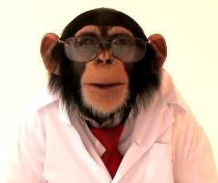 February 2012: Bluestone is still
monkeying with the CKCS.
Dr. Charles D. Bluestone,
renowned self-styled chimpanzee descendant, is still monkeying with the
Eustachian tubes of our cavaliers. (See
December 2010 entry and November
2011 entry.) In
a brief resume released by the University of Pittsburgh Medical
Center, he states:
February 2012: Bluestone is still
monkeying with the CKCS.
Dr. Charles D. Bluestone,
renowned self-styled chimpanzee descendant, is still monkeying with the
Eustachian tubes of our cavaliers. (See
December 2010 entry and November
2011 entry.) In
a brief resume released by the University of Pittsburgh Medical
Center, he states:
“Another ongoing research project involves a potential animal model, Cavalier King Charles Spaniel, which has a greater than 50% incidence of chronic otitis media effusion. Research being conducted with his colleagues in this animal involves histopathology of the middle-ear cleft in cadavers, and Eustachian tube function tests in live animals, conducted at the Ohio State University School of Veterinary Medicine in Columbus Ohio. ”
So now he tells us that the prevalence of PSOM in the CKCS is "greater than 50%". Each time he makes a public statement, the ante goes up, without ever providing any statistical data to support his assertions. This is much like the non-existent data supporting his claim to be descended from chimpanzees.*
* The notion of man's alleged descent from chimpanzees has long been rejected by evolutionists and creationists, alike. See, e.g.: "The common chimp (Pan troglodytes) and human Y chromosomes are 'horrendously different from each other', says David Page of the Whitehead Institute for Biomedical Research in Cambridge, Massachusetts." The fickle Y chromosome, Nature, Jan. 2010;463(14):149.
November 2011:
Bluestone keeps mucking up research of PSOM in the cavalier. In a video
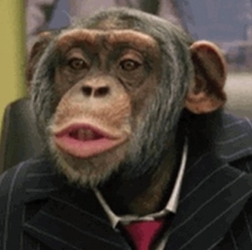 released in
November 2011 by the University of Pittsburgh Medical Center,
Dr. Charles D. Bluestone,
noted self-styled chimpanzee descendant, insists he still is
researching PSOM in the cavalier King Charles spaniel. (See
December 2010 entry.) He claims to be
basing his research on what in fact is the patently false premise that
the cavalier "has been bred over 300 years to have a short snout." He
goes on:
released in
November 2011 by the University of Pittsburgh Medical Center,
Dr. Charles D. Bluestone,
noted self-styled chimpanzee descendant, insists he still is
researching PSOM in the cavalier King Charles spaniel. (See
December 2010 entry.) He claims to be
basing his research on what in fact is the patently false premise that
the cavalier "has been bred over 300 years to have a short snout." He
goes on:
"So how did the Cavalier King Charles develop chronic persistent [sic] secretory otitis media? Their short snout changed the palatal anatomy. So the question is does the middle ear effusion with this very thick mucus in that Cavalier, it causes trouble by the way with hearing loss, have a consequence? There are two muscles that are related to this issue, the muscles that come from the palate, the tensor veli palatini, TVP, and the LVP muscles are altered causing the eustachian tube dysfunction, that’s our hypothesis. It’s a current research project which we are undergoing now."
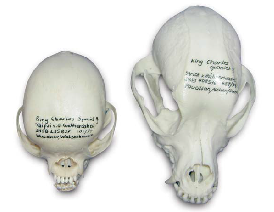 As
noted above, Dr. Bluestone also mis-names the disorder he claims to be
researching. PSOM stands for "primary secretory otitis media". He oddly
and repeatedly calls it "persistent" secretory otitis media. So
he does not even know the name of the disorder, and he is under the
abject mistaken belief that the cavalier "has been bred over 300 years
to have a short snout" when, in fact,
the breed was created less than 85 years ago, from the King Charles
spaniel, and has been bred to have a longer snout. Note in the
comparison of skulls at left: the King Charles spaniel skull is at the
far left, and the skull of the cavalier King Charles spaniel is to its
right.
As
noted above, Dr. Bluestone also mis-names the disorder he claims to be
researching. PSOM stands for "primary secretory otitis media". He oddly
and repeatedly calls it "persistent" secretory otitis media. So
he does not even know the name of the disorder, and he is under the
abject mistaken belief that the cavalier "has been bred over 300 years
to have a short snout" when, in fact,
the breed was created less than 85 years ago, from the King Charles
spaniel, and has been bred to have a longer snout. Note in the
comparison of skulls at left: the King Charles spaniel skull is at the
far left, and the skull of the cavalier King Charles spaniel is to its
right.
(Specimens from the collections of the Albert Heim Foundation, Museum of Natural History, Bern.)
He also has upped his stated prevalence of PSOM in the breed from "up to 40%" to a flat "50%", all without providing any statistical data to support either assertion.* Once again, we can only hope that this misguided gentleman's federal funding runs out as quickly as possible.
* Click here for some more interesting reading about Dr. Bluestone's credibility.
December
2010: Pitt Medical
Center's self-styled chimpanzee descendants muck up
PSOM study of cavaliers on taxpayers' dime.
Drs. Charles D. Bluestone
(left),
a human ear, nose, and
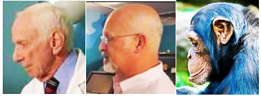 throat specialist, and J.
Douglas Swarts (middle), an anthropologist, both at the University of Pittsburgh
Medical Center, who apparently still harbor the disproven belief that
humans evolved from chimpanzees (They even include a tiresome skull
comparison of the two completely separate species, in profile, and
therefore, so do we; the chimpanzee is at far right), have also
gratuitously and erroneously concluded in their federally-funded
"research" article, Human
evolutionary history: Consequences for the pathogenesis of otitis media,
that "up to 40%" of cavalier King Charles spaniels have PSOM because
they have been "artificially selected to have a shortened front-to-back
diameter of the skull, a term called brachycephaly". This is an
utter (and easily provable) falsehood. So much for their
scientific method. It is more like their feces-tossing ancestors.
throat specialist, and J.
Douglas Swarts (middle), an anthropologist, both at the University of Pittsburgh
Medical Center, who apparently still harbor the disproven belief that
humans evolved from chimpanzees (They even include a tiresome skull
comparison of the two completely separate species, in profile, and
therefore, so do we; the chimpanzee is at far right), have also
gratuitously and erroneously concluded in their federally-funded
"research" article, Human
evolutionary history: Consequences for the pathogenesis of otitis media,
that "up to 40%" of cavalier King Charles spaniels have PSOM because
they have been "artificially selected to have a shortened front-to-back
diameter of the skull, a term called brachycephaly". This is an
utter (and easily provable) falsehood. So much for their
scientific method. It is more like their feces-tossing ancestors.
We suppose that if one must "publish or perish" (and with a taxpayer-funded grant, to boot), these two had to come up with something no one else would dare think to write about. But how do they get around the fact that PSOM is essentially unique to the cavalier, while many other canine breeds are brachycephalic, and some even more so? The answer is, they do not. There is a little value in their article, however, because Dr. Lynette Cole provided them with some quite accurate (albeit not new) information about PSOM in the cavalier, which is summarized below. Dr. Cole's input makes that part of the article worth reading.
But the scariest part of the article is their statement (threat?) near the end that:
"The underlying pathogenesis of the Cavalier's ME disease is currently under investigation in our laboratory."
We sure hope not. Leave that to Dr. Cole, please.
 October
2010: UK surgeon opines CKCS's brachycephalic disorders
and PSOM are related to Chiari-like malformation. Robert N.
White (right), a board certified veterinary soft tissue surgeon
practicing at Willows Veterinary Centre and Referral Service in
Solihull, West Midlands, observed in an
October 2010 presentation before a meeting of the UK's Association
of Veterinary Soft Tissue Surgeons, that the cavalier King Charles
spaniel does not appear to be a classically brachycephalic breed,
despite the extent of BAOS in the cavalier, and that the extent of both
PSOM and SM in the breed suggests that the CKCS may suffer from a
combination syndrome of the three disorders, all associated with
Chiari-like malformation.
October
2010: UK surgeon opines CKCS's brachycephalic disorders
and PSOM are related to Chiari-like malformation. Robert N.
White (right), a board certified veterinary soft tissue surgeon
practicing at Willows Veterinary Centre and Referral Service in
Solihull, West Midlands, observed in an
October 2010 presentation before a meeting of the UK's Association
of Veterinary Soft Tissue Surgeons, that the cavalier King Charles
spaniel does not appear to be a classically brachycephalic breed,
despite the extent of BAOS in the cavalier, and that the extent of both
PSOM and SM in the breed suggests that the CKCS may suffer from a
combination syndrome of the three disorders, all associated with
Chiari-like malformation.
September
2010:
UK
researchers find PSOM (OME) to be progressive.
 A team of UK
veterinary researchers (S.J. McGuinness, E.J. Friend, C.
Rusbridge (right), P. Knowler and N. D. Jeffery)
examined MRI scans of the skulls of 34 cavalier King Charles spaniels,
each of which had been scanned twice over periods from one month to 46
months. They concluded in their
report that PSOM (OME) is a progressive condition in the CKCS and
can progress from none to unilateral or bilateral; or from unilateral to
bilateral on sequential scans, and that OME is an acquired condition in
the CKCS and will not resolve spontaneously once it has developed. (See
also a
February 2013 entry on this same study.)
A team of UK
veterinary researchers (S.J. McGuinness, E.J. Friend, C.
Rusbridge (right), P. Knowler and N. D. Jeffery)
examined MRI scans of the skulls of 34 cavalier King Charles spaniels,
each of which had been scanned twice over periods from one month to 46
months. They concluded in their
report that PSOM (OME) is a progressive condition in the CKCS and
can progress from none to unilateral or bilateral; or from unilateral to
bilateral on sequential scans, and that OME is an acquired condition in
the CKCS and will not resolve spontaneously once it has developed. (See
also a
February 2013 entry on this same study.)
July 2010: UK researchers find association between PSOM and brachycephalic conformation in cavaliers. In a July 2010 article, UK investigators (Hayes GM, Friend EJ, Jeffery ND) studied the MRI scans of 68 cavaliers, 54% of them also being diagnosed with PSOM. They also observed that the brachycephalic characteristics of an abnormally thick palate and reduced naso-pharyngeal aperature were "significantly associated" with PSOM. They concluded that:
"These results suggest that auditory tube dysfunction and OME may represent a previously overlooked consequence of brachycephalic conformation in dogs."
March
2010: Ohio State Vet School needs cavalier cadavers for
Eustachian tube study. Dr. Lynette Cole of the
Dermatology Service at the Ohio State University's veterinary teaching
hospital is seeking deceased cavaliers and candidates for euthanasia,
with or without PSOM, for a study of the muscles which open the
Eustachian tube. The study will include dissections of the Eustachian
tube, its muscles, and the inner ear, and imaging with advanced MRI. The
veterinary research is in conjunction with an OSU medical school
otolaryngology study of secretory otitis media in humans. The
otolaryngologists are looking for abnormalities within the muscles that
open Eustachian tube, that prevent secretions from being removed from
the tube, thereby causing a build-up of the mucus causing secretory
otitis media.
Dr. Cole asks: "If at all possible, I would prefer
to have the cavalier brought to me at the Ohio State University for
euthanasia. Please contact me prior to make proper arrangements. If that
is not possible, please have your veterinarian refrigerate the dog after
euthanasia, and then have your veterinarian contact me." Contact
Dr. Cole at telephone number 614-292-3551, email
cole.143@osu.edu
September
2009:
Drs. Andrew Hillier and Lynette Cole
(right) and others of
the Dermatology Service at the Ohio State University's veterinary
teaching hospital are conducting research into the prevalence of PSOM in
the cavalier King Charles spaniel breed in the United States, as well as
the mode of inheritance, data about the clinical signs of the disorder,
alternative methods of diagnosing it (CT scan, tympanic bulla
ultrasonography, BAER test, impedance audiometry, otoscopic examination,
and pneumotoscopy), and methods of treatment. The diagnostic tests will
be compared to the results of the CT scan for the diagnosis of PSOM, to
determine which test or group of tests are the best to use for the
diagnosis of PSOM.
In their August 2007 interim report to the American Cavalier King Charles Spaniel Club's charitable trust, Dr. Cole reports that 75 Cavaliers are to be "enrolled, placed under general anesthesia, and the following diagnostics performed: CT scan, tympanic bulla ultrasonography, BAER test, impedance audiometry, otoscopic examination, and pneumotoscopy. If the CT scan is suggestive of otitis media (i.e. a soft tissue density present in the tympanic bulla-the bony part of the middle ear), then a myringotomy (incision into the eardrum) will be performed and the mucus flushed out of the middle ear. Cytology and bacterial cultures will be performed on the mucus from the middle ear. A BAER and CT scan will be performed post-middle ear flush on those CKCS with PSOM."
Dr. Cole states that as of August 2007, 13 CKCS have been examined, with ages ranging from 5 months to 9 years of age. Of those, 8 (62%) cavaliers had PSOM (6 had PSOM in both ears, 2 had PSOM in one ear) and 5 (38%) did not have PSOM.
The doctors still are seeking cavaliers to participate in this study. They may be reached by telephone at 614-292-3551 and email Dr. Hillier at hillier.4@osu.edu and Dr. Cole at cole.143@osu.edu and website www.vet.ohio-state.edu/876.htm The American Cavalier King Charles Spaniel Club's charitable trust has contributed a grant to help underwrite this project.
RETURN TO TOP
Related Links
RETURN TO TOP
Veterinary Resources
Insertion of a transtympanic ventilation tube for the teatment of otitis media with effusion. Cox C,J., Slack R.W.T., Cox G.J. J.Sm.Anim.Prac., Sept. 1989;30(9):517-519. Quote: In this paper, a case of otitis media with effusion ("glue ear") is described in a [2-year old] Cavalier King Charles spaniel. Its presentation, diagnosis and surgical management by the insertion of a trans-tympanic ventilation tube (grommet) is discussed. ... The diagnosis of glue ear in the absence of otitis externa in the dog described here is highly suggestive of eustachian tube dysfunction. Eustachian tube dysfunction prevents normal aeration of the middle ear cavity during swallowing. Thus the normal soluble gaseous contents of the middle ear cavity are absorbed, generating a subatmospheric pressure. A seromucinous effusion (glue) is formed which fills the middle ear cavity, and may become secondarily infected. This was the reason for the prolonged course of antibiotics at the start of the treatment. The grommet used in this case is retained in the tympanic membrane by its flanges, although in time the tympanic membrane heals, and extrudes the grommet into the external ear canal. This usually occurs between six and 18 months in man with this type of grommet. The recurrence of clinical signs following grommet extrusion in the dog indicates that the primary causative factor still remains. As symptomatic treatment was achieved with a grommet, a second one was inserted. Should the same sequence of events occur, the authors would suggest the insertion of a long term grommet, although this carries a higher risk of infection. The complications of grommet insertion include the development of otorrhoea, as occurred in this case.
Evaluation of radiography, otoscopy, pneumotoscopy, impedance audiometry and endoscopy for the diagnosis of otitis media in the dog. Cole LK, Kwochka KW, Podell M et al. August 2002. In: Advances in Veterinary Dermatology, volume 4. Thoday KL, Foil CS, Bond R, eds. Oxford: Blackwell Science; pp. 49–55. Quote: "45 dogs with chronic recurrent otitis externa were evaluated for otitis media using radiography, otoscopy, pneumotoscopy, tympanometry and the acoustic reflex. The results of these diagnostic tests were compared with the results of culture and cytology from the middle ear obtained by myringotomy. The diagnosis of otitis media was made if infection was detected by either method. Of the 90 ears evaluated, 80 (88.9%) ears had concurrent otitis media and the tympanic membrane was intact in 58 of 80 (72.5%) ears. 17 different organisms (Staphylococcus intermedius, Pseudomonas aeruginosa, Enterococcus, Proteus, Corynebacterium, Escherichia coli, Moraxella, Pseudomonas, Staphylococcus aureus, Pasteurella, Citrobacter, Lactobacillus, β-haemolytic streptococcus, group D non-enterococcus, α-haemolytic streptococcus, coagulase-negative staphylococcus and non-group D streptococcus) were isolated from the horizontal ear canal and 16 organisms from the middle ear. The high false-negative rates of all the tests resulted in low sensitivities and negative predictive values; therefore, many ears with otitis media were not identified and myringotomy appears to be the optimum technique for the diagnosis of otitis media. However, in the ears with an abnormal test, over 90% had otitis media. Therefore, otitis media should be suspected when an abnormal test is obtained radiographically, otoscopically, pneumotoscopically or by impedance audiometry in a dog with chronic recurrent otitis externa."
Primary secretory otitis media in the Cavalier King Charles spaniel: a review of 61 cases. Stern-Bertholtz W.; Sjöström L.; Wallin Håkanson N. J Small Animal Practice, 30 June 2003, 44(6): 253-256(4). Quote: "Sixty-one episodes of primary secretory otitis media (PSOM) were diagnosed in 43 Cavalier King Charles spaniels over a 10-year period. The principal findings were signs of moderate to severe pain localised to the head or cervical area, and/or neurological signs. Diagnosis was made by examination of the tympanic membrane and middle ear with the aid of an operating microscope under general anaesthesia. A bulging, but intact, tympanic membrane was found in most cases. he cytological and bacteriological examinations that were carried out in a few cases, and the macroscopic appearance of the contents of the middle ear, suggest that infection is not a part of the pathogenesis of PSOM. Following myringotomy, a highly viscous mucus plug was found filling the middle ear. Treatment, consisting of removal of the mucus plug, flushing of the middle ear, and local and systemic medical therapy, had to be repeated between one and five times. The prognosis was good in all cases. PSOM is an important differential diagnosis in Cavalier King Charles spaniels with signs of pain involving the head and neck, and/or neurological signs."
Neurological signs and results of magnetic resonance imaging in 40 cavalier King Charles spaniels with Chiari type 1-like malformations. Lu D, Lamb CR, Pfeiffer DU, Targett MP. Vet Rec. Aug 2003;153(9):260-3. Quote: "In human beings a Chiari type 1 malformation is a developmental condition characterised by cerebellar herniation and syringohydromyelia. Abnormalities compatible with such a malformation were identified by magnetic resonance imaging in 39 cavalier King Charles spaniels with neurological signs and in one neurologically normal cavalier King Charles spaniel that was examined postmortem. The dogs with these abnormalities had a wide variety of neurological signs, but there was no apparent correlation between the neurological signs and the severity of cerebellar herniation, syringohydromyelia or hydrocephalus."
Material in the middle ear of dogs having magnetic resonance imaging for investigation of neurologic signs. Owen MC, Lamb CR, Targett MP. Vet. Radiology & Ultrasound, Mar 2004, 45(2):149-155. Quote: "The aim of this study was to determine the prevalence and potential significance of finding material in the middle ear of dogs having magnetic resonance (MR) imaging. Of 466 MR studies reviewed, an increased signal was identified in the tympanic bulla in 32 (7%) dogs. Cavalier King Charles spaniels, Cocker spaniels, Bulldogs, and Boxers were over-represented compared to the population of dogs having MR imaging. Five (16%) dogs had definite otitis media and one (3%) had a meningioma invading the middle ear. Of the remaining dogs, 13 (41%) had possible otitis media and 13 (41%) had neurologic conditions apparently unrelated to otitis media. The most common appearance of material in the middle ear was isointense in T1-weighted images and hyperintense in T2-weighted images. There was no apparent correlation between the signal characteristics of the material and the diagnosis. Enhanced signal after gadolinium administration was observed affecting the lining of the bulla in dogs with otitis media and in dogs with unrelated neurologic conditions. In dogs without clinical signs of otitis media, finding an increased signal in the middle ear during MR imaging may reflect subclinical otitis media or fluid accumulation unrelated to inflammation. Brachycephalic dogs may be predisposed to this condition."
Primary secretory otitis media in Cavalier King Charles spaniels. Clare Rusbridge. J Small Anim Pract. 2004 Apr; 45:222.
Secretory otitis media in cavalier King Charles spaniels. Advances in Sm. Anim. Med. & Surg. August 2004;17(8):6-7.
Primary Secretory Otitis Media (Glue Ear). Hillier A., Cole L. CKCSC,USA Bulletin, Fall/Winter 2005:16.
Diagnosis and management of otitis media. Thomas, Randall C. Proceedings, No. Am. Vet. Conf., Vol. 20, Jan. 2006: 979.
Diagnostic imaging of ear disease in the dog and cat. Livia Benigni, Chris Lamb. In Practice. March 2006;28:122-130. Quote: Most dogs and cats with common aural conditions, such as otitis externa or aural haematoma, may be treated satisfactorily without the need for diagnostic imaging. However, animals with recurrent or severe otitis, and those with more marked signs, such as para-aural swelling, pain on opening the mouth, vestibular syndrome or facial paralysis, may benefit from a more thorough work-up to examine the middle ear and adjacent structures. In such cases, the anatomical complexity and relative inaccessibility of these structures is best addressed by diagnostic imaging – involving, as appropriate, radiography, computed tomography, magnetic resonance imaging or ultrasonography. This article reviews the use of these imaging techniques in dogs and cats with clinical signs of ear disease and illustrates some of the more typical findings. ... In dogs, the tympanic cavity is usually quite large and rounded, although in some breeds (eg, Cavalier King Charles spaniel, bulldog) it is smaller and flatter.
Contrast-enhanced Computed Tomographic Imaging of the Auditory Tube in Mesaticephalic Dogs. Lynette K. Cole, Valerie F. Samii. Vet. Radiology & Ultrasound, Vol 48(2): 125-128, Mar 2007. Quote: "Auditory tube dysfunction has been speculated as the cause of primary secretory otitis media (PSOM), reported recently in the Cavalier King Charles spaniel. A simple, noninvasive technique is needed for evaluation of the canine auditory tube."
The method of application and short term results of tympanostomy tubes for the treatment of primary secretory otitis media in three Cavalier King Charles Spaniel dogs. Corfield GS, Burrows AK, Imani P, Bryden SL. Aust Vet J. March 2008; doi: 10.1111/j.1751-0813.2008.00254.x. Quote: Primary secretory otitis media is an uncommon disease affecting predominantly Cavalier King Charles Spaniel dogs. Current treatment recommendations include repeated manual removal of the mucoid effusion from the tympanic cavity through a myringotomy incision and topical or systemic corticosteroids. The aim of this study was to assess the efficacy of tympanostomy tubes to provide continual tympanic cavity ventilation and drainage for the treatment of primary secretory otitis media in three dogs. Tympanostomy tubes were placed within a myringotomy incision in the pars tensa with the aid of an operating microscope. Clinical signs resolved rapidly in all cases following the procedure and all cases were asymptomatic at the time of follow-up, 8, 6 and 4 months later. Results of this study indicate that tympanostomy tubes provide continual tympanic cavity ventilation and drainage and may be an acceptable alternative to repeated myringotomy for the treatment of primary secretory otitis media.
A Practical Guide to Canine and Feline Neurology. Curtis W. Dewey. John Wiley & Sons; 2008; 273.
Secretory otitis media in cavalier King Charles spaniels. Advances in Sm. Anim. Med. & Surg. February 2009;22(2):8. Quote: "Primary secretory otitis media (PSOM) is an uncommon idiopathic disease of cavalier King Charles spaniels. Clinical signs include pain of the head or neck, or both. Episodes of vocalization and a guarded neck carriage can occur. Some cases have pruritus of the pinnae and external ear canals without signs of otitis externa. Hearing may be impaired, and head tilt, nystagmus, ataxia, facial paralysis, and fatigue may be present. Myringotomy with removal of a typically viscous, opaque, mucous effusion from the tympanic bulla is required for a definitive diagnosis."
Effect of Middle Ear Effusion On The Brainstem Auditory Response Of Cavalier King Charles Spaniels. T. R. Harcourt-Brown, J. E. Parker, N. D. Jeffery. 22nd ESVN Annual Symposium, Sept. 2009; J Vet Intern Med, Jan/Feb 2010;24(1):242. Quote: "The purpose of this study was to investigate the effect of middle ear effusion on the Brainstem Auditory Evoked Reponses (BAER) of [23] cavalier king charles spaniels. BAER’s were obtained from dogs following Magnetic Resonance (MR) imaging screening for syringomyelia. Middle ear effusion was diagnosed if the auditory bulla was completely filled with material that was isontinense to brain parenchyma on T1 weighted images and hyperintense on T2 weighted images. Dogs with otitis externa were excluded from the study. BAER’s were obtained at sound intensities ranging from 10 to 100 dB nHL (normal hearing level). The BAER threshold was determined for each ear as the last trace that showed a recognisable wave V. The latency of wave V was recorded for each intensity where it was identified and the interwave latency between waves I and V was calculated at 90 dB nHL. ... Each dog’s hearing was considered normal by their owner. The median BAER threshold was 60 dB for ears with effusion and 30 dB for those without. The proportion of ears with abnormal BAER thresholds (4 30 dB nHL) was greater for ears with effusion (11/16) than those without (8/30) (Fishers exact test, p = 0.011). Severity of hearing loss for ears with effusion was calculated by linear regression of wave V latencies to be 23 dB (95% confidence 18 to 31 dB). The mean interwave latency between waves I and V at 90 dB for ears with and without effusion was not significantly different (Students t-test, p > 0.05). These data show that middle ear effusion is associated with conductive hearing loss of 21–30 dB in affected ears."
Relationship between pharyngeal conformation and otitis media with effusion in Cavalier King Charles spaniels. Hayes GM, Friend EJ, Jeffery ND. Vet Rec. July 2010;167(2):55-8. Quote: "Otitis media with effusion (OME) is a common incidental finding in otherwise normal Cavalier King Charles spaniels (CKCS). ... The incidence of OME as an incidental finding in a sample of 68 CKCS undergoing MRI was 54 per cent in this study, which is comparable to previously reported incidences of 47 per cent ... and 28 per cent ... in this breed. The CKCS in this study were reported to be ‘clinically normal’ by their owners, who did not report clinical signs of neurological disease or otitis in these dogs. ... In this study, measurements made on MRI were used to determine whether there was an association between OME and brachycephalic conformation. The results confirm that association and also demonstrate that, in CKCS, greater thickness of the soft palate and reduced naso-pharyngeal aperture are significantly associated with OME. ... An overlong soft palate has long been accepted as contributing to the [brachycephalic airway] syndrome, but more recently the importance of abnormally thick soft palates has also been recognised ... [B]rachycephalic airway syndrome may occur as a consequence of the selection for morphological neotony in this breed ... Although the aetiology is probably multifactorial, OME is more frequently found in patients with more severe anomalies of nasopharyngeal conformation. Changes within the nasopharynx may impair auditory tube drainage. ... These results suggest that auditory tube dysfunction and OME may represent a previously overlooked consequence of brachycephalic conformation in dogs."
Breed Predispositions to Disease in Dogs & Cats (2d Ed.). Alex Gough, Alison Thomas. 2010; Wiley-Blackwell Publ. 53.
Progression of Otitis Media with Effusion in the Cavalier King Charles Spaniel. S.J. McGuinness, E.J. Friend, C. Rusbridge, P. Knowler and N. D. Jeffery. Abstract at 23d ECVN symposium, Sept. 2010. Investigation of 34 MRI scans of CKCS having 2 scans as part of CMSM screening. Interpreted by the same author (S.J. McGuinness). The scans were classified according to negative, unilateral or bilateral OME based on the absence or presence of hyperintense material on T2 weighted MRI images. ... A total of 34 dogs had 2 MRI scans a median of 20 months apart (range 1 to 46); the incidence of OME at the first MRI was 32%, which increased to 50% at the time of the second MRI, which is comparable with previous studies. At the time of the first MRI, 23 CKCS had no effusion, 10 had unilateral effusion and 1 had bilateral effusion. By the second MRI, 3 cases had progressed from unilateral to bilateral; 5 had progressed from no effusion to effusion being present (4 negative to unilateral, 1 negative to bilateral) and 8 of those having effusion at the first scan remained the same. No cases that had OME at the first MRI had resolved at the time of the second MRI and no case had clinical signs that could be associated with OME. This study demonstrates that OME is a progressive condition in the CKCS and can progress from absent to unilateral or bilateral; or from unilateral to bilateral on sequential scans. It also suggests that OME is an acquired condition in the CKCS and will not resolve spontaneously once it has developed."
Brachycephalic Airway Obstructing Syndrome: Some Further Controversies. Robert N. White. Assn. of Veterinary Soft Tissue Surgeons. Oct. 2010. Quote: "The Cavalier King Charles spaniel also warrants further discussion. Previous reports from the UK and Australia would suggest that this breed in commonly presented for the investigation and management of BAOS (Lorinson and others 1997, Torrez and Hunt 2006) in both these countries. Surprisingly, a recent retrospective series of 62 dogs from North America (Riecks and others 2007) contained no individuals of the breed. Like the three breeds discussed above [Yorkshire terrier, the Norfolk terrier and the Norwich terrier], it is my experience that the Cavalier King Charles spaniel presented for the further investigation of ʻBAOSʼ commonly does not have the classic findings seen in the bulldog, French bulldog, Pekinese, Pug, etc. Their nasopharyngeal obstruction is often characterised by a subjectively narrow (smaller than expected for a breed of their size) nasopharyngeal space and it is quite common that they do not show evidence of overlength of the soft palate. Interestingly, a recent report (Hayes and others 2010) confirmed a significant association between the presence of otitis media with effusion (on MRI) and an increase thickness of the soft palate and reduced nasopharyngeal aperture. Many of the cases show evidence of otitis media with effusion (OME) on either otoscopic examination or other imaging studies of the tympanic bullae. Malformation of the nasopharynx and soft palate is recognised to be associated with the formation of otitis media with effusion in the dog (White and others 2009). The prevalence of caudal fossa (craniocervical junction abnormalities including occipital hypoplasia) malformations is high in the Cavalier King Charles spaniel and is considered to be associated with the presence of neurological signs observed in individuals suffering from cerebellar herniation and syringohydromyelia (Cerda-Gonzalez and others 2009). It would seem reasonable to hypothesise that the Cavalier King Charles spaniel might suffer from a syndrome of conditions (syringomyelia, OME, nasopharyngeal airway obstruction, etc.) that are all associated with the malformation of the caudal aspect of the skull."
Human evolutionary history: Consequences for the pathogenesis of otitis media. Charles D Bluestone, J. Douglas Swarts. Otolaryngology -- Head & Neck Surgery; Dec 2010; 143(6):739-744. Quote: "Among veterinarians, chronic ME effusion (termed primary secretory otitis media) is a well-known disease in the Cavalier King Charles Spaniel. It has been reported to be present in up to 40 percent of these animals. The effusion is mucoid and fills the entire ME. Diagnosis is made by operating microscopic examination, computed tomography scanning, or magnetic resonance imaging (MRI), and has been confirmed at the time of myringotomy. Myringotomy and tympanostomy tube placement has been recommended for treatment. This breed has been artificially selected to have a shortened front-to-back diameter of the skull, a shape termed brachycephaly, which arises due to premature fusion of the coronal sutures. The term neotenous (retention of juvenile characteristics into adulthood) is also appropriate for these breeds. The Cavalier snores habitually like other brachycephalic dogs, including the English Bulldog, a breed that has been reported to be the only animal known to develop obstructive sleep apnea. The snoring is undoubtedly secondary to its constricted pharynx, a consequence of the shortening of the snout. ... Figure 5 compares the head shape of a Cavalier King Charles Spaniel, with its extremely short face, to that of a Golden Retriever, which has a classic prognathic snout. The Cavalier King Charles Spaniel is an animal model of chronic OM with effusion. It has been “artificially selected” (Charles Darwin's term) for its short snout and globular head, but an unintended consequence of breeding for this characteristic is the propensity for chronic OM with effusion. In a recently reported study using MRI, veterinarians from England found that not only did the Cavalier have OM (54%), but another brachycephalic breed, the Boxer, also had ME disease (32%), which was not present in Cocker Spaniels, a mesaticephalic breed. The investigators suggested that the reduced nasopharyngeal space in the Cavalier and Boxers, when compared with the Cocker Spaniel, predisposed them to OM. It might be that one or both of the paratubal muscles is dysfunctional due to the abnormal palatal anatomy in these breeds and is the cause of their OM. The underlying pathogenesis of the Cavalier's ME disease is currently under investigation in our laboratory. Analogously, one could speculate that with the loss of their prognathic face, humans became susceptible to OM, an unintended consequence of “natural selection” for another adaptation (again, Darwin's term), as described above."
Middle ear effusions in dogs: An incidental finding? H.A. Volk, E.S. Davies. Vet.J. June 2011; 188(3):256-257
Effect of middle ear effusion on the brain-stem auditory evoked response of Cavalier King Charles Spaniels. Thomas R. Harcourt-Brown, John E. Parker, Nicolas Granger, and Nick D. Jeffery. Vet.J. June 2011; 188(3):341-345. Quote: "Brain-stem auditory evoked responses (BAER) were assessed in 23 Cavalier King Charles Spaniels with and without middle ear effusion at sound intensities ranging from 10 to 100 dB nHL. Significant differences were found between the median BAER threshold for ears where effusions were present (60 dB nHL), compared to those without (30 dB nHL) (P = 0.001). The slopes of latency–intensity functions from both groups did not differ, but the y-axis intercept when the x value was zero was greater in dogs with effusions (P = 0.009), consistent with conductive hearing loss. Analysis of latency–intensity functions suggested the degree of hearing loss due to middle ear effusion was 21 dB (95% confidence between 10 and 33 dB). Waves I–V inter-wave latency at 90 dB nHL was not significantly different between the two groups. These findings demonstrate that middle ear effusion is associated with a conductive hearing loss of 10–33 dB in affected dogs despite the fact that all animals studied were considered to have normal hearing by their owners."
Diagnosis of primary secretory otitis media in the cavalier King Charles spaniel. VCole LK, Samii VF, Wagner S, and PJ Rajala-Schultz. Dermatol April 2011;22:297.
Genetic Connection: A Guide to Health Problems in Purebred Dogs, Second Edition. Lowell Ackerman. July 2011; AAHA Press; pg 59. Quote: "Primary secretory otitis media (PSOM), sometimes referred to as glue ear, is a poorly defined disorder of the middle ear that is most commonly reported in the Cavalier King Charles spaniel. ... The cause of PSOM has not been determined, but because of the predominance of Cavalier King Charles spaniels affected, a genetic link is suspected."
Primary Secretory Otitis Media in Cavalier King Charles Spaniels. Lynette K. Cole. Vet Clin Small Anim; Oct. 2012.;42(6):1137-1142. Quote: "Primary secretory otitis media (PSOM) is a disease that has been described in the Cavalier King Charles spaniel (CKCS). Signs suggestive of primary secretory otitis media include hearing loss, neck scratching, otic pruritus, head shaking, abnormal yawning, head tilt, facial paralysis, or vestibular disturbances, with an individual Cavalier King Charles spaniel presenting with one or multiple signs. A large, bulging pars flaccida identified on otoscopic examination confirms the diagnosis. However, in many CKCS with PSOM the pars flaccida is flat, and radiographic imaging is needed to confirm the diagnosis. Current treatment for PSOM includes performing a myringotomy into the caudal-ventral quadrant of the pars tensa with subsequent flushing of the mucus out of the bulla using a video otoscope. Until a treatment is found to prevent the mucus from recurring or the cause of the disease is identified and controlled, repeat myringotomies and flushing of the middle ear will be needed to keep the middle ear free of mucus. At present, the cause of PSOM is unknown; however, it has been speculated to be due to a dysfunction of the middle ear or auditory tube (increased production of mucus in the middle ear) or decreased drainage of the middle ear through the auditory tube, or both. Until a treatment is found to prevent the mucus from recurring or the cause of the disease is identified and controlled, the fact that these CKCS had a relapse of PSOM is not surprising."
Electrodiagnostic Evaluation of Auditory Function in the Dog. Peter M. Scheifele, John Greer Clark. Vet.Clinics of N.A.:Sm.Anim.Prac. Nov. 2012; 42(6):1241-1257. Quote: "Given the high incidence of deafness within several breeds of dogs, accurate hearing screening and assessment is essential. In addition to brainstem auditory evoked response (BAER) testing, 2 other electrophysiologic tests are now being examined as audiologic tools for use in veterinary medicine: otoacoustic emissions and the auditory steady state response (ASSR). To improve BAER testing of animals and ensure an accurate interpretation of test findings from one test site to another, the establishment of and adherence to clear protocols is essential. The ASSR holds promise as an objective test for rapid testing of multiple frequencies in both ears simultaneously."
Progression of otitis media with effusion in the Cavalier King Charles spaniel. S. J. McGuinness, E. J. Friend, S. P. Knowler, N. D. Jeffery, C. Rusbridge. Vety.Rec. Feb. 2013. Quote: "Previously, the term ‘Primary secretory otitis media’ (PSOM) has been used to describe an effusion present in the tympanic bullae of Cavalier King Charles Spaniels (CKCS) (Stern-Bertholtz and others 2003). All these cases were associated with moderate to severe pain and/or neurological signs. As no cases in our study had associated pain, and due to the chronicity of the condition, the authors have classified the condition described in this study as OME [otitis media with effusion]. This study aimed to determine if, once present, OME resolved without treatment; and also if OME in the CKCS was a progressive condition. ... At the time of the first MRI scan, 23 CKCS had no effusion, 10 had unilateral effusion and one had bilateral effusion. By the second MRI scan, nine cases had progressed (26.5 per cent), three from unilateral to bilateral effusion; six to acquire an effusion (five negative to unilateral effusion; one negative to bilateral effusion). Eight CKCS having effusion in the bullae at the first MRI scan remained with this effusion (either unilateral or bilateral) at the time of the second MRI scan (Fig 2). No cases that had OME at the first MRI had resolved at the time of the second MRI. No dog had a hearing test as part of the investigation. ... The incidence of OME in CKCS in this study was 32 per cent at the first MRI scan and 50 per cent at the second MRI scan and is comparable with previous studies (Owen and others 2004, Hayes and others 2010). In this study, six cases progressed from having no effusion to either unilateral (five cases) or bilateral (one case) OME. ... The dogs reported in [previous] studies were described as having severe clinical signs including vocalising and pain localised to the head or cervical area; none of these cases underwent an MRI scan at the time of treatment, and it is possible that the clinical signs seen in these cases could be explained by undiagnosed syringomyelia. ... The possibility of extrusion of the tympanostomy tube (Cox and others 1989), the inability to allow the dog’s ear to become wet and the possibility for tracking infections make the tympanostomy tube less suitable for long-term use in the CKCS, as the anatomical changes that are the likely cause of the OME are present in the adult (Hayes and others 2010). In conclusion, this study suggests that OME is an acquired and progressive condition in the CKCS and will not resolve spontaneously once it has developed. As the clinical signs associated with OME are minimal, it is unclear if treatment is required. If treatment was required in any one case, an alternative method to repeated myringotomies or tympanostomy tube placement needs to be developed in order to treat the hearing loss associated with this condition.
Neck Stiffness & Head Tilt in a Young Spaniel. Louis N. Gotthelf. Clinician's Brief. March 2013: 23-25. Quote: "A 9-month-old female Cavalier King Charles spaniel presented for apparent neck pain when walked on a leash. History: The owner reported neck stiffness and head tilt. After the leash had been removed, the signs continued to persist for more than 24 hours. The puppy appeared bright, alert, and responded when called. Examination: Physical examination did not elicit a painful response when the head and neck were manipulated in any direction. A slight head tilt was observed when the puppy walked freely in the clinic; however, laboratory diagnostics were not performed. Evaluation may reveal a bulging eardrum, indicative of primary secretory otitis media (PSOM). PSOM of Cavalier King Charles spaniels (CKCSs) commonly occurs as head and neck pain that may be difficult to localize. With pressure in the bulla, Horner syndrome or facial nerve palsy may be evident. Vestibular disease may be present with increased bulla pressure and can manifest as nystagmus or head tilt. Breed-Specific Considerations: Careful examination can reveal an abnormal or bulging eardrum. Clinicians adept at visualizing a normal eardrum with a handheld otoscope can detect a bulging eardrum; the tip of the cone has to be small yet long enough to make the bend in the ear canal. The tip of the otoscope cone should be placed in the horizontal ear canal close enough to the eardrum to focus on the tympanic membrane. A highly magnified, well illuminated examination should be done with a video otoscope to determine eardrum status. Bottom Line: CKCS eardrums should always be evaluated. If PSOM is suspected, a high-magnification eardrum examination may require referral. PSOM can be asymptomatic in CKCSs, in which case myringotomy is not required; however, the owner should be educated to take appropriate steps if signs are displayed. The Take-Home: CKCSs often have a genetic defect of their eustachian tube that results in PSOM, which may be asymptomatic. Bulging eardrum and ear pain are typical of PSOM in CKCSs. PSOM should be differentiated from SM via eardrum evaluation and MRi (if necessary). Myringotomy can relieve PSOM signs. Antibiotics will not be helpful. Periodic treatment is required."
Use of a hearing loss grading system and an owner-based hearing questionnaire to assess hearing loss in pet dogs with chronic otitis externa or otitis media. Carly L. Mason, Susan Paterson1, Peter J. Cripps. Vet. Dermatology; Oct. 2013;24(5):512–e121. Quote: "Background: Hearing loss is important when assessing the suitability of dogs with otitis externa/media for medical or surgical therapy. Hypothesis/objectives: To assess an owner-completed questionnaire as an indicator of hearing loss and a canine hearing loss scoring system in chronic canine otitis. Animals: One hundred hospital population dogs referred for chronic otitis investigation [including 6 cavalier King Charles spaniels]. Methods: Owners completed a questionnaire to assess their dog's response to common household noises. The presence of otitis externa or media was determined and brainstem auditory-evoked response measurements were performed on each dog. The minimal hearing threshold (MHT) in decibels normal hearing level (dB NHL) was recorded and categorized according to the human World Health Organization grading system into five grades from 0 to 4 with cut-off values of ≤25 dB NHL, 26–40 dB NHL, 41–60 dB NHL, 60–80 dB NHL and ≥81 dB NHL. Results: The questionnaire correctly determined normal hearing in grade 0 cases, but did not reliably detect unilateral or grade 1 bilateral hearing loss. For dogs with bilateral hearing loss ≥ grade 2, questionnaire sensitivity was 83% [24 of 29, 95% confidence interval, (CI) 64–94%] and specificity was 94% (67 of 71, 95% CI 86–98%). Higher grades of hearing loss were significantly associated with the presence of otitis media (P < 0.01). Conclusions and clinical importance: The questionnaire may be a useful in-practice screening tool in chronic canine otitis for moderate to severe bilateral hearing deficits (MHT ≥41 dB NHL). The hearing loss grading system may help clinicians make therapeutic decisions. Chronic otitis media may be associated with higher grades of hearing loss."
CASE REPORT: Primary Sectetory Otitis Media in a Cavalier King Charles Spaniel. Downs Veterinary Practice. Quote: "Bilateral myringotomy was performed and the bullae were flushed with sterile saline; large amounts of yellow tinged mucus were removed from both bulla and sent for cytology and bacteriology. The dog was treated post operatively with meloxicam and amoxiclav pending culture results. The owners were contacted one week after surgery and reported an improvement in the dog’s hearing. ... PSOM can cause deafness, neck pain and vestibular disease. It occurs predominantly in Cavalier King Charles Spaniels, and should be considered as a differential diagnosis along with syringomyelia, cervical disc disease and progressive hereditary deafness. ... Diagnosis of PSOM can be challenging. In one report an operating microscope was used to examine the tympanic membrane as it can be difficult to accurately assess opacity of the tympanic membrane or changes in pressure with an auroscope. In this case, MRI clearly revealed bilateral fluid accumulation in the bullae. When presenting signs include head and neck pain, this imaging modality is invaluable in differentiating PSOM from syringomyelia secondary to Chiari malformation. Treatment of PSOM is surgical; myringotomy with subsequent flushing of the bulla is the standard recommendation. In addition, steroids have reportedly been used in conjunction with lavage of the bullae. In some cases, repeated surgeries may be required as the underlying abnormality leads to a potential recurrence of signs."
A Case of Primary Secretory Otitis Media in a Cavalier King Charles Spaniel. Takehiro Hasegawa. Jpn. J. Vet. Dermatol. March 2014; doi: 10.2736/jjvd.20.91. Quote: A 1-year-old spayed female Cavalier King Charles spaniel (CKCS) presented with right aural pruritis. A bulging pars flaccida was identified on otoscopic examination with no evidence of otitis externa noted. Hyperintense tissue was identified in the right bulla on T2 weighted MRI images. A myringotomy and video otoscopy were performed, and highly viscous, gray mucous discharged from the right tympanic bulla. Two weeks following the procedure, the pruritic behavior was decreased, and based on this progress, the dog was diagnosed with primary secretory otitis media (PSOM).
Intermittent Pain & Scratching in a Cavalier King Charles Spaniel. Megan Stout Steele, Jonathan Levine. Clinicians Brief. May 2014. Quote: "Lucy, a 7-year-old spayed Cavalier King Charles spaniel, was presented for chronic intermittent cervical pain and scratching of the head and neck. History: ... Lucy had a 2- to 3-year history of intermittent episodes of vocalization and reluctance to be touched around the head and neck. In addition, the dog frequently attempted to scratch her head, neck, muzzle, and ears, although rarely did the paws come into contact with the skin surface. During the past year, the dog had progressive difficulty using her pelvic limbs on slippery surfaces. The owners also suspected some hearing loss. Examination: ... Bilateral mild ceruminous otic debris was noted. The dog’s mentation appeared normal and no cranial nerve deficits were noted; however, response to auditory stimuli was absent bilaterally. On gait analysis, mild generalized proprioceptive ataxia, cerebellar ataxia, and tetraparesis were noted. Myotatic reflexes were increased in the pelvic limbs, and thoracic limb flexor withdrawal reflexes were diminished bilaterally. Cervical pain and phantom scratching were elicited during vertebral column palpation. Lesions were localized to the C6-T2 spinal cord segments, cerebellum, and auditory apparatus. Diagnostics: CBC, serum chemistry panel, and urinalysis results were within reference ranges. Three-view thoracic radiographs showed no abnormalities. MRI of the brain and cranial cervical vertebral column showed cerebellar compression and coning secondary to the malformed caudal occipital bone, with lack of normal cerebrospinal fluid (CSF) surrounding the caudal cerebellum. A T2 hyperintense–T1 hypointense lesion within the spinal cord parenchyma from the level of C1 caudally was noted. The tympanic bullae contained T2-hyperintense and T1-isointense material. No contrast enhancement was identified. Diagnosis: Chiari-like malformation (CLM) and syringohydromyelia (SHM); bilateral primary secretory otitis media (PSOM). Treatment: Lucy was admitted for additional diagnostic testing and treatment and underwent myringotomy and bilateral deep ear flushing. Large, clear mucus plugs were removed from both middle ears; culture of the plugs was negative for bacteria. These findings and the MRI results were most consistent with PSOM. The patient recovered well from anesthesia and the next day was reanesthetized for foramen magnum decompression and C1 dorsal laminectomy with durotomy. Surgical recovery was uncomplicated. After 3 days, the patient was discharged with pregabalin at 2 mg/kg PO q12h for neuropathic pain associated with SHM, omeprazole at 1 mg/kg PO q24h to decrease CSF production, and tramadol at 4 mg/kg PO q8h for 5 days to address temporary soft tissue pain. In addition, N-acetylcysteine at 600 mg PO q24h was prescribed as a mucolytic to lower the risk for PSOM recurrence. Outcome: At the 1-month reevaluation, the dog’s hearing seemed to have improved, but her gait abnormalities and phantom scratching persisted."
Handheld tympanometer measurements in conscious dogs for the
evaluation of the middle ear and auditory tube. GM Strain,
AJ Fernandes. Vet. Dermatology. June 2015;26(3):193-e40. Quote: "Background:
... Evaluation of the patency of the tympanum can be very difficult
in infected ears; assessment of the middle ear and auditory tube are
difficult without expensive imaging techniques. The availability of
a low-cost method of assessing the tympanum, middle ear and auditory
tube would be a significant tool for practitioners. Primary
secretory otitis media (PSOM), also known as glue ear, is a
condition observed primarily in the Cavalier King Charles
spaniel (CKCS) dog breed, but also reported in the
dachshund, boxer and shih tzu. Incidence in clinically normal
CKCS may be as high as 54%. Primary secretory
otitis media is similar to the condition of otitis media with
effusion that is frequently seen in young children. With PSOM, the
middle ear becomes filled with a sterile, viscous plug of mucus that
typically must be removed by myringotomy and middle ear flushing,
often repeatedly, and in some cases by bulla osteotomy. Aetiologies
of PSOM are unknown; a pharyngeal conformational predisposition has
been suggested, and auditory (Eustachian) tube dysfunction has also
been proposed. Due to its nearly exclusive occurrence in
CKCS, there may also be a genetic component. Deafness may
result from loss of neurons in the cochlea (sensorineural deafness)
or from obstruction of sound transmission into the cochlea
(conductive deafness). Conductive deafness may result from a variety
of causes, including otitis externa and/or media, cerumen impaction,
ear canal inflammation, ear canal foreign bodies (awns) and middle
ear polyps. Tympanometry, or impedance audiometry, is a noninvasive
method of examining the function of the middle ear while varying the
atmospheric pressure in the external ear canal and inferring the
amount of sound energy that is transmitted through the tympanum by
measuring the reflected sound energy. ... Hypothesis/Objectives: The
objective was to develop techniques for tympanometry testing in
conscious dogs and to
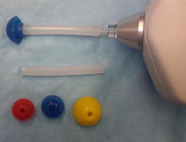 present normative data for clinical use of
this equipment to enable assessment of the tympanum, middle ear and
auditory tube. Animals: Sixteen hounds (14 female) from a school
teaching colony. Methods: Dogs were gently restrained in a standing
position. After cleaning of the ear canal, a tympanometer probe tip
extension was placed in the vertical canal and automated testing
performed using a handheld device. Both ears were tested in all
dogs. [See, at right, Otowave 102 tympanometer with eartip
modified for dog use, showing 14, 15 and 19 mm mushroom-shaped
eartips.] Results: Acceptable recordings were obtained from both ears of
13 dogs, from one ear in each of two dogs and from neither ear of
one dog, resulting in data from 28 of 32 (88%) ears. Otoscopic
examination confirmed the absence of inflammation or any other
obvious explanation for the noncompliant dogs. No significant
differences were seen between ears for any measure. Normative data
are reported for peak compliance, peak compliance pressure, gradient
and ear canal volume. ... The sensitivity and specificity of
tympanometry for the diagnosis of PSOM in Cavalier King
Charles spaniels were 84 and 47%, respectively (L. Cole,
personal communication), results that are better than those for
suspected otitis media cases. ... Conclusions and clinical
importance: Tympanograms can be recorded in conscious dogs to assist
in the evaluation of the middle ear structures. ... Clinical studies
of conscious dogs with otitis media, perforated tympanums, PSOM or
auditory tube disorders need to be performed to validate the
clinical usefulness of these recordings."
present normative data for clinical use of
this equipment to enable assessment of the tympanum, middle ear and
auditory tube. Animals: Sixteen hounds (14 female) from a school
teaching colony. Methods: Dogs were gently restrained in a standing
position. After cleaning of the ear canal, a tympanometer probe tip
extension was placed in the vertical canal and automated testing
performed using a handheld device. Both ears were tested in all
dogs. [See, at right, Otowave 102 tympanometer with eartip
modified for dog use, showing 14, 15 and 19 mm mushroom-shaped
eartips.] Results: Acceptable recordings were obtained from both ears of
13 dogs, from one ear in each of two dogs and from neither ear of
one dog, resulting in data from 28 of 32 (88%) ears. Otoscopic
examination confirmed the absence of inflammation or any other
obvious explanation for the noncompliant dogs. No significant
differences were seen between ears for any measure. Normative data
are reported for peak compliance, peak compliance pressure, gradient
and ear canal volume. ... The sensitivity and specificity of
tympanometry for the diagnosis of PSOM in Cavalier King
Charles spaniels were 84 and 47%, respectively (L. Cole,
personal communication), results that are better than those for
suspected otitis media cases. ... Conclusions and clinical
importance: Tympanograms can be recorded in conscious dogs to assist
in the evaluation of the middle ear structures. ... Clinical studies
of conscious dogs with otitis media, perforated tympanums, PSOM or
auditory tube disorders need to be performed to validate the
clinical usefulness of these recordings."
Primary Secretory Otitis Media in the Cavalier King Charles spaniel: Outcomes and recurrence of clinical signs after myringotomy and tympanostomy procedures. Bolder, N. Utrecht University. April 2015. Quote: "In this study, the data of 27 Cavalier King Charles spaniels diagnosed with Primary Secretory Otitis Media (PSOM) and treated at the Department of Clinical Sciences of Companion Animals at the Faculty of Veterinary Medicine of Utrecht University were collected and analyzed. The aim of this study was to assess the time interval in which recurrence of clinical signs occurs after treatment of PSOM with a myringotomy or tympanostomy procedure and to examine if there was any progression of this disease over time or affecting sides. Clinical signs, diagnosis, treatment outcomes and follow-up information of these dogs were collected by using patient records in Vetware and contacting the owners by phone. Results were presented using Kaplan-Meier survival analysis. Results of this study indicate that after a single myringotomy procedure the mean recurrence time is 19.9 months with a median of 13 months and a recurrence rate of 61%. After tympanostomy the time to recurrence was shorter then after myringotomy (p = 0.022), which is contrary to the theory which describes that the continual tympanic cavity ventilation by using ventilation tubes may provide a longer symptom-free period. No signs of progression from unilateral to bilateral PSOM were seen."
Video-otoscopy-guided tympanostomy tube placement for treatment of middle ear effusion. V. Guerin, R. Hampel, G. Ter Haar. J. Small Animal Prac. October 2015;56(10):606-612. Quote: "Objective: To describe video-otoscopy-guided tympanostomy tube placement in 12 cavalier King Charles spaniels with middle ear effusion and assess the clinical outcome. Methods: A retrospective review of medical records of cavalier King Charles spaniels diagnosed with middle ear effusion and treated with tympanostomy tubes placement between 2012 and 2014 was performed. Outcome was assessed based on a telephone questionnaire. Results: Twenty-two tympanostomy tubes were successfully placed in the tympanic membrane in 12 cavalier King Charles spaniels under video-otoscopic guidance using a rigid endoscope and grasping forceps. Follow-up based on an owner questionnaire was available for 11/12 dogs. Subjective improvement in hearing was observed in 9/11 dogs with three dogs achieving normal hearing, according to the owners, and six demonstrating partial improvements. Out of 11 dogs, 10 dogs were reported with improved quality of life. Pruritus of the ears resolved in 3/9 dogs. Clinical signs recurred in four dogs because of tube dislodgement. Clinical Significance: Video-otoscopic tympanostomy tube placement appeared to be indicated as a treatment for middle ear effusion in cavalier King Charles spaniels. It subjectively improved hearing, pruritus and quality of life in most dogs. The tympanostomy tubes dislodged in some cases, leading to recurrence of clinical signs, which were effectively eliminated by replacement of a fresh tube."
The genetics of deafness in domestic animals. George M. Strain. Frontiers in Vet. Sci. September 2015. Quote: "Although deafness can be acquired throughout an animal’s life from a variety of causes, hereditary deafness, especially congenital hereditary deafness, is a significant problem in several species. Extensive reviews exist of the genetics of deafness in humans and mice, but not for deafness in domestic animals. Hereditary deafness in many species and breeds is associated with loci for white pigmentation, where the cochlear pathology is cochleo-saccular. In other cases, there is no pigmentation association and the cochlear pathology is neuroepithelial. Late onset hereditary deafness has recently been identified in dogs and may be present but not yet recognized in other species. Few genes responsible for deafness have been identified in animals, but progress has been made for identifying genes responsible for the associated pigmentation phenotypes. Across species, the genes identified with deafness or white pigmentation patterns include MITF, PMEL, KIT, EDNRB, CDH23, TYR, and TRPM1 in dog, cat, horse, cow, pig, sheep, ferret, mink, camelid, and rabbit. Multiple causative genes are present in some species. Significant work remains in many cases to identify specific chromosomal deafness genes so that DNA testing can be used to identify carriers of the mutated genes and thereby reduce deafness prevalence. ... Cavalier King Charles spaniels: A form of late onset conductive hearing loss known as primary secretory otitis media (PSOM) appears in Cavalier King Charles spaniels, although at least one case each has also been reported in a Dachshund, a boxer, and a Shih Tzû. No proven hereditary cause has been identified, but the preponderance of occurrence in the one breed supports this as a possible factor. Several single-gene otitis media-causing mouse mutants have been identified and there is a suggestion of a hereditary component in OM in humans. A correlation between PSOM and pharyngeal conformation has been suggested, possibly from a thick soft palate and reduced nasopharyngeal aperture, which might argue against a hereditary basis. Because the phenotype of the Cavalier includes white pigment that probably results from a recessive allele of the S locus, affected dogs may incorrectly be thought to exhibit the congenital CS pathology. Primary secretory otitis media, also known as glue ear, results from the development of a solid viscous plug in the middle ear due to either increased production of mucus by the middle ear lining or reduced drainage through the auditory (Eustachian) tube. Signs include hearing loss, neck scratching, otic pruritus, head shaking, abnormal yawning, head tilt, facial paralysis, or vestibular disturbances. Testing has confirmed that the hearing loss is conductive. Implantation of tympanostomy tubes has been successful in a small number of cases, but treatment is typically by myringotomy followed by flushing of the mucus plug, repeated as needed. No studies have been reported of genetic association studies."
Diagnosis of primary secretory otitis media in the cavalier King Charles spaniel. Lynette K. Cole, Valerie F. Samii, Susan O. Wagner, Päivi J. Rajala-Schultz. Vet. Dermatology. December 2015;26(6):459-e107. Quote: "Background: Primary secretory otitis media (PSOM) is a disease reported in the cavalier King Charles spaniel (CKCS). The diagnosis of PSOM has been made based only on visualization of a bulging tympanic membrane and mucus in the middle ear post-myringotomy. No additional tests have been evaluated for the diagnosis of PSOM; CKCSs with early disease may have been missed. Hypothesis/Objectives: The objective of this study was to compare otoscopy, tympanometry, pneumotoscopy and tympanic bulla ultrasonography, using computed tomography (CT) as the gold standard for the diagnosis of PSOM in the CKCS. Animals: Sixty CKCSs with clinical signs suggestive of PSOM. Methods: Otoscopy, CT scan, tympanic bulla ultrasonography, tympanometry and pneumotoscopy were performed; those CKCSs with a soft tissue density in the middle ear identified on CT had a myringotomy and middle ear flush. Results: Forty-three (72%) CKCSs had PSOM (30 bilateral, 13 unilateral). A large bulging pars flaccida was identified in only those CKCS with PSOM (specificity of 100%); however, only 21 of 73 ears with PSOM had a large bulging pars flaccida (sensitivity of 29%). Sensitivity and specificity for tympanometry, pneumotoscopy and tympanic bulla ultrasonography were (84%, 47%), (75%, 79%) and (67%, 47%), respectively. Conclusions and clinical importance: Based on these results a large bulging pars flaccida indicates the presence of PSOM, whereas a flat pars flaccida may be present in CKCS that have PSOM as well as those that do not. In CKCSs with a flat pars flaccida none of the above diagnostic tests can be recommended in place of CT scan for the diagnosis of PSOM."
What is your diagnosis? Middle ear material from a dog. Nicole M. Weinstein, Katie M. Boes, Elizabeth Mauldin, John Rossmeisl. Vet. Clinical Pathology. March 2016;45(1):195-196. Quote: “Case Presentation: A 13-year-old, male castrated Cavalier King Charles Spaniel (CKCS) was referred ... for suspected vestibular disease of 2 days duration. The clinical history included a left head tilt and circling to the left, which progressed to left lateral recumbency within approximately 12 hours. Upon admission, respiratory distress was also noted. Relevant physical and neurologic examination findings included moderate bilateral otic discharge, positional strabismus (OS), rotary nystagmus (OU) with a fast phase to the right, absence of postural reactions in all limbs, and exaggerated spinal reflexes in all limbs. The clinical examination findings supported a multifocal neurologic condition that included dysfunction of both peripheral and central vestibular systems. Magnetic resonance imaging (MRI) and an otoscopic examination were performed. MRI revealed material in both the left and right ear bullae as well as caudal occipital malformation syndrome–syringohydromyelia (COMS-SM). During the otoscopic exam, the left tympanic membrane was bulging, but intact, while the right was ruptured. Slides of material obtained during myringotomy on the left ear were submitted for cytologic evaluation. Interpretation: Primary secretory otitis media. ... No evidence of inflammation or infection was identified. The combination of patient breed, gross appearance, and cytologic findings was compatible with primary secretory otitis media (PSOM). While the peripheral vestibular signs were explained by PSOM, the central nervous system signs were attributed to COMS and syrinx formation. Both brachycephalic airway syndrome and laryngeal paralysis were also diagnosed and may have contributed to the patient’s inability to ambulate given persistent hypoxemia that required intubation and oxygen supplementation. Surgery to alleviate the upper airway disease was discussed, but the owner elected euthanasia. Discussion: ... Common clinical signs in dogs with PSOM include head and cervical pain, and vocalization. Some dogs also exhibit aural pruritus with or without obvious otitis externa. Neurologic signs are present less often and may include ataxia, nystagmus, head tilt, facial paralysis, or seizures. The tympanic membrane is often intact but may be bulging and appear opaque. A white to yellow mucus plug can be visualized during myringotomy or if the tympanic membrane is not intact. Recurrence following ear flushing is common, but the prognosis is considered good, including in dogs with progressive disease. In approximately 50% of CKCS dogs undergoing imaging, PSOM is an incidental finding. The resolution of vestibular signs following removal of the mucus plug, even in dogs with COMS with or without syringomyelia, supports PSOM as a distinct disease entity rather than as an entirely incidental finding. The exact cause of PSOM in dogs is unknown.”
Comparison between computed tomographic characteristics of the middle ear in nonbrachycephalic and brachycephalic dogs with obstructive airway syndrome. Raquel Salgüero, Michael Herrtage, Mark Holmes, Paddy Mannion, Jane Ladlow. Vet. Radiology & Ultrasound. March 2016;57(2):137-143. Quote: “Prevalence of subclinical middle ear lesions in dogs that undergo computed tomography (CT) and magnetic resonance imaging of the head has been reported up to 41%. ... Eustachian tube dysfunction has been postulated to be the cause of the high prevalence (54%) of middle ear effusions in Cavalier King Charles spaniels. ... A predisposition in brachycephalics has been suggested, however evidence-based studies are lacking. Aims of this retrospective cross-sectional study were to compare CT characteristics of the middle ear in groups of nonbrachycephalic and brachycephalic dogs that underwent CT of the head for conditions unrelated to ear disease, and test associations between thickness of the soft palate and presence of subclinical middle ear lesions. One observer recorded CT findings for each dog without knowledge of group status. A total of 65 dogs met inclusion criteria (25 brachycephalic, 40 nonbrachycephalic). Brachycephalic dogs had a significantly thicker bulla wall (P = 2.38 × 10⁻²⁶) and smaller luminal volume (P = 5.74 × 10⁻²⁰), when compared to nonbrachycephalic dogs. Soft palate thickness was significantly greater in the brachycephalic group (P = 2.76 × 10⁻⁹). Nine of 25 brachy-cephalic dogs had material in the lumen of the tympanic cavity, compared to zero of 45 of non-brachycephalics. Within the brachycephalic group, a significant difference in mean soft palate thickness was identified for dogs with material in the middle ear (12.2 mm) vs. air-filled bullae (9 mm; P = 0.016). Findings from the current study supported previous theories that brachycephalic dogs have a greater prevalence of subclinical middle ear effusion and smaller bulla luminal size than nonbrachycephalic dogs. ... When the Eustachian tube function is impaired, middle ear pressure becomes more negative, drawing extracellular fluid into the bulla and producing serous effusion. 12 Similar changes were found in Cavalier King Charles spaniels, where a greater thickness of the soft palate and reduced nasopharyngeal aperture were significantly associated with subclinical otitis media ... Authors recommend that the bulla lumen volume formula previously developed for mesaticephalic dogs, (−0.612 + 0.757 [lnBW]) be adjusted to 1/3(−0.612 + 0.757 [lnBW]) for brachycephalic breeds. ... Further prospective studies are needed with a larger number of brachycephalic dogs and to compare our breeds with Cavalier King Charles spaniels as previous publications have already shown the presence of subclinical otitis media in this breed.”
Bacteriology and cytology of otic exudates in 41 cavalier King Charles spaniels with primary secretory otitis media. L. K. Cole, P. Rajala-Schultz, G. Lorch. Vet. Dermatology. June 2016;27(Suppl s1):38-39(#FC47). Quote: Primary secretory otitis media (PSOM) in the Cavalier King Charles spaniel (CKCS) is thought to be similar to otitis media with effusion (OME) in humans. One proposed aetiology of OME is inflammation of the middle ear mucosa, usually due to bacterial infection, leading to auditory tube dysfunction. Our objective was to characterize the bacteriology and cytologic findings of otic exudates from the external ear canal (68 ears) and middle ear (69 middle ears) in 41 CKCS with PSOM. Swab samples from the external ear canals and from mucus aspirated from the middle ears after performing a myringotomy were obtained for bacterial culture and cytologic analysis. There was no bacterial growth from the otic exudates in 55/68 (81%) external ear canals and 46/69 (67%) middle ears. Culture results of the 68 ears revealed 38 (56%) had no bacterial growth in both the external ear canals and middle ears, seven (10%) had bacteria identified in the external ear canals only, 17 (25%) had bacteria identified in the middle ears only, and six (8%) had bacteria in both the external ear canals and middle ears. The most common bacteria isolated were coagulase-negative staphylococci. Evaluation of otic cytology identified coccoid organisms in only three of 68 external ear canals and four of 69 middle ears, and in those middle ears with a positive culture, cytology was a poor indicator of a bacterial infection. Based on the results of this study, bacterial infections are unlikely to be the cause of PSOM in the CKCS. [See also this April 2019 article.]
Comparison of ultrasound imaging and video otoscopy with cross-sectional imaging for the diagnosis of canine otitis media. J. Classen, A. Bruehschwein, A. Meyer-Lindenberg, R.S. Mueller. Vet. J. November 2016;217:68-71. Quote: Ultrasound imaging (US) of the tympanic bulla (TB) for diagnosis of canine otitis media (OM) is less expensive and less invasive than cross-sectional imaging techniques including computed tomography (CT) and magnetic resonance imaging (MRI). Video otoscopy (VO) is used to clean inflamed ears. The objective of this study was to investigate the diagnostic value of US and VO in OM using cross-sectional imaging as the reference standard. Client owned dogs with clinical signs of OE and/or OM were recruited for the study. Physical, neurological, otoscopic and otic cytological examinations were performed on each dog and both TB were evaluated using US with an 8 MHz micro convex probe, cross-sectional imaging (CT or MRI) and VO. Of 32 dogs enrolled [one cavalier King Charles spaniel], 24 had chronic otitis externa (OE; five also had clinical signs of OM), four had acute OE without clinical signs of OM, and four had OM without OE. Ultrasound imaging was positive in three of 14 ears, with OM identified on cross-sectional imaging. One US was false positive. Sensitivity, specificity, positive and negative predictive values and accuracy of US were 21%, 98%, 75%, 81% and 81%, respectively. The corresponding values of VO were 91%, 98%, 91%, 98% and 97%, respectively. Video otoscopy could not identify OM in one case, while in another case, although the tympanum was ruptured, the CT was negative. ... This study compared the diagnostic value of US and VO in canine OM. Video otoscopy has a high diagnostic value and in the author’s hands was much more reliable than US. Ultrasound imaging can be used as a less invasive screening tool in nonsedated dogs to provide evidence of OM. ... Highlights: • Video otoscopy and ultrasound of the tympanic bulla were compared to CT. • Ultrasound cannot replace CT in the diagnosis of canine otitis media. • Video otoscopy is more reliable for the diagnosis of canine otitis media.
Computed tomographic morphometry of tympanic bulla shape and
position in brachycephalic and mesaticephalic dog breeds.
Ben Mielke, Richard Lam, Gert Ter Haar. Vet. Rad. & Ultra. July
2017. Quote: Anatomic variations in skull morphology have been
previously described for brachycephalic dogs; however there is
little published information on interbreed variations in tympanic
bulla morphology. This retrospective observational study aimed to
(1) provide detailed descriptions of the computed tomographic (CT)
morphology of tympanic bullae in a sample of dogs representing four
brachycephalic breeds (Pugs, French Bulldogs, English Bulldog, and
Cavalier King Charles Spaniels) versus two
mesaticephalic breeds (Labrador retrievers and Jack Russell
Terriers); and (2) test associations between tympanic bulla
morphology and presence of middle ear effusion. Archived head CT
scans for the above dog breeds were retrieved and a single observer
measured tympanic bulla shape (width:height ratio), wall thickness,
position relative to the temporomandibular joint, and relative
volume (volume:body weight ratio). A total of 127 dogs were sampled
[including 25 cavaliers]. Cavalier King
Charles Spaniels had significantly flatter tympanic bullae
(greater width:height ratios) versus Pugs, English Bulldogs,
Labrador retrievers, and Jack Russell terriers. French Bulldogs and
Pugs had significantly more overlap between tympanic bullae and
temporomandibular joints versus
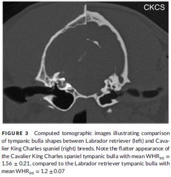 other breeds.
[See Figure 3 at right.] All brachycephalic
breeds had significantly lower tympanic bulla volume:weight ratios
versus Labrador retrievers. Soft tissue attenuating material (middle
ear effusion) was present in the middle ear of 48/100 (48%) of
brachycephalic breeds, but no significant association was found
between tympanic bulla CT measurements and presence of this
material. Findings indicated that there are significant interbreed
variations in tympanic bulla morphology, however no significant
relationship between tympanic bulla morphology and presence of
middle ear effusion could be identified. ... Cavalier King
Charles Spaniels represent a unique subset of
brachycephalic animals as they were found to have significantly
flatter tympanic bullae (defined by width: height ratios) than other
brachycephalic breeds. There was a high percentage of
Cavalier King Charles Spaniels with soft tissue attenuating
material in the middle ear (68%), which may have falsely reduced the
internal measurements obtained, increasing width: height ratio
(internal) artifactually. However, given the fact that the lower
bound of the 95% confidence interval measurements for
Cavalier King Charles Spaniels (width:height ratio
(internal) 1.47−1.65)) was greater than the upper bound for all
breeds (except the French Bulldog which also had a high percentage
of material in middle ear (80%)), authors believe it is likely that
these measurements represented real changes present in this breed.
Cavalier King Charles Spaniels have been described
to have a unique disease resulting in the formation of a buildup of
highly viscous mucus within the middle ear (primary secretory otitis
media or otitis media with effusion) of dogs without clinical
evidence of otitis externa. It is thought that, due to a lack of
inflammation or signs of infection in the middle ear, the disease is
due to auditory tube dysfunction based on possible anatomical
changes of the middle ear or the auditory tube. The finding of an
anatomical variation in the shape of the tympanic bulla of
Cavalier King Charles Spaniels may offer a potential
explanation for the pathogenesis of this disease in addition to the
previously suggested changes in the orientation and function of the
auditory tube. However, given the lack of histopathology and
contrast-enhanced CT scan evaluations in the current study, this
hypothesis is speculative. Further investigation to identify
potential pathway alteration of the auditory tube in the
Cavalier King Charles Spaniel would be an interesting
addition for further clarification of this disease process.
other breeds.
[See Figure 3 at right.] All brachycephalic
breeds had significantly lower tympanic bulla volume:weight ratios
versus Labrador retrievers. Soft tissue attenuating material (middle
ear effusion) was present in the middle ear of 48/100 (48%) of
brachycephalic breeds, but no significant association was found
between tympanic bulla CT measurements and presence of this
material. Findings indicated that there are significant interbreed
variations in tympanic bulla morphology, however no significant
relationship between tympanic bulla morphology and presence of
middle ear effusion could be identified. ... Cavalier King
Charles Spaniels represent a unique subset of
brachycephalic animals as they were found to have significantly
flatter tympanic bullae (defined by width: height ratios) than other
brachycephalic breeds. There was a high percentage of
Cavalier King Charles Spaniels with soft tissue attenuating
material in the middle ear (68%), which may have falsely reduced the
internal measurements obtained, increasing width: height ratio
(internal) artifactually. However, given the fact that the lower
bound of the 95% confidence interval measurements for
Cavalier King Charles Spaniels (width:height ratio
(internal) 1.47−1.65)) was greater than the upper bound for all
breeds (except the French Bulldog which also had a high percentage
of material in middle ear (80%)), authors believe it is likely that
these measurements represented real changes present in this breed.
Cavalier King Charles Spaniels have been described
to have a unique disease resulting in the formation of a buildup of
highly viscous mucus within the middle ear (primary secretory otitis
media or otitis media with effusion) of dogs without clinical
evidence of otitis externa. It is thought that, due to a lack of
inflammation or signs of infection in the middle ear, the disease is
due to auditory tube dysfunction based on possible anatomical
changes of the middle ear or the auditory tube. The finding of an
anatomical variation in the shape of the tympanic bulla of
Cavalier King Charles Spaniels may offer a potential
explanation for the pathogenesis of this disease in addition to the
previously suggested changes in the orientation and function of the
auditory tube. However, given the lack of histopathology and
contrast-enhanced CT scan evaluations in the current study, this
hypothesis is speculative. Further investigation to identify
potential pathway alteration of the auditory tube in the
Cavalier King Charles Spaniel would be an interesting
addition for further clarification of this disease process.
Transoral approach for ventral tympanic bulla osteotomy in the dog: A descriptive cadaveric study. Maria Manou, Pierre H. M. Moissonnier, Nicolas Jardel, Aymeric Tissier, Rosario Vallefuoco. Vet. Surgery. August 2017;46(6):773-779. Quote: Objective: To describe a transoral approach for tympanic bulla osteotomy in the dog. ... A lateral or ventral approach to the tympanic bulla ... can be used to treat primary secretory otitis media (PSOM) of the Cavalier King Charles spaniel. ... In cases of otitis media, the oral approach could enhance drainage from the bulla into the nasopharynx, which may prevent the surgical site infections often encountered after traditional approaches to the middle ear. Improved drainage would also be expected to limit local accumulation of erythrocytes and associated cholesterol deposition. PSOM of the Cavalier King Charles spaniel has been attributed to decreased drainage of the middle ear through the Eustachian tube. This condition is treated by repetitive myringotomies or by tympanostomy tube (TT) placement, in order to restore drainage. The opening created after transoral osteotomy could have the same effect. Study design: Anatomic cadaveric study. Sample population: Fifteen canine cadavers (n = 29 tympanic bullae), including mesaticephalic, dolichocephalic, and brachycephalic breeds [none were CKCSs]. Materials and methods: The oral surface of the tympanic bulla was identified during an anatomical study (3 canine cadavers) and the ventral approach to the tympanic bulla was described (3 canine cadavers). The safety of the technique was assessed (9 canine cadavers, n = 17 bullae) during further anatomical dissections, where a complete approach and drilling of the tympanic bulla were performed. Results: In all cases, tympanic bulla osteotomy was performed without damaging the inner ear, the epitympanic recess contents, and the neurovascular structures. The oral approach to the tympanic bulla was easier in mesaticephalic and dolichocephalic dogs than in brachycephalic breeds. Conclusion: This study defines anatomical landmarks for transoral bulla osteotomy, without a high risk of damage to neurovascular and anatomical structures within and/or surrounding the tympanic cavity. This minimally invasive approach to the tympanic bulla is performed via a natural opening, and does not require simultaneous access through the ear canal. In vivo evaluation of this technique is required to verify its safety in clinical cases prior to large scale application. ... The transoral approach to the tympanic bulla could also be considered in Cavalier King Charles Spaniel with PSOM. Although the pathogenesis of this condition remains unclear, ventilation and drainage of the tympanic bulla through auditory (Eustachian) tube have been proposed as causative factors for this auditory tube dysfunction.16-18 Recent studies have reported the successful treatment of the PSOM in the Cavalier King Charles spaniel via TT instead of repeated opening, flushing, and curettage of the TC. In spite of its small diameter, TT provides constant ventilation and drainage of the middle ear cavity, resulting in subjectively improved hearing, pruritus, and quality of life. We speculate that the transoral approach of the tympanic bulla could be used in cases of refractory PSOM treated with TT, or in cases where repeated TT placement and associated anesthetic episodes, are contraindicated due to the general condition of the patient. Removal of the entire ventral portion of the TB creates a larger opening than the TT and should therefore result in superior drainage.
Twelve years of chiari-like malformation and syringomyelia scanning in Cavalier King Charles Spaniels in the Netherlands: Towards a more precise phenotype. Wijnrocx K, Van Bruggen LWL, Eggelmeijer W, Noorman E, Jacques A, Buys N, Janssens S, Mandigers PJJ. PLoS One. September 2017;12(9). Quote: Chiari-like malformation (CM), syringomyelia (SM) and middle ear effusion (also called PSOM) are three conditions that frequently occur in Cavalier King Charles Spaniels (CKCS). ... This study evaluated the prevalence and estimated genetic parameter of CM, SM and middle ear effusion from 12 years of screening results. ... 1249 screening of 1020 dogs were re-evaluated. ... The presence of middle ear effusion in this study was 19%-21% for dogs younger than 3 years, and 32%-38% for dogs older than 3 years. ... Middle ear effusion: The presence of middle ear effusion was recorded in all dogs. For the group of dogs younger than 3 years of age (809 out of 1020 dogs), 19% presented middle ear effusion in the left bullae and 22% in their right bullae. This percentage had risen to 32% (left bullae) and 38% (right bullae) for dogs older than three years of age (211 out of 1020 dogs). Interestingly the same increase was noted for the dogs that were scanned multiple times. The number of “middle-ear effusion free” dogs decreased from 82% to 72% (left middle ear) and 79% to 70% (right middle ear) respectively. There was no correlation for the CM grading and the presence of either left (p = 0.47) or right middle ear effusion (p = 0.22) or for dogs that had in both middle ears middle ear effusion (p = 0.96). However a small statistical significant correlation of 0.1 was observed for CCD [central canal dilation] and both left and right middle ear effusion (p = 0.000). ... In this study, the prevalence of middle ear effusion was assessed and estimated at 19%–21% for dogs younger than 3 years, and 32%–38% for dogs older than 3. The condition has been diagnosed with various clinical signs, and it has been associated with brachycephalic conformation. It is currently under debate whether the behaviors that are associated with pain due to SM in the CKCS are related to middle ear effusion. In other CKCS populations, estimates of the presence of middle ear effusion reach about 32%–70%. There was no correlation, in this study, for CM and the presence of middle ear effusion, which seems to suggest that middle ear effusion is more associated with a brachycephalic conformation rather than the actual CM grading. This is remarkable as CM is clearly associated with a brachycephalic conformation. Remarkably, a small but statistically significant correlation was found for CDD and middle ear effusion. If this observation is correct it merits further research, as it has not been investigated before. Furthermore it must be noted that in a CKCS, it might be difficult to notice clear signs that could be caused by the middle ear effusion as most of them suffer from CM as well.
Computed tomographic findings in 205 dogs with clinical signs compatible with middle ear disease: a retrospective study. Audrey Belmudes, Charline Pressanti, Paul Y. Barthez, Eloy Castilla-Castaño, Lionel Fabries, Marie C. Cadiergues. Vet. Derm. February 2018;29(1):45-e20. Quote: Background: Computed tomography (CT) is considered to be the reference method to evaluate middle ear structures. Objectives: To evaluate the presence and severity of CT changes in the middle ear and establish if any specific clinical presentations are associated with otitis media. Animals: Medical records of animals referred for CT with history and clinical signs consistent with middle ear disease. [Of 205 dogs, 13 (6.5%) were cavalier King Charles spaniels.] Methods: Retrospective evaluation of CT examinations of tympanic bullae performed over a six year period. Medical records were reviewed for signalment, clinical signs and cytological evaluation of the external ear canal. Dogs were divided into three clinical groups: chronic otitis externa (Group 1), peripheral vestibular disorder (Group 2) and other clinical presentations (Group 3). Results: Group 1 – Of 214 ears, 87 (40.7%) had CT abnormalities: 38 of 87 (17.7%) had material-filled bullae, 42 of 87 (19.6%) had thickened bullae walls and seven of 87 (3.2%) had lysis of the bulla. Abnormalities were significantly more frequent in dogs with suppurative otitis than in erythemato–ceruminous otitis (57% and 23%, respectively; P = 0.003). Proliferative otitis, particularly in French bulldogs, was associated with severe otitis media. Group 2 – Of the 106 ears, 91 (85.8%) had normal tympanic bullae. Group 3 – Of the 26 ears from deaf dogs, 16 had fluid-filled bullae and were from dogs bilaterally affected, one had only one fluid-filled bulla and all nine dogs were Cavalier King Charles spaniels. ... Deafness in CKCS was identified in Group 3 as a specific entity. It was associated with fluid-filled bullae in all cases and was presumed to be due to primary secretory otitis media which is a sterile effusion in the tympanic cavity presumably secondary to a Eustachian tube dysfunction. The presence of material in the bulla may be considered as an incidental finding but can also be accompanied by deafness (conductive hearing loss) or with signs of pain involving the head and neck, and/or neurological signs, and therefore should be differentiated from syringomyelia. ... All dogs with Claude Bernard Horner syndrome or head tilt had normal tympanic bullae. Clinical significance: CT is useful for canine chronic otitis externa, particularly in suppurative or proliferative otitis, even in the absence of associated neurological signs.
Otitis media with effusion in the boxer: a report of seven cases. S. Paterson. J. Sm. Anim. Pract. December 2017. DOI: 10.1111/jsap.12801. Quote: The aim of this study was to describe otitis media with effusion in seven boxers. All dogs presented with a range of clinical signs, which included head shaking, neurological dysfunction, pain on opening of the mouth and reduction in hearing ability. Otitis media was confirmed under general anaesthesia in each case by video-otoscopic identification of a bulging pars tensa and subsequent myringotomy, which revealed a tenacious mucus plug within the middle ear. Brainstem auditory evoked response thresholds were elevated in all affected ears. In three cases, CT revealed soft tissue opacity in the affected bulla. All of the affected middle ears were flushed using warm sterile saline to remove the mucus. A combination of glucocorticoid and antibiotic in EDTA tris was instilled into the middle ears. After the initial middle ear flush under general anaesthesia, topical therapy was applied into the ear canals daily by the owners using the same combination of drugs. Dembrexine, a systemic mucolytic, was administered with food daily. Six out of seven dogs were also prescribed oral prednisolone. In each case, the middle ear effusion was sterile. All clinical signs resolved with treatment, with the exception of facial paralysis in two dogs. Otitis media with effusion should be considered a cause of otitis media in boxers.
Facial paralysis associated with primary secretory otitis media in Cavalier King Charles Spaniels: Study of 2 cases. M. Debuigne, A. Drut, N. Soetart, D. Guillemaille, T. Brément, M. Fusellier, D. Fanuel, V. Bruet. Revue Vétérinaire Clinique. February 2018. DOI: 10.1016/j.anicom.2018.01.002 Quote: Primary secretory otitis media is a disease due to an accumulation of mucus in the tympanic bulla, mostly described in the Cavalier King Charles Spaniel. Clinical signs are numerous and can be subtle; facial paralysis is possible but signs of pruritus or pain are most commonly reported. A 4-year-old male and a 7-year-old spayed female Cavalier King Charles have been presented for lip drooping. Neurologic exam showed a generalized decrease in facial muscle motricity. Because of the history of these animals, a primary otitis media was firstly suspected. Imaging of tympanic bullae showed a unilateral or bilateral otitis media. Video-otoscopic examinations and myringotomy revealed acellular mucus in the concerned tympanic bullae. The myringotomy and the flus h of tympanic bullae, associated with corticosteroids, improved the clinical signs for several months. These two cases highlight the importance of otitis media in the differential diagnosis of facial paralysis and focus on the originality of primary secretory otitis media in the Cavalier King Charles Spaniels.
Sleep disordered breathing in the Cavalier King Charles Spaniel: a case series. Tom Hinchliffe, Nai-Chieh Liu, Jane Ladlow. BSAVA Congress 2018 Proceedings. April 2018;pp. 479-480. Quote: Objectives: Obstructive sleep apnoea has been widely investigated in human medicine, but relatively few studies have described this condition in veterinary species; to date it has only been reported in English Bulldogs, French Bulldogs and Pugs. The aim of this study was to report the clinical features, diagnosis, management and outcome of five cases of obstructive sleep apnoea in the Cavalier King Charles Spaniel (CKCS). Methods: A retrospective case series was created using hospital records over five years, followed up via recheck appointments and a telephone questionnaire. The inclusion criteria consisted of CKCSs presenting primarily for sleep disorders, which were treated surgically for obstructive anatomical pathology. Results: All five cases presented with stertorous breathing, choking and apnoea during sleep. All cases had a number of co-morbidities: 5/5 had eosinophilic stomatitis and otitis media, 4/5 had mitral valve insufficiency and 3/5 had syringomyelia. CT and rhinoscopy revealed that 5/5 had aberrant nasal turbinates, nasal septal deviation and soft palate thickening and 3/5 had nasopharyngeal thickening and tracheal collapse. Treatment consisted of a combination of laser turbinectomy, folding flap palatoplasty, tonsillectomy, laryngeal sacculectomy and cuneiform process resection. All cases had an improvement in both the incidence and severity of sleep apnoea within a week, with 4/5 cases achieving complete resolution. Conclusions: This study concludes that obstructive sleep apnoea is a condition that may affect the CKCS and all five cases described here improved following surgical intervention. Consequently, sleep disordered breathing is a presentation that veterinary surgeons should be aware of in this breed.
Bacteriology and cytology of otic exudates in 41 cavalier King Charles spaniels with primary secretory otitis media. Lynette K. Cole, Päivi J. Rajala-Schultz, Gwendolen Lorch, Joshua B. Daniels. Vet. Derm. April 2019;30(2):151-e44. Quote: Background: Primary secretory otitis media (PSOM) in the cavalier King Charles spaniel (CKCS) is similar to otitis media with effusion (OME) in humans. A proposed aetiology of OME is inflammation of the middle ear mucosa, usually due to bacterial infection, leading to auditory tube dysfunction. Hypothesis/Objectives: Our objective was to characterize the microbiological and cytological findings of otic exudates from the external ear canal (EEC) (n = 68) and middle ear (ME) (n = 69) from 41 CKCSs with PSOM. Methods and materials: Swab samples from the EEC and mucus aspirated from the ME after performing a myringotomy were obtained for bacterial culture and cytological analysis. Results: Fifty-five of 68 (81%) EEC and 46 of 69 (67%) ME yielded no bacterial growth. Thirty-eight of the 68 (56%) ears had no microbial growth from neither the EEC nor ME; seven (10%) had bacteria isolated from the EEC only; 17 (25%) had bacteria isolated from the ME only, and six (8%) had bacteria isolated from both EEC and ME. Thirty-four total bacterial isolates were cultured from ME. The most common bacterial species isolated were coagulase-negative staphylococci, followed by Staphylococcus pseudintermedius. Otic cytology identified coccoid organisms in only three of 68 EEC and four of 69 ME. Conclusions: The role of bacteria in the pathogenesis of PSOM in CKCS is unclear. The majority of the EEC and ME of the CKCS with PSOM were negative by conventional bacterial culture and the cytological presence of bacteria was not correlated with culture positives. The potential role of noncultivable microbiota in PSOM requires exploration using molecular methods. ... In conclusion, bacterial otitis externa is an uncommon finding in CKCS with PSOM and the majority of ME with PSOM are culture-negative, with the cytological presence of bacteria in mucous being poorly correlated with conventional bacterial culture results. Additional studies evaluating the microbiome as well as additional exploration of the potential role of staphylococci in CKCS with PSOM are needed to further understand the pathogenesis of PSOM. [See also this June 2016 abstract.]
Occult otitis media in dogs with chronic otitis externa – magnetic resonance imaging and association with otoscopic and cytological findings. Andrea Lorek, Ruth Dennis, Jan van Dijk, Jeanette Bannoehr. Vet. Derm. December 2019;doi: 10.1111/vde.12817. Quote: Background: Identification of perpetuating factors, such as otitis media (OM), is important for the successful management of canine chronic otitis externa (OE). Hypothesis/Objectives: Occult OM can occur in cases of chronic OE; a focused magnetic resonance imaging (MRI) examination is a useful tool in their management. Animals One hundred twenty one client‐owned dogs presented for investigation and treatment of chronic OE between 2009 and 2018. Methods and materials: Mixed retrospective (74 dogs) and prospective (47 dogs) study of chronic OE cases without neurological signs, describing the MRI, otoscopic and cytological findings; comparing cases with and without MRI evidence of OM. Results: A total of 123 MRI studies were analysed (two dogs scanned twice). A short, focused MRI scan allowed detection of inflammation of the mucosal bulla lining as well as excellent discrimination between avascular material and vascularised soft tissue in the tympanic cavity. OM was found in 41 of 197 (21%) ears with chronic otitis externa. On otoscopy, the tympanic membrane was intact in six of 41 ears (15%), ruptured in 16 of 41 (39%) and not visible in 14 of 41 (34%) [no data in five of 41 (12%)]. Analysis of cytological findings showed that the presence of rods was only associated with an increased likelihood of OM when found together with inflammatory cells. ... The CKCS in the present study had unilateral middle ear disease with a fluid-filled tympanic cavity, but also showed a thin rim of contrast enhancement along the bulla wall, as did all the other affected cases with a fluid-filled tympanic cavity. Inflammatory mediators are capable of affecting auditory tube function in dogs,24 so the cases in the present study could represent genuine inflammatory OM rather than PSOM. ... Conclusions and clinical importance: Occult OM is a not uncommon finding on MRI of dogs with chronic OE. A targeted MRI study (“bulla mini‐scan”) may be useful as part of the clinical investigations.
The effect of phenotypic selection on fourteen years of chiari-like malformation and syringomyelia MRI scanning in Cavalier King Charles Spaniels in the Netherlands. Laterveer, M. Utrecht Univ. master thesis. March 2020. Quote: Chiari-like malformation (CM) and Syringomyelia (SM) is an inherited disease complex which is common in the Cavalier King Charles Spaniels (CKCS). Since 2004 a law-enforced obligatory screening program prior to breeding is used to achieve a healthier population of Dutch CKCS. This study evaluated the effect of phenotypic selection by combining pedigree data and screening results of two to three generations of dogs to see if there is any improvement in the breeding stock. In total, 572 MRI scans from 518 dogs were prospectively enrolled if they were screened between January 1st 2016 and December 31st 2018. CM and PSOM (primary secretory otitis media) were scored and SM was defined as unaffected (CCD (central canal dilation) of 0 mm) or affected (CCD of >0 mm). The prevalence of SM in this study was 22.7% and increased with age clearly demonstrating the age-effect of SM. The prevalence of middle ear effusion in the left ear was 19.4% and in the right ear 20.0%. Results indicated that selecting unaffected parents leads to a lower CCD in offspring. This reduction in CCD is even more if parents with healthier grandparents are given priority. Conversely, the offspring of affected parents was more likely to have an increase in CCD. CM and PSOM did not improve upon screening for unaffected parents. It is concluded that, the obligatory screening for and subsequent selection for older unaffected CKCS, the offspring indeed improved each generation and as indicated by a reduced prevalence of Syringomyelia.
Cytological and microbiological characteristics of middle ear effusions in brachycephalic dogs. Elspeth Milne, Tim Nuttall, Katia Marioni-Henry, Chiara Piccinelli, Tobias Schwarz, Ali Azar, Jennifer Harris, Juliet Duncan, Michael Cheeseman. J. Vet. Intern. Med. May 2020; doi: 10.1111/jvim.15792. Quote: Background: Middle ear effusion [MEE] is common in brachycephalic dogs with similarities to otitis media with effusion in children. ... MEE is most common in Cavalier King Charles spaniels (CKCS) but occurs in other brachycephalic breeds, for example, boxers and bulldogs. ... The condition is also referred to as primary secretory otitis media (PSOM) or otitis media with effusion (OME), but the term MEE has been advocated for dogs as inflammation is not invariably present. The nomenclature is also influenced by the human manifestation of OME, which is most prevalent in children and commonly known as “glue ear.” This shares many of the features of MEE in dogs. ... Association with the cranial and eustachian tube morphology and bacterial infection is suspected in both species. Hypothesis/objectives: To determine cytological and bacteriological features of middle ear effusions in dogs, provide information on histological features, and further assess the dog as a model of the human disease. Animals: Sixteen live dogs [including 8 CKCSs], 3 postmortem cases of middle ear effusion, and 2 postmortem controls. Methods: Prospective; clinical investigation using computed tomography, magnetic resonance imaging, video‐otoscopy, myringotomy; cytological assessment of 30 and bacteriology of 28 effusions; histology and immunohistochemistry (CD3 for T-lymphocytes, Pax5 for B lymphocytes and MAC387 for macrophages) of 10 middle ear sections. Results: Effusions were associated with neurological deficits in 6/16 (38%) and concurrent atopic dermatitis and otitis externa in 9/16 (56%) of live cases. Neutrophils and macrophages predominated on cytology (median 60 [range 2%-95.5%] and 27 [2%-96.5%]) whether culture of effusions was positive or not. ... In the live cases, the final diagnoses in the 8 CKCS were MEE only in 1 case, and MEE with concurrent Chiari-like malformation and syringomyelia in 7 cases; 2 of these cases were also diagnosed with idiopathic epilepsy, 2 with diabetes mellitus, and 1 with brachycephalic obstructive airway syndrome (BOAS). ... In histology sections, the mucosa was thickened in affected dogs but submucosal gland dilatation occurred in affected and unaffected dogs. There was no bacterial growth from 22/28 (79%) of effusions. Bacteria isolated from the other 6 (21%) were predominantly Staphylococcus pseudintermedius (4/6, 67%). Conclusions and Clinical Importance: Clinical, morphological, and cytological findings in middle ear effusions of dogs and people suggest similar pathogeneses. Middle ear effusion of dogs could be a useful model of human otitis media with effusion. Such comparisons can improve understanding and management across species.
When and how to do a myringotomy – a practical guide. Lynette K Cole, Tim Nuttall. Vet. Dermatology. May 2021; doi: 10.1111/vde.12966. Quote: A myringotomy is a surgical incision made in the tympanic membrane (TM). This gives access to the middle ear for sampling, flushing and instilling topical therapy. It should be considered whenever the TM is intact and there is clinical evidence of otitis media, abnormal TMs and/or abnormal diagnostic imaging. ... A sterile form, primary secretory otitis media (PSOM) or otitis media with effusion (OME),was recognized in cavalier King Charles spaniels (CKCS) initially, although it can affect any brachycephalic breed. It may be present in ≤70% of CKCS with or without associated clinical signs. ... In the CKCS a bulging pars flaccida obscuring some or all of the pars tensa is diagnostic for PSOM (Figure at left), although a flat pars flaccida does not rule it out (Figure at right). ... In some CKCS with PSOM the pars flaccida is so large that it completely obscures the pars tensa. ... Samples should be collected for cytological investigation and culture, and then the external ear should be cleaned and dried (if required). Myringotomies should be performed under general anaesthesia and, wherever possible, using a video otoscope; the enhanced view and instrument ports facilitate the technique and reduce the risk of complications. The myringotomy incision should be made in the caudoventral quadrant of the TM using an appropriately sized urinary catheter to collect samples and flush the middle ear cavity. A thorough understanding of the anatomy, technique and potential ototoxicity of topical therapy is needed to minimize the risk of neurological and other complications. The TM usually heals within 35 days if kept free of infection.
Eustachian Tube Formation and Angulation in Dogs Affected by Primary Secretory Otitis Media. F. Possiel, S. De Decker, H.A. Volk, A.V. Volk. 33d ESVN-ECVN Symposium 2021, #73. September 2021. Quote: Primary secretory otitis media (PSOM) is common especially in brachycephalic dogs. Various aetiologies have been discussed, including infectious, inflammatory and morphological causes. However, there remains a lack of data supporting any of the current hypothesis. The aim of the current study was to elucidate the role of the eustachian tube [ET] in PSOM. Computer tomography images of 72 dogs with or without PSOM were evaluated in the study. Morphological measurements (Eustachian tube length and width) and angulation of the Eustachian tube of 97 control ears [of Jack Russell terriers, Cocker Spaniels] were compared to 47 PSOM affected ears [of French bulldogs, Cavalier King Charles Spaniels, Pugs]. Data are reported as Median with 25-75 percentiles. Groups were compared with a Mann Whitney U-test and a P-value of less than 0.05 was deemed significant. Eustachian width was significant smaller in width in affected cases (1.02 (0.86-1.46)) compared to controls (1.29 (0.71-1.52)), as was angulation wider in affected (42.22 (33.91-44.43)) versus non-affected (35.64 (31.91-40.12)) respectively. ... The ET prime function is ventilation and pressure equalisation of the tympanic bullae. As far as currently known, when that fails, PSOM arises. The analysis of our data supported that theory, showing a shorter, and significant flatter ET with a significant different angulation in brachycephalic compared to mesencephalic dogs. In summary a marked reduction of tympanic bulla volume, as shown by Mielke and us, and known thicker muscles in the nasopharynx, as well as, cartilage weakness in brachycephalic dogs suggests that the ventilation and pressure equalisation system of the ET in brachycephalic canines is a very dysfunctional, weak system. ... This study demonstrates that Eustachian tube width and angulation might contribute to development of PSOM, which is similar to what has been shown in children. Future studies are needed to explore if the morphological changes have functional consequences and therefore lead to PSOM.
Investigations on the epidemiology and pathophysiology of primary secretory otitis media in canine brachycephalene. Sarah-Fabienne Charlott Possiel. Thesis, Univ. Vet. Med. Hannover. June 2022. Quote: Background: The Eustachian tube connects the middle ear with the nasopharynx, regulates the drainage of middle ear secretions and provides a protective function of the middle ear against bacterial entry. It regulates ventilation between the tympanic cavity and the pharynx and also provides pressure equalization between the compartments. Experimental obstruction of the auditory tube may result in tympanic cavity effusion. This tympanic cavity effusion is diagnosed with progressive frequency in brachycephalic breeds as a type of "incidental finding." This effusion presents in most cases as a sterile, mucous secretion and when present is referred to as primary secretory otitis media- PSOM. ... Changes in the growth of the skull and the auditory tube are associated with the development of tympanic effusion. Since multiple, breeding-related malformations of the skull are already known in brachycephalic dogs, positional changes and shortening of structures of the tuba auditiva as well as possible morphometric changes of the middle ear could have occurred simultaneously. The aim of this work is to increase the understanding of the etiology and pathogenesis of PSOM in brachycephalic dog breeds. In this regard, special attention is paid to the anatomy of the auditory tube and middle ear of these particular breeds by performing measurements using computed tomographic studies on the 3D reconstruction model. The data obtained were compared with a normocephalic control group of 24 animals (9 Jack Russell terriers, 15 Cocker Spaniels), whose data were also obtained retrospectively. Material and Methods: In this retrospective study, CT images of 40 dogs of brachycephalic breeds (15 Cavalier King Charles Spaniels, 15 French Bulldogs, 10 Pugs) and 24 dogs of mesocephalic breeds (9 Jack-Russel Terriers, 15 Cocker Spaniels) were measured ... . During the evaluations, attention was paid to an existing bulla filling, the anatomical representation of the middle ear and the auditory tube. Bulla volume, bulla wall thickness, the bony portion of the auditory tube with height, width, and length, and the total length of the auditory tube and its angulation to the rostrocaudal median of the skull were measured. The dogs were further divided into three groups based on their bulla filling. Each ear/bulla was considered individually and thus two ears were always reported per dog (n= 2). The group of non-sufferers was formed by dogs without a bulla filling and consisted mostly of dogs of the mesocephalic breed [and 2 of the 15 CKCSs]. The PSOM-unilateral group, except for one mesocephalic dog (Cocker Spaniel), consisted of only brachycephalic dogs [including 7 of the 15 CKCSs]. The unaffected ear in these dogs was included in this group. The third group, PSOM-bilateral, consisted of dogs with bilateral middle ear filling and included only brachycephalic dogs, as before, except for one Cocker Spaniel [and 6 of the 15 CKCSs]. Data were tested for normal distribution using four different tests, Anderson-Darling, D'Agostino & Pearson, Shapiro-Wilk, and Kolmogorov-Smirnov test. Because the data were mainly nonparametrically distributed, the median, 25% and 75% percentiles, interquartile range (IQR), and range adjacent to minimum and maximum were mathematically determined descriptively; for normally distributed data, the mean, standard deviation, and standard error were more accurate descriptions. ... Results: Of the dogs in the brachycephalic breeds, 60% (24/40) had tympanic sinus effusion. Overall, 25% (10/40) of brachycephalic dogs were diagnosed with unilateral tympanic cavity effusion and 35% (14/40) of dogs were diagnosed with bilateral tympanic cavity effusion. Dogs of the mesocephalic breeds rarely had tympanic sinus effusion at 8% (2/24), resulting in clustered effusions in dogs of the brachycephalic group. Dogs in the brachycephalic group with a bilateral tympanic sinus effusion had only about half the bulla volume bilaterally compared with dogs without an effusion. The bulla wall was also significantly thickened in dogs with bilateral effusion compared to dogs without an effusion. There was a significant shortening as well as flattening of the bony portion of the auditory tuba in dogs with bilateral effusion, compared to dogs without an effusion. The width of the bony portion showed no difference in measurement in dogs with and without effusion. The angle of the auditory tube was significantly greater in dogs with a bilateral bulla filling compared with dogs without a bulla filling. ... The bulla in the Cavalier King Charles Spaniel also was ventrally flattened. The sagittal cross-sectional images showed visually in the French bulldog and the Cavalier King Charles Spaniel has a significant narrowing of the bony part of the auditory tube. The opening of the bony part in the Cavalier King Charles Spaniel and the French Bulldog pointed ventrally, those of the Jack Russel Terrier, the Cocker Spaniel and of the pug remained horizontal and then bent ventrally. ... Thus, these dogs show a much flatter tuba auditive compared with dogs without a bulla filling. The lengths of the hard and soft palate did not differ in their measured values in dogs with and without a bulla filling. With regard to the thickness of the soft palate, dogs with a bilateral tympanic effusion showed a thickened soft palate compared with dogs without an effusion. Conclusion: This study shows for the first time that brachycephaly in dogs leads to specific changes in the skull with a possible predisposition to PSOM. It appears that brachycephaly has resulted in multiple changes in tubal morphology, affecting the bony portion as well as the location of the entire auditory tube. These changes in the morphology of the auditory tube predispose to tube dysfunction with subsequent effusion formation in brachycephalic dogs. In addition, the smaller bulla volume and thickened bulla wall of these dogs also appear to play a major role in tubal dysfunction.
Prevalence and characterization of middle ear effusion in 55 brachycephalic dogs. Riccarda Schuenemann, Anne Kamradt, Katrin Truar, Gerhard Oechtering. Tierarztl Prax Ausg K Kleintiere Heimtiere. September 2022; doi: 10.1055/a-1913-7216. Quote: Objective: Multiple, breeding-related malformations of the skull of brachycephalic dogs are well-known. Whereas the eye-catching deformities of the nose that lead to dramatic respiratory problems are obvious, changes of the middle ear are often an incidental finding on CT examinations and usually clinically inapparent. The objectives of this work were to investigate the prevalence and characteristics of middle ear effusion in brachycephalic dog breeds presented for multilevel surgery of upper airway obstructions. Material and methods: Brachycephalic dogs with incidental middle ear effusion detected on CT scans obtained prior to surgical treatment of brachycephalic airway syndrome were prospectively enrolled. A perendoscopic tympanocentesis followed by macroscopic description, microscopic cytology and bacteriological analysis of the fluid was performed. Results: Prevalence of middle ear effusion in all dogs presented to the department during the study period was 55/170 (32%) in 86 middle ears. The only breeds suffering from MEE were French Bulldogs (FB) with a prevalence of 35/66 (53%) and Pugs with a prevalence of 20/79 (25%). Tympanocentesis was performed in 80 ears. In the majority of cases the effusion was either mucoid or serous, with a honey-like or ochre colour. Bacteriology was available for 76 ears and tested positive in 34 (45%) cases. Cytology was performed in 73 ears and revealed all effusions to contain inflammatory cells with a high concentration in 23 (31.5%) cases. Conclusions and clinical relevance: Brachycephalic dogs presented for surgical treatment of brachycephalic airway syndrome have a high prevalence of incidental middle ear effusions. Cytological findings differ from previously reported analyses of effusions in Cavalier King Charles spaniels with clinical symptoms of primary secretory otitis media, where usually cell-free effusions are found. A study comparing effusions of brachycephalic dogs with vestibular disease to those found as an incidental condition is warranted.
Protocol for Diagnosis, Treatment and Subsequent Care of Primary
Secretory Otitis Media in Cavalier King Charles Spaniels.
Ivelina
Vacheva. Tradition & Modernity in Vet. Med. March 2023; doi:
10.5281/zenodo.7707497. Quote: Primary Secretory Otitis Media in the
Cavalier King Charles Spaniel is a rare and complex
disorder. However, using a well established protocol, including well
established procedures, modern day imaging technology, precise
treatment and adequate post-treatment care can lead to a high rate
of successfully dissipated symptoms and long-term well-being our the
patients. The suggested protocol is based on 14 examined [CKCS]
patients in a 1 year time period. It includes otoscopic examination,
cytology, culture and sensitivity testing, magnetic resonance
imaging, viodeootoscopy, deep ear cleaning, myringotomy, educating
the owners, owners’ feedback, subsequent therapy and long-term
follow up. The study concludes that MRI and educating the owners are
two extremely important tools that should not be overlooked.
Additionally, further research with the addition of DNA analysis is
needed. ... Conclusion and recommendations: To summarise, my
protocol for diagnosis, treatment and subsequent care is a
following: 1. Complete exam including otoscopy. 2. Ear Cytology and
Culture and sensitivity testing. 3. MRI. 4. Videootoscopy. 5. Deep
Ear Cleaning. 6. Myringotomy. 7. Subsequent care including therapy,
control examinations and owner education.
Structure and scaling of the middle ear in domestic dog breeds. Matthew J. Mason, Madaleine A. Lewis. J. Anatomy. April 2024; doi: 10.1111/joa.14049. Quote: Although domestic dogs vary considerably in both body size and skull morphology, behavioural audiograms have previously been found to be similar in breeds as distinct as a Chihuahua and a St Bernard. In this study, we created micro-CT reconstructions of the middle ears and bony labyrinths from the skulls of 17 dog breeds, including both Chihuahua and St Bernard, plus a mongrel and a wolf. From these reconstructions, we measured middle ear cavity and ossicular volumes, eardrum and stapes footplate areas and bony labyrinth volumes. All of these ear structures scaled with skull size with negative allometry and generally correlated better with condylobasal length than with maximum or interaural skull widths. Larger dogs have larger ear structures in absolute terms: the volume of the St Bernard's middle ear cavity was 14 times that of the Chihuahua. The middle and inner ears are otherwise very similar in morphology, the ossicular structure being particularly well-conserved across breeds. ... The Cavalier King Charles Spaniel has relatively large ossicles. ... The expectation that larger ear structures in larger dogs would translate into hearing ranges shifted towards lower frequencies is not consistent with the existing audiogram data. Assuming that the audiograms accurately reflect the hearing of the breeds in question, oversimplifications in existing models of middle ear function or limitations imposed by other parts of the auditory system may be responsible for this paradox. ... From our regression analyses, the breed that stood out most was the Cavalier King Charles Spaniel, which had larger ossicular and labyrinth volumes than expected for its skull length. Its ossicles considered collectively were 60% larger in volume than expected, its labyrinth just over 35% larger. The ossicles did not, however, differ noticeably in morphology to those of the other dogs. Since only one specimen was investigated we cannot be certain that this is typical of the breed, but it is interesting to note that cases of primary secretory otitis media are disproportionately common in Cavalier King Charles Spaniels (Cole, 2012; Stern-Bertholtz et al., 2003), suggesting that there may be something unusual about their middle ears. Cole et al. (2015) commented that these dogs appeared to have ‘small and shallow’ bullae, which could potentially be linked to the condition if drainage of fluids from the Eustachian tube is impeded. Relative to skull length, our specimen had a middle ear cavity volume that fell slightly below the regression line (Figure 6a), but nothing stands out regarding its cavity morphology (Figure 4p). Other authors have found a link between primary secretory otitis media and abnormal soft-tissue morphology in the nasopharyngeal region (Hayes et al., 2010), which would not be visible in our scans.
Clinical characterisation of a novel idiopathic episodic mandibular tremor in dogs and cats. Theofanis Liatis, Sofie F.M. Bhatti, Albert Aguilera, Nikoleta Makri, Amit Batla, Elena Scarpante, Joon Park, Steven De Decker. Recent Advances in Tremors in Dogs and Cats. Chapter 5:80-100. Ghent Univ. May 2024; doi: 10.12968/coan.2023.0031. Quote: Episodic mandibular tremors (EMT), manifested as teeth chattering, are not well described in dogs and cats. The aim of this study is to describe semiology, MRI findings and outcome of dogs and cats with presumptive idiopathic episodic mandibular tremor (IEMT). We hypothesized that EMT represents a benign movement disorder. This is a retrospective, multi-center, study of dogs and cats with EMT between 2018-2023 and prospective online questionnaire open to owners with pets with teeth chattering. Eleven dogs and 1 cat met the inclusion criteria in the retrospective part; 31 dogs and 1 cat were recruited from the online survey. ... Breeds included Cavalier King Charles Spaniel (CKCS; 4/11; 36.4%), CKCS-Bichon Frise cross (Cavachon; 2/11; 18.2%) and 1 of each (9.1%): Northern Inuit, Little lion dog, Springer Spaniel, Labrador retriever, West Highland White Terrier. ... All dogs and cats had rapid and short-lasting (< 1 minute) episodes of EMT in absence of other neurologic signs. Lip smacking occasionally accompanied the tremor in 5/11 (45.5%) hospital dog cases. Excitement was a common trigger in 14/31 (45.2%) dogs from the survey. Cavalier King Charles Spaniel was the most common breed in both clinical and survey populations. Median age at presentation was 3 years for both hospital cases and the survey dogs. A concurrent medical condition was present in 8/10 (80%) hospital cases and 20/31 (64.5%) survey dogs but no causal relationship with EMT was noticed. ... Concurrent diseases were present in 8/10 (80%) dogs including Chiari-like malformation (n=3), inflammatory bowel disease (n=2), PSOM (n=2), atlanto-axial dorsal band (n=1), atlanto-occipital overlap (n=1), eosinophilic oral granuloma (in remission; n=1), eosinophilic bronchopneumopathy (n=1), syringomyelia (n=1), fractured tooth (n=1), pododermatitis (n=1), chronic meningomyelitis of unknown origin in remission (patient received a combination of cytarabine and prednisolone; n=1). ... In 2 CKCS, PSOM was considered to have caused the EMT. In the first PSOM case, EMT ceased shortly after myringotomy and middle ear flush. However, 2 weeks post-treatment, EMT recured with the repeat CT revealing normal middle ears. In the second PSOM case, EMT along with head shaking ceased shortly after myringotomy, middle ear flush and administration of anti-inflammatory prednisolone. ... In 2 dogs with PSOM, myringotomy and middle ear flush were also performed. ... Given the over-representation of CKCS and their crossbreeds, an idiopathic movement disorder, IEMT, was suspected. A genetic underlying etiology might be present and worthy of future investigation. In 3 hospital dogs that underwent further investigations no brain disease was present. ... Conscious electroencephalography was performed in one dog (Cavalier King Charles Spaniel) for 15 minutes; this did not reveal any epileptogenic activity, however no episode occurred during the test. ... In conclusion, IEMT might be an idiopathic movement disorder of dogs, such as essential tremor,presenting with teeth chattering. It is usually manifested in young dogs. Cavalier King Charles Spaniels might be over-represented, which raises the possibility of a genetic background. A manifestation of pain though cannot be completely ruled out.
CONNECT WITH US