
Eosinophilic Stomatitis and the
Cavalier King Charles Spaniel

Cavalier King Charles spaniels are predisposed to some eosinophilic syndromes, especially eosinophilic stomatitis, an autoimmune disorder.* Eosinophilic stomatitis may also be referred to as eosinophilic granuloma.
* See Eosinophilic Diseases of Dogs.
What It Is
Stomatitis is an inflammation of the mucous lining of any parts of the mouth, such as the tongue, palate, and gums. It usually appears as ulcers and lesions on the surfaces within the mouth. If the ulcers are located on the palate, the disorder may be called ulcerative eosinophilic palatitis.
Eosinophils, short for eosinophil granulocytes, are white blood cells which are a component of the immune system. These cells normally are found in parts of the brain, gastrointestinal tract, and some lower body organs and serve to combat certain allergens, parasites, and infections. When eosinophils are activated by an immune stimulus, they release cell-killing proteins capable of damaging tissues, ideally those of the parasites and infecting pathogen. When they infiltrate and accumulate in the lungs, esophagus, or respiratory tract, they often are associated with an immune dysfunction and may damage the dog's own tissues.
Other forms of eosinophilic syndromes found in Cavaliers are eosinophilic bronchopneumonopathy (airway disease) and eosinophilic enteritis (intestinal).
RETURN TO TOP
Symptoms
 The common symptoms are ulcers - which appear as appear as inflamed
crater-like sores - on the surfaces of the mouth, such as the gums and soft
palate (see photo at right by Dr. Gregg A. DuPont). Lesions - such as bumps or blisters - also may be present
(see photo below).
The common symptoms are ulcers - which appear as appear as inflamed
crater-like sores - on the surfaces of the mouth, such as the gums and soft
palate (see photo at right by Dr. Gregg A. DuPont). Lesions - such as bumps or blisters - also may be present
(see photo below).
The inflamed areas will be sensitive and painful, so the dog may exhibit difficulty in eating or a loss of appetite. Other clinical signs include:
• halitosis
• swallowing problems
• chattering lower jaw
• coughing during meals
• clearing the throat.
Often, the dog shows no visible symptoms, and the disorder is not discovered unless the dog's mouth is examined.
Other disorders could have similar symptoms, such as mucous membrane pemphigoid (MMP), although there are no known reports of cavaliers having been diagnosed with MMP.
RETURN TO TOP
Diagnosis
The appearance of the sores could be due to any of several causes. Thorough visual examination and a biopsy are standard procedure. A complete blood count, viral panel, and chemistry panel likely will be performed. Blood panel results usually are normal, except for elevated immunoglobulins reflecting chronic stimulation. Radiographs of the jaw may also be taken. The dog may be placed under anesthesia for some of these procedures.
RETURN TO TOP
Treatment
Allopathic (conventional) veterinarians often assume that eosinophilic stomatitis is the result of an inappropriate, exceptionally active immune response. Therefore, their recommended treatment usually includes immunosuppressive doses of corticosteriods and/or other immune-suppressing drugs. They include prednisolone (Prednidale), azathioprine (Imuran, Azasan), cyclosporine (Sandimmune, Neoral, Cicloral, Gengraf), dexamethasone (Decadron, Dexasone, Diodex, Hexadrol, Maxidex), and glucocorticoid (cortisol).
However, since eosinophil granulocytes are blood cells which are activated by an immune stimulus to combat certain allergens, parasites, and infections, it would be advisable to test for the probable presence of an allergen, parasite, or infection, and these invaders should be treated accordingly, rather than to suppress the immune system which is trying to combat the invaders.
Antibiotics, especially clavamox and clindamycin, but also metronidazole* (Flagyl), doxycycline, minocycline, and azithromycin, may be prescribed to offset any bacterial infection due to the side effects of the immune suppressing drugs.
* Metronidazole (Flagyl) may cause adverse events in dogs hypersensitive to it. See Metronidazole Risks.
Non-steroidal anti-inflammatory agents (NSAIDs) may provide both pain killing and anti-inflammatory relief.
Tooth removal may also be necessary. Laser therapy, such as CO2 laser treatment, has had inconsistent results in initiating a healing process of the ulcerated lesions, with any even limited success ususally only after full-mouth extractions.
In view of the limited treatment options offered by conventional veterinary practice, owners may want to consider contacting well-qualified licensed veterinarians who practice holistic care and treat auto-immune disorders. Search webpages for finding holistic veterinarians in the United States are located here and here.
RETURN TO TOP
Research News
May 2019:
US Study finds cavaliers are most commonly affected by
eosinophilic oral disease.
 In a
March 2019 article, a team of US veterinarians (Danielle Mendelsohn
[right],
John R. Lewis, Kristin Iglesias Scott, Dorothy C. Brown, Alexander M.
Reiter) studied the reports of eosinophilic oral disease in 24 dogs at
NorthStar Vets in New Jersey and the University of Pennsylvania
veterinary hospital in Philadelphia over a 17-year period,. Fifteen
breeds were represented, and the cavalier King Charles spaniel was the
most commonly affected breed (16.7%). All four cavaliers had lesions on
their palate. All dogs with lesions of the palate wered asymptomatic
after treatment with a combination of antibiotics and prednisone. The
authors cautioned that:
In a
March 2019 article, a team of US veterinarians (Danielle Mendelsohn
[right],
John R. Lewis, Kristin Iglesias Scott, Dorothy C. Brown, Alexander M.
Reiter) studied the reports of eosinophilic oral disease in 24 dogs at
NorthStar Vets in New Jersey and the University of Pennsylvania
veterinary hospital in Philadelphia over a 17-year period,. Fifteen
breeds were represented, and the cavalier King Charles spaniel was the
most commonly affected breed (16.7%). All four cavaliers had lesions on
their palate. All dogs with lesions of the palate wered asymptomatic
after treatment with a combination of antibiotics and prednisone. The
authors cautioned that:
"If immunosuppressive therapy is chosen as treatment, it is important to attempt identification of an underlying or predisposing cause. In particular, the risk of using glucocorticoids must be weighed against the benefits gained. Immunosuppressive treatments are not generally considered cures and may carry undesirable side effects with symptoms recurring once therapy is interrupted or discontinued. Alternatively, while some lesions may regress spontaneously without treatment, systemic glucocorticoid therapy has been shown to hasten the regression. Combining medications, such as administering a low dose of prednisone together with an antihistamine, can balance achieving clinical improvement while minimizing the risk of immunosuppression."
May 2018:
Mississippi State Univ. student reports a case study of a
cavalier with oral eosinophilic granuloma.
 In
a
May 2018 presentation at a cinicopathological conference,
Mississippi State Univ. veterinary student Amanda Rigney-Alden
(right) reported on a case study of a year old cavalier King
Charles spaniel with a 5-day history of lower jaw chattering. The dog,
Sven, had two smooth dime sized circular lesions located side by side
in the rear portion of the palate. The lesions were ulcerated and
surrounded by reddened raised nodules. The pharynx was engorged with
blood and inflamed and the nodules coalesced from the two lesions to the
back of the throat. Punch biopsies of the lesions were found to contain
a heavy predominance of eosinophils and diagnosed the lesions to be a
variation of oral eosinophilic granuloma. The pathogenesis of CEG is
presumed to occur following a Type 1 hypersensitivity immune-mediated
reaction. When in the face of an environmental or food allergen,
eosinophils are involved in the inflammatory response to the foreign
material and respond to the antigens inappropriately producing a
hypersensitive reaction. Once an eosinophil is activated the cells
phagocytize foreign material or degranulate, as well as synthesize
cytokines that are involved in the inflammatory response. Thes cascade
of eosinophilic degranulation and production of inflammatory cytokines
results in a profound tissue reaction. A genetic component to CEG is
also suspected to occur in Siberian Husky and cavalier breeds.
In
a
May 2018 presentation at a cinicopathological conference,
Mississippi State Univ. veterinary student Amanda Rigney-Alden
(right) reported on a case study of a year old cavalier King
Charles spaniel with a 5-day history of lower jaw chattering. The dog,
Sven, had two smooth dime sized circular lesions located side by side
in the rear portion of the palate. The lesions were ulcerated and
surrounded by reddened raised nodules. The pharynx was engorged with
blood and inflamed and the nodules coalesced from the two lesions to the
back of the throat. Punch biopsies of the lesions were found to contain
a heavy predominance of eosinophils and diagnosed the lesions to be a
variation of oral eosinophilic granuloma. The pathogenesis of CEG is
presumed to occur following a Type 1 hypersensitivity immune-mediated
reaction. When in the face of an environmental or food allergen,
eosinophils are involved in the inflammatory response to the foreign
material and respond to the antigens inappropriately producing a
hypersensitive reaction. Once an eosinophil is activated the cells
phagocytize foreign material or degranulate, as well as synthesize
cytokines that are involved in the inflammatory response. Thes cascade
of eosinophilic degranulation and production of inflammatory cytokines
results in a profound tissue reaction. A genetic component to CEG is
also suspected to occur in Siberian Husky and cavalier breeds.
 August
2015:
US vet reports a case of eosinophilic stomatitis in a cavalier.
In a
July 2015 article, veterinary dental surgeon John R. Lewis
(right) of NorthStar Vets in New Jersey reports discovering a
cavalier King Charles spaniel with multiple areas of ulceration on her
soft palate, including areas of yellow punctate raised plaques. He
diagnosed eosinophilic stomatitis. He adds, "My personal experiences
with use of amoxicillin-clavulanate or other antibiotics for
eosinophilic diseases have not shown marked improvement."
August
2015:
US vet reports a case of eosinophilic stomatitis in a cavalier.
In a
July 2015 article, veterinary dental surgeon John R. Lewis
(right) of NorthStar Vets in New Jersey reports discovering a
cavalier King Charles spaniel with multiple areas of ulceration on her
soft palate, including areas of yellow punctate raised plaques. He
diagnosed eosinophilic stomatitis. He adds, "My personal experiences
with use of amoxicillin-clavulanate or other antibiotics for
eosinophilic diseases have not shown marked improvement."
RETURN TO TOP
Related Links
RETURN TO TOP
Veterinary Resources
Ulcerative eosinophilic stomatitis in three
Cavalier King Charles spaniels.
Joffe DJ, Allen AL. J Am Anim Hosp Assoc 1995 Jan-Feb; 31(1):34-7. Quote:
Ulcerative eosinophilic
 stomatitis affecting three Cavalier King
Charles spaniels is described. The lesions are
similar in gross appearance to previously reported palatine eosinophilic
granulomas, but histologically they lack granuloma formation. The cause of the
lesions is not known. Treatment with corticosteroids led to the resolution of
one case and partial resolution of a second. A third case resolved spontaneously
without therapy.
stomatitis affecting three Cavalier King
Charles spaniels is described. The lesions are
similar in gross appearance to previously reported palatine eosinophilic
granulomas, but histologically they lack granuloma formation. The cause of the
lesions is not known. Treatment with corticosteroids led to the resolution of
one case and partial resolution of a second. A third case resolved spontaneously
without therapy.
Oral eosinophilic granuloma in three cavalier King Charles spaniels. Bredal WP, Gunnes G, Vollset I, Ulstein TL. J Small Anim Pract. October 1996;37(10):499-504. Quote: Oral eosinophilic granuloma is a rare and enigmatic disease in dogs. The clinical, haematological, cytological and histopathological features of three unrelated Cavalier King Charles spaniels with oral ulcers are presented. The disease was characterised by granuloma or plaque formation in the oral cavity. Common clinical signs were clearing the throat, swallowing problems, coughing during and after meals, reduced appetite and difficulty in eating. Haematological findings were not specific. Cytology was considered easier to perform than tissue biopsy due to friability of the tissue, but could not be used to confirm a granuloma diagnosis. The diagnosis of oral eosinophilic granuloma was verified histopathologically in each case. Response to glucocorticoid therapy varied, from complete remission to lack of any visible effect, leading to a guarded prognosis. The aetiology of the disease was not determined; however, the gross and microscopic morphologies of the lesions, their location and the response to corticosteroid therapy was suggestive of hypersensitivity.
Eosinophilic diseases in two Cavalier King Charles spaniels. A. J. German, D. J. Holden, E. J. Hall, M. J. Day. J Small Anim Pract. 2002 Dec;43(12):533-8. Quote: This report describes the clinical presentation of two Cavalier King Charles spaniels with different eosinophilic diseases. The first case presented with dyspnoea and a non-productive cough, and investigations demonstrated eosinophilic bronchopneumonopathy. The second dog was referred for the investigation of haemorrhagic vomiting and diarrhoea and was eventually diagnosed with eosinophilic enteritis. Both dogs had concurrent eosinophilic stomatitis, and both responded completely to immunosuppressive glucocorticoid therapy. This report is the first to describe the concurrence of eosinophilic stomatitis and systemic eosinophilic disease in Cavalier King Charles spaniels, and suggest that this breed may be predisposed to eosinophilic syndromes.
Use of CO2 Laser as an Adjunctive Treatment for Caudal Stomatitis in a Cat. John R. Lewis, Anson J. Tsugawa, Alexander M. Reiter. J Vet Dent 2007; 24(4):240-249.
Eosinophilic Diseases of Dogs. Caroline Mansfield. 2008 WSAVA Congress. Quote: Cavalier King Charles spaniels, Alaskan malamutes and Siberian huskies appear predisposed to eosinophilic stomatitis, intestinal and airway disease.
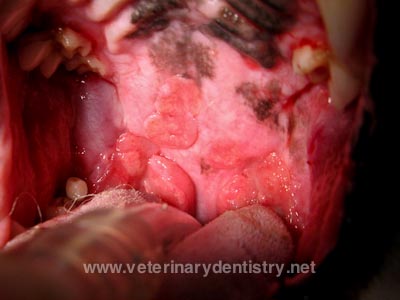 Eosinophilic Stomatitis, Granuloma and CUPS in a Cavalier
King Charles Spaniel. Brett Beckman. veterinarydentistry.net
April 2011. Quote: Eosinophilic stomatitis and eosoniphilic granuloma are common names
associated with raised ulcerative oral lesions in dogs mainly associated with
the palate and occasionally the tongue. The Cavalier King Charles
Spaniel is particularly susceptable. Antigenic stimulation is thought
to be the cause however often specific etiology cannot be determined. ... Biopsy
is indicated to confirm eosinophilic stomatitis. Treatment involves
corticosteroids or cyclosporin and some have advocated hypoallergenic foods.
Some patients do not respond to any of the above recommendations. A
determination must be made to assess patient pain. Some of these patients
appear not to be painful, however careful evaluation is needed to determine oral
pain for this condition in dogs. Palpation under light sedation may reveal jaw
chattering.
Eosinophilic Stomatitis, Granuloma and CUPS in a Cavalier
King Charles Spaniel. Brett Beckman. veterinarydentistry.net
April 2011. Quote: Eosinophilic stomatitis and eosoniphilic granuloma are common names
associated with raised ulcerative oral lesions in dogs mainly associated with
the palate and occasionally the tongue. The Cavalier King Charles
Spaniel is particularly susceptable. Antigenic stimulation is thought
to be the cause however often specific etiology cannot be determined. ... Biopsy
is indicated to confirm eosinophilic stomatitis. Treatment involves
corticosteroids or cyclosporin and some have advocated hypoallergenic foods.
Some patients do not respond to any of the above recommendations. A
determination must be made to assess patient pain. Some of these patients
appear not to be painful, however careful evaluation is needed to determine oral
pain for this condition in dogs. Palpation under light sedation may reveal jaw
chattering.
Keratoconjunctivitis associated with eosinophils in dogs: A retrospective study of 35 cases (2004-2009). G. de Geyera, I. Raymond-Letronb. Pratique Medicale et Chirurgicale de l'Animal de Compagnie. doi:10.1016/j.anicom.2011.09.002. Quote: The objective of this study is to present the clinical and histopathologic features of dogs with keratoconjunctivitis selected based on eosinophils detected in corneal histopathology. Thirty-five cases were reviewed focusing on breed, history, ophthalmic lesions, results of cytology and intradermal allergy testing for 19 allergens, and response to treatment which included keratectomy, topical antibiotics, and corticosteroids in variable conjunction with cyclosporine. Results are: patients included 18 males and 17 females, 9 months to 12 years of age (mean 6.8 years). Among the 34 pure bred dogs were seven Boxers, five French Bulldogs and four Labrador Retrievers. History was that of uni- or bilateral chronic or recurrent corneal ulcers or chronic keratitis. Lesions most commonly were located in the temporal cornea with vessels extending from the limbus centrally to the mid-periphery to form a dense meshwork of thin vessels with an associated superficial stromal infiltrate and a superficial ulcer and associated corneal edema. Conjunctival inflammation and follicular hyperplasia of the bulbar surface of the third eyelid were a consistent finding. Ocular surface cytology showed a predominance of neutrophils and lymphocytes and infrequently eosinophils. Intradermal allergy testing showed a positive reaction to injected aeroallergens in 23 of 26 tested dogs with house dust mite the most common positive allergen. Corneal histopathology showed a hyperplasic epithelium, a lacking basal membrane in the area of corneal defect, an epitheliostromal clivage, a hyalinized acellular zone on the superficial stroma, and corneal infiltrate with neutrophils, monocytes and variable eosinophils. Treatment was effective in all dogs with complete resolution of the ulcers; variable recurrence was successfully managed by topical corticosteroids. In conclusion, this study indicates that eosinophils may participate to the corneal infiltrate of dogs with keratitis associated or not with chronic or recurrent ulcer. Hypotheses include an allergy.
What You Need to Know About Oral Eosinophilic Diseases in Dogs and Cats. There are many appearances to canine and feline oral eosinophilic disease, so know what to look for. John R. Lewis. Vet. Practice News. August 2015. Quote: I recently saw a 5-year-old, spayed Cavalier King Charles spaniel named Kaycee. Kaycee was presenting for a dental cleaning and possible extractions. Due to Kaycee's signalment, I knew I should take a look at her soft palate during her conscious and anesthetized examinations. When I opened Kaycee's mouth, I could see irregularity to the entire surface of the soft palate, with multiple areas of ulceration. Some of the areas of ulceration had areas of yellow punctate raised plaques. Cavaliers are commonly affected by a condition called eosinophilic stomatitis, which often manifests on the soft palate. This inflammatory condition is similar to eosinophilic granulomas in cats, but histologically, the disease in Cavaliers often lacks granuloma formation. ... Diagnosis: Diagnosis can sometimes be made with fine-needle aspirate and cytology. However, patients requiring diagnosis of an oral swelling often require sedation or anesthesia due to the challenges of obtaining a fine-needle aspirate in the conscious patient, and concerns for patient movement causing the needle to poke an undesired structure, such as the eye. If sedation or anesthesia is necessary, it may be best to forego cytology in lieu of an incisional biopsy to provide the pathologist with more tissue to make an accurate diagnosis. Histopathology may show infiltration of eosinophils, neutrophils and flame figures, which are aggregates of eosinophilic granules, nuclear debris and collagen. ... Treatment: In one of the earlier published case series of three Cavalier King Charles spaniels affected by eosinophilic stomatitis, treatment with corticosteroids led to resolution of one case and partial resolution of a second. A third case resolved spontaneously without therapy. The potential for spontaneous resolution makes it difficult for clinicians to know which therapies truly work, versus the waxing and waning nature of the disease. Treatment often revolves around removal of offending allergens. ... My personal experiences with use of amoxicillin-clavulanate or other antibiotics for eosinophilic diseases have not shown marked improvement.
Gastro-oesophageal reflux disease in 20 dogs (2012 to 2014). Muenster, M., Hoerauf, A. and Vieth, M. Small Anim Pract. May 2017;58(5):276-283. Quote: Objectives: To describe the clinical features of canine gastro-oesophageal reflux disease. Materials and Methods: A search of our medical records produced 20 dogs [including one cavalier King Charles spaniel] with clinical signs attributable to oesophageal disease, hyper-regeneratory oesophagopathy and no other oesophageal disorders. The clinical, endoscopic and histological findings of the dogs were analysed. Results: The 3-year incidence of gastro-oesophageal reflux disease was 0·9% of our referral dog population. Main clinical signs were regurgitation, discomfort or pain (each, 20/20 dogs) and ptyalism (18/20 dogs). Oesophagoscopy showed no (5/20 dogs) or minimal (13/20 dogs) mucosal lesions. In oesophageal mucosal biopsy specimens, there were hyperplastic changes of the basal cell layer (13/20 dogs), stromal papillae (14/20 dogs) and entire epithelium (9/20 dogs). ... Intraepithelial neutrophils and eosinophilic infiltration were not observed, and none of the dogs had active oesophagitis. ... Eleven dogs received omeprazole or pantoprazole and regurgitation and ptyalism improved in eight and pain diminished in six of these dogs within three to six weeks. Clinical Significance: Our findings suggest that canine gastro-oesophageal reflux disease is a more common clinical problem than hitherto suspected.
Sleep disordered breathing in the Cavalier King Charles Spaniel: a case series. Tom Hinchliffe, Nai-Chieh Liu, Jane Ladlow. BSAVA Congress 2018 Proceedings. April 2018;pp. 479-480. Quote: Objectives: Obstructive sleep apnoea has been widely investigated in human medicine, but relatively few studies have described this condition in veterinary species; to date it has only been reported in English Bulldogs, French Bulldogs and Pugs. The aim of this study was to report the clinical features, diagnosis, management and outcome of five cases of obstructive sleep apnoea in the Cavalier King Charles Spaniel (CKCS). Methods: A retrospective case series was created using hospital records over five years, followed up via recheck appointments and a telephone questionnaire. The inclusion criteria consisted of CKCSs presenting primarily for sleep disorders, which were treated surgically for obstructive anatomical pathology. Results: All five cases presented with stertorous breathing, choking and apnoea during sleep. All cases had a number of co-morbidities: 5/5 had eosinophilic stomatitis and otitis media, 4/5 had mitral valve insufficiency and 3/5 had syringomyelia. CT and rhinoscopy revealed that 5/5 had aberrant nasal turbinates, nasal septal deviation and soft palate thickening and 3/5 had nasopharyngeal thickening and tracheal collapse. Treatment consisted of a combination of laser turbinectomy, folding flap palatoplasty, tonsillectomy, laryngeal sacculectomy and cuneiform process resection. All cases had an improvement in both the incidence and severity of sleep apnoea within a week, with 4/5 cases achieving complete resolution. Conclusions: This study concludes that obstructive sleep apnoea is a condition that may affect the CKCS and all five cases described here improved following surgical intervention. Consequently, sleep disordered breathing is a presentation that veterinary surgeons should be aware of in this breed.
Oral eosinophilic granuloma in one Cavalier King Charles Spaniel. Amanda Rigney-Alden. Mississippi State Univ. Clinicopathological Conference. May 2018. Quote: Canine eosinophilic granuloma (CEG) is rare disease that occurs in dogs. This skin disease usually presents in the oral cavity as papules, nodules, or plaques located on the palate or tonsils, however, case reports have documented CEG lesions in other cutaneous sites such as the nasal planum, ventral abdomen, thorax, metatarsus, prepuce, flank, digit, eyelid, external ear canal, and cheek region.4 Lesions in the oral cavity may or may not be painful and the most common sign owners report in their pet is halitosis. CEG is verified based on histologic diagnosis. Histopathological findings in cases of CEG include granulomatous inflammation surrounding areas of degenerative collagen, but the essential finding is a predominance of eosinophils.2 While the exact cause is unknown, the proposed pathogenesis of CEG is a hypersensitivity reaction to infectious agents, environmental allergies or food allergies.6 Since case reports of CEG frequently involve Siberian Huskies and Cavalier King Charles Spaniel dogs, there is reason to believe that there is a genetic involvement in these breeds. An immunosuppressive dose of corticosteroids at a tapering dose is commonly successful at resolving the lesions, however, antibiotics and chemotherapy drugs have been used in reoccurring lesions.6 In patients with recurring CEG, determining the allergen that provoked the immune response is fundamental in preventing future lesions from developing. The purpose of this case report is to describe the management of one case of CEG in a Cavalier King Charles Spaniel using oral corticosteroids and a food allergy trial. ... The pathogenesis of CEG is presumed to occur following a Type 1 Hypersensitivity immune-mediated reaction.1 When in the face of an environmental or food allergen, eosinophils are involved in the inflammatory response to the foreign material and respond to the antigens inappropriately producing a hypersensitive reaction. Once an eosinophil is activated the cells phagocytize foreign material or degranulate, as well as synthesize cytokines that are involved in the inflammatory response.1 This cascade of eosinophilic degranulation and production of inflammatory cytokines results in a profound tissue reaction. A genetic component to CEG is also suspected to occur in Siberian Husky and Cavalier King Charles Spaniel dog breeds.
Clinicopathological Features, Risk Factors and Predispositions, and Response to Treatment of Eosinophilic Oral Disease in 24 Dogs (2000-2016). Danielle Mendelsohn, John R. Lewis, Kristin Iglesias Scott, Dorothy C. Brown, Alexander M. Reiter. J Vet Dent. March 2019;36(1):25-31. Quote: The objectives of this study were to retrospectively describe clinico-pathological features of eosinophilic oral disease in dogs, to identify possible risk factors or predispositions to the condition, and to report overall treatment response. Canine medical records from a veterinary teaching hospital and private referral practice over a 17-year period were reviewed for a diagnosis of eosinophilic oral disease. Twenty-four dogs with 26 lesions met the inclusion criteria. Patient mean age and body weight were 6.8 (3.8) years and 13.4 kg, respectively. Fifteen breeds were represented including Cavalier King Charles spaniel (16.7%), Labrador retriever (12.5%), and West Highland white terrier (12.5%). Eosinophilic lesions were found in the palate (65.4%), tongue (26.9%), and other oral locations (7.7%). ... The typical presentation of eosinophilic disease in dogs involves only one body system, but there have been reports of multiple systems involved in Cavalier King Charles spaniels. ... Median follow-up time was 5 months. Analysis revealed statistically significant associations between lesion location and body weight (palatal and tongue lesions were more likely in smaller dogs, whereas lesions in the other category [lip or mucosa] were more likely in larger dogs). ... Although all 4 Cavalier King Charles spaniels in this study had lesions on their palate, this was not found to be statistically significant, which may be due to the low sample size. These Cavalier King Charles spaniels, however, all fell within the body weight parameter significantly correlated with palatal lesions. The lesion location/body weight correlation is further supported by 2 case reports of Cavalier King Charles spaniels where the lesions were located to the palate. ... There was a correlation in lesion location and resolution (all dogs with palatal lesions became asymptomatic at their last recheck), and resolution and the use of antibiotics plus prednisone (greater likelihood of resolution without the use of this combination). Seventy percent of asymptomatic dogs resolved without medication or with allergen therapy alone, suggesting that asymptomatic dogs may respond well to conservative management. No associations were found between lesion location and breed, signalment and response to therapy, lesion resolution and the use of glucocorticoids, or significance of peripheral eosinophilia.
CONNECT WITH US