
Patellar Luxation (Loose Knees) in the
Cavalier King Charles Spaniel
-
 What It Is
 - Possible Cause
- Symptoms
- Diagnosis
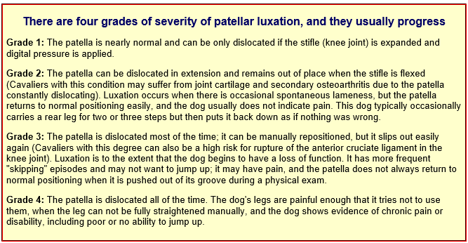
- Grades of Severity
- Treatment
- -- medications
- -- surgery
- -- alternative therapies
- Breeders' Responsibilities
- What You Can Do
- Research News
- Related Links
- Veterinary Resources
Cavalier King Charles spaniels may suffer from a recurring hereditary condition which causes luxating patellas -- loose knee caps -- due to conformational defects (see below). The disorder is believed to affect as many as 20% of cavaliers, therefore the breed is considered to be "at high risk".
What It Is
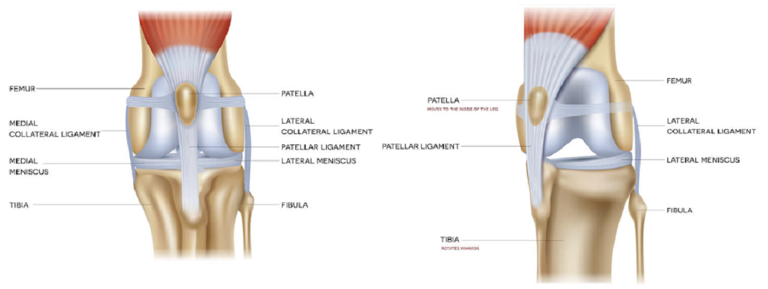
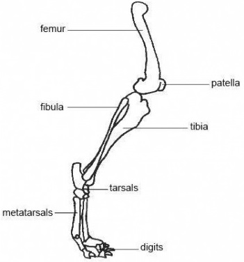
The patella is the dog's knee cap. It should be located in its groove in the center of the stifle (knee joint) of the femur (upper leg bone). A luxating patella is a knee cap that moves out of its groove. It also may be called a floating kneecap. Genetic conditions which cause patellar luxation are a shallow groove, weak ligaments, and misalignment of the muscles and ligaments between the femur, patella, and tibia (lower leg bone).
There are three types of patella luxation: medial (the inner side), lateral (the outer side), and bidirectional (both sides). Medial luxation is the most common form in cavaliers and other small breeds, with bidirectional being second most common. Lateral luxation is more common among larger breeds. A comparison between a normal knee (left) and a medial luxated knee (right) is below. Click on the image to see an enlarged version.
In some cases of this disorder, there are no symptoms and therefore pose no physical problem. These cases are called "occult" conditions. In others, the symptoms are apparent. These cases are referred to as "clinical". In these clinical cases, if the condition is not corrected, it may degenerate: the patella's ridges will wear, its groove will become shallower, and the cavalier will become progressively more lame. Arthritis will prematurely affect the joint, causing a permanently swollen knee with poor mobility.
RETURN TO TOP
Possible Cause
It is generally accepted that luxating patellas are an hereditary condition in the CKCS and some other canine breeds. In veterinary research studies, the disorder has been associated with an hereditary lack of sufficient, normal collagen fibers in the dogs' ligaments and tendons. Ehlers-Danlos syndrome (EDS) is an hereditary connective tissue disease in which the skin is easily torn and there is excessive laxity in the ligaments of joints connecting the dogs' bones, such as the ligaments between the patella and the femor.
In a July 2018 article in which nine dogs (none were cavaliers) with patellar luxation were studied with five normal dogs as the control group. The researchers concluded that patellar luxation in dogs is associated with collagen fiber abnormalities similar to those observed in humans with EDS. See also this 1987 study of a CKCS-mix dog with a similar diagnosis of EDS, although luxaing patellas were not reported among the indications.
RETURN TO TOP
 Symptoms
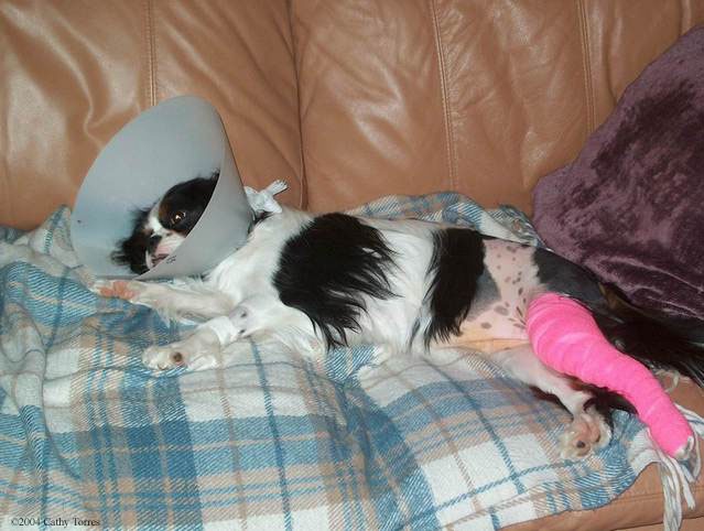
When the loose knee cap pops out of its socket, the dog may hold up its affected hind leg or stretch the leg out or just stop moving and sit down. The dog may also whimper or cry out in pain. Signs vary, and in some cases, the knee cap will re-insert itself, and the dog will move on as if nothing has happened. In other cases, the knee cap will not return spontaneousy, and manual pressure may be required. In the most severe cases, the knee cap will not return despite manual manipulation. See the Grades of Severity below.
RETURN TO TOP
Diagnosis
 Manipulation: Veterinarians can check for patellar luxation by manipulating it. The dog is
examined awake and the veterinarian classifies its degree of luxation. Adult
cavaliers should be evaluated annually, and puppies should be examined at 6-8
weeks of age prior to their release to the new owners.
Manipulation: Veterinarians can check for patellar luxation by manipulating it. The dog is
examined awake and the veterinarian classifies its degree of luxation. Adult
cavaliers should be evaluated annually, and puppies should be examined at 6-8
weeks of age prior to their release to the new owners.
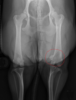
X-ray: X-rays (radiographs) can confirm a displaced patella. The relocation of the patella can be observed in the x-rays as being either towards the center-line of the dog's body (called medial patellar luxation or MPL) or in the opposite direction (called lateral patellar luxation).
In the x-ray image here, the 12-month-old cavalier's right patella, outlined in red, can be seen to be displaced medially (towards the center of the dog's body), when compared with the left patella, which is in its proper location in the center groove of the upper leg bone, the femur.
The Orthopedic Foundation for Animals (OFA) has a diagnosis protocol and registration for examining dogs for patellar luxation.
• Grades of Severity
Treatment
-- medications
Dogs with milder grades of luxation often are prescribed non-steroidal anti-inflammatory drugs (NSAIDs), such as such as carprofen* (Rimadyl, Quellin), meloxicam (Metacam), firocoxib (Previcox), mavacoxib (Trocoxil), deramaxx (Deracoxib), and aspirin** to relieve the pain but not any but not the deterioration. NSAIDs should not be administered without supervision by a veterinarian. The US Food & Drug Administration (FDA) warns about the dangers of using NSAIDs here on its website.
*Carprofen
(Rimadyl, Quellin)
may have serious side effects and should not be given without a
veterinarian's close guidance and monitoring.
**Aspirin
may also have serious side effects and should not be given without a
veterinarian's close guidance and monitoring.
The anticonvulsant (antiepileptic) gabapentin (Neurontin, Gabarone), has been successful in relieving neuropathic pain. Gabapentin works through a receptor on the membranes of brain and peripheral nerve cells. It binds to calcium channels and modulates calcium influx as well as influences GABergic neurotransmission. Its effect is to deaden the irritated nerve impulses in the dog's neck. In humans, gabapentin reportedly does not interact with any other medications, and it is not metabolized, so it is fully excreted in the urine and has no affect upon the liver. However, in dogs, gabapentin is partially metabolized in the liver, and therefore the prescribing neurologist may be expected to order periodic blood tests to check the liver enzymes.
In an August 2020 article, researchers studied the treatment records of 240 dogs suffering chronic pain due to a variety of diagnosed causes, including osteoarthritis (84.6%), generalized, nonspecific back pain (9.2%), intervertebral disk disease (7.5%), luxating patellas (6.67%), and 56.67% not having a definitive anatomical cause for their pain. They report finding that:
"The results from this case series suggest that gabapentin is well-tolerated at much higher doses than what is typically prescribed. Side effects were uncommon, with no clear pattern based on dose, or dog size or age. Therefore, like many other analgesic medications, the efficacy of gabapentin appears patient-specific and should be dosed to effect until side effects are noted or analgesia is achieved.
-- surgery
Veterinary orthopedic surgery usually is required to correct more severe
stages of the condition, such as
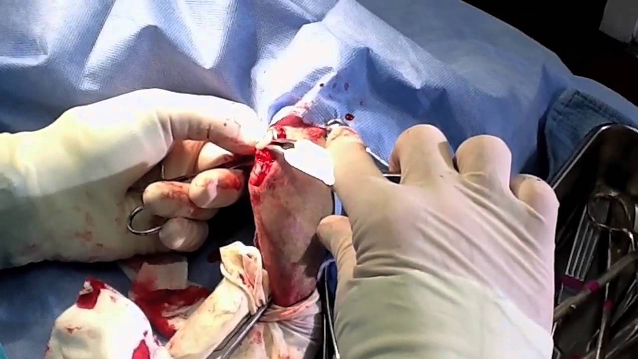 Grade 3 or 4. The groove may be surgically deepened to better
contain the patella. The patella itself may be tied down laterally (on its
outside), to prevent it from deviating medially (toward the inside). The bony
protuberance at the point the quadriceps tendon attaches to the tibia may be cut
off and then re-attached in a more lateral position.
Grade 3 or 4. The groove may be surgically deepened to better
contain the patella. The patella itself may be tied down laterally (on its
outside), to prevent it from deviating medially (toward the inside). The bony
protuberance at the point the quadriceps tendon attaches to the tibia may be cut
off and then re-attached in a more lateral position.
Recovery from patella surgery includes a lot of downtime, resting in a crate, with very limited physical activity, for as long as the surgeon recommends.
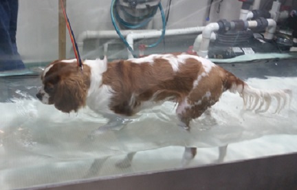 -- alternative therapies
In some cases, non-traditional therapies such as holistic healing or homeopathy, including acupuncture, and joint supplements such as Glyco-Flex II by VetriScience, may be useful alternatives to conventional medicine and surgery. Some physical therapy, such as hydrotherapy (swimming) (right), may also be prescribed. Dogs with patellar luxation should not be over-weight.
RETURN TO TOP
 Breeders' Responsibilities
The Canine Inherited Disorders Database recommends that any dogs with patellar luxation not be bred, nor should their parents or littermates. Because of the strong hereditary relationship, all cavalier King Charles spaniel breeding stock should be examined by qualified veterinarians at least annually and cleared for patellar luxation, the closer the examination to the breeding the better.
The Cavalier King Charles Spaniel Club, USA (CKCSC,USA) recommends that, prior to breeding any cavalier, the dog have no evidence of patellar luxation from an evaluation by a licensed veterinarian. The American Cavalier King Charles Spaniel Club (ACKCSC) states that "Cavaliers used for breeding should have within normal limits patellas as determined by an OFA examination at age one. The patellas should be reevaluated as the Cavalier ages."
The Canine Health Information Center (CHIC) is a centralized canine health database sponsored by the AKC/Canine Health Foundation (AKC/CHF) and OFA. The CHIC, working with participating parent clubs, provides a resource for breeders and owners of purebred dogs to research and maintain information on the health issues prevalent in specific breeds.
AKC's national breed clubs establish the breed specific testing protocols. Dogs complying with the breed specific testing requirements are issued CHIC numbers. The ACKCSC requires that, to qualify for CHIC certification, cavaliers must be screened for patellar luxation (OFA).
RETURN TO TOP
What You Can Do
Exercise your dog regularly. Short walks at first may become longer ones as the hind leg muscles are strengthened over time. All dogs physically capable of walking should be walked to keep their legs healthy.
In this case, the "What You Can Do" section really should be labeled "What You Should Not Do":
• Do not over-feed your cavalier. Do not allow your cavalier to become obese. Follow the Body Condition Scoring Charts on our Diets webpage.
• Do not allow cavalier puppies to gain weight rapidly.
• Do not over-supplement your cavalier with calcium.
• Do not neuter your cavaliers before they reach physical maturity. The reproductive hormones have a great influence on bone development. They initiate growth plate closure in long bones. They are responsible for maintaining tendon, ligament, and muscle integrity. There is a current theory that their abrupt removal may lead to weakening of joints and musculature as well as changes in joint conformation.
RETURN TO TOP
Research News
 December
2023:
Cavaliers ranked second in most common breeds diagnosed with
bidirectional patellar luxation in Japanese study. In a
December 2023 article, Japanese investigators (Itaru Mizutani, Reo
Nishi, Masahiro Murakami [right]) reported finding that among
130 dogs diagnosed with medial (MPL) or bidirectional patellar luxation
(BPL), the toy poodle was the most commonly diagnosed breed in the BPL
group, representing 53.5% of BPL dogs, and the cavalier King Charles
spaniel ranked second, representing 50.0% of BPL dogs.
December
2023:
Cavaliers ranked second in most common breeds diagnosed with
bidirectional patellar luxation in Japanese study. In a
December 2023 article, Japanese investigators (Itaru Mizutani, Reo
Nishi, Masahiro Murakami [right]) reported finding that among
130 dogs diagnosed with medial (MPL) or bidirectional patellar luxation
(BPL), the toy poodle was the most commonly diagnosed breed in the BPL
group, representing 53.5% of BPL dogs, and the cavalier King Charles
spaniel ranked second, representing 50.0% of BPL dogs.
August 2020:
Colorado State Univ. researchers find gabapentin is
well-tolerated at high dosages for dogs in chronic pain.
 In
an
August 2020 article, a team of Colorado State University researchers
(Lily V Davis, Peter W Hellyer, Robin A Downing, Lori R Kogan [right])
studied the treatment records of 240 dogs suffering chronic pain due to
a variety of diagnosed causes, including osteoarthritis (84.6%),
generalized, nonspecific back pain (9.2%), intervertebral disk disease
(7.5%), luxating patellas (6.67%), and 56.67% not having a
definitive anatomical cause for their pain. They
report finding that:
In
an
August 2020 article, a team of Colorado State University researchers
(Lily V Davis, Peter W Hellyer, Robin A Downing, Lori R Kogan [right])
studied the treatment records of 240 dogs suffering chronic pain due to
a variety of diagnosed causes, including osteoarthritis (84.6%),
generalized, nonspecific back pain (9.2%), intervertebral disk disease
(7.5%), luxating patellas (6.67%), and 56.67% not having a
definitive anatomical cause for their pain. They
report finding that:
"The results from this case series suggest that gabapentin is well-tolerated at much higher doses than what is typically prescribed. Side effects were uncommon, with no clear pattern based on dose, or dog size or age. Therefore, like many other analgesic medications, the efficacy of gabapentin appears patient-specific and should be dosed to effect until side effects are noted or analgesia is achieved.
February 2020:
Cavaliers were second-most-common breed in UK study of Grade II
patellar luxation study.
 In a
February 2020 article, a team of UK orthopedic surgeons (L.
Hamilton, Mike Farrell [right], B. Mielke, M. Solano, S. Silva, I. Calvo) studied
the risk of lameness due to patellar luxation (looseness of the kneecap)
and the rate of surgery required to correct the disorder. A total of 38
dogs were included in the study, including 6 cavaliers (16%) -- the
second most common breed in the study, behind Staffordshire bull
terriers. All dogs were diagnosed with
Grade II patellar luxation, meaning that the patella may luxate
during stifle flexion or manual manipulation and remains luxated until
stifle extension or manual replacement occurs. Grade II is considered
border-line as far as the need for surgery to correct spontaneous
luxation, depending upon the frequency of the instances. Of the 38 dogs
in the study, 17 (including one CKCS) required unscheduled surgeries.
While the average time period between first diagnosis and the need for
surgery was 15 months, for the cavalier, the time period was 7 years.
One other cavalier had chronic lameness after 4 months following
diagnosis, but surgery was not performed due to financial reasons. The
other 4 cavaliers did not experience sufficient long term lameness to
require surgery.
In a
February 2020 article, a team of UK orthopedic surgeons (L.
Hamilton, Mike Farrell [right], B. Mielke, M. Solano, S. Silva, I. Calvo) studied
the risk of lameness due to patellar luxation (looseness of the kneecap)
and the rate of surgery required to correct the disorder. A total of 38
dogs were included in the study, including 6 cavaliers (16%) -- the
second most common breed in the study, behind Staffordshire bull
terriers. All dogs were diagnosed with
Grade II patellar luxation, meaning that the patella may luxate
during stifle flexion or manual manipulation and remains luxated until
stifle extension or manual replacement occurs. Grade II is considered
border-line as far as the need for surgery to correct spontaneous
luxation, depending upon the frequency of the instances. Of the 38 dogs
in the study, 17 (including one CKCS) required unscheduled surgeries.
While the average time period between first diagnosis and the need for
surgery was 15 months, for the cavalier, the time period was 7 years.
One other cavalier had chronic lameness after 4 months following
diagnosis, but surgery was not performed due to financial reasons. The
other 4 cavaliers did not experience sufficient long term lameness to
require surgery.
August 2016:
Cavaliers were 10% of 5-year study of bilateral surgeries for patellar
luxation.
 In an
August 2016 article, veterinary surgeons (Javier Gallegos [right],
Marcos Unis, James K. Roush, Lori
Agulian) studied the results of 50
single-session bilateral corrective surgeries for congenital medial
patellar luxations. Five of the 50 dogs were cavalier King Charles
spaniels (CKCS). All dogs were under 33 lbs. Of 11 dogs with
post-operative complications, one was a CKCS. The surgeries were
performed at the Kansas State University's veterinary school and at a
Fountain Valley, California VCA referral center. The authors concluded
that:
In an
August 2016 article, veterinary surgeons (Javier Gallegos [right],
Marcos Unis, James K. Roush, Lori
Agulian) studied the results of 50
single-session bilateral corrective surgeries for congenital medial
patellar luxations. Five of the 50 dogs were cavalier King Charles
spaniels (CKCS). All dogs were under 33 lbs. Of 11 dogs with
post-operative complications, one was a CKCS. The surgeries were
performed at the Kansas State University's veterinary school and at a
Fountain Valley, California VCA referral center. The authors concluded
that:
"Single-session bilateral corrective surgery for MPL [medial patellar luxation] is well tolerated in small dogs with complication rates historically similar to dogs undergoing unilateral or staged bilateral surgery. With individual case assessment, single-session surgery could be offered to owners, allowing one anesthetic episode and potentially lowering the chance for morbidity. Further studies, ideally prospective studies, are needed to assess long-term outcome following single-session bilateral corrective surgery for MPL."
 March
2016:
UK orthopods report CKCSs are over-represented for
medial patella luxation. In a
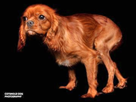
March 2016 article, UK veterinary orthopedists (Albane Fauron, Karen
Perry) report that cavalier King Charles spaniels are "consistently
over-represented" for medial patella luxation. In their article, they
summarize the pathophysiology and diagnosis of patella luxation (PL) and
use one of their cavalier patients (at right) as an example of a dog
affected with the medial version (near the mid-line of the knee) of PL.
March
2016:
UK orthopods report CKCSs are over-represented for
medial patella luxation. In a
March 2016 article, UK veterinary orthopedists (Albane Fauron, Karen
Perry) report that cavalier King Charles spaniels are "consistently
over-represented" for medial patella luxation. In their article, they
summarize the pathophysiology and diagnosis of patella luxation (PL) and
use one of their cavalier patients (at right) as an example of a dog
affected with the medial version (near the mid-line of the knee) of PL.
RETURN TO TOP
Related Links
RETURN TO TOP
Veterinary Resources
Ehlers-Danlos syndrome in a dog: ocular, cutaneous and articular abnormalities. K. C . Barnett, Beverley D. Cottrell. J. Sm. Anim. Pract. October 1987;8(10):941-946. Quote: Ehlers-Danlos syndrome (EDS) is an hereditary connective tissue disease in man in which the skin is easily torn. A similar condition has been described in dogs and other animals. This case report records a case in the United Kingdom in which the whole syndrome was exhibited: skin fragility, joint laxity and ocular signs of bilateral lens luxation, cataract and corneal oedema. It is the first report of ocular signs in EDS in the dog and joint laxity has been reported only rarely. ... A crossbred bitch (Cavalier King Charles Spaniel x Rorder Collie bitch) was presented at the age of 12 months for investigation of failing vision. ... In addition, excessive joint laxity was present, both flexion and extension, in the carpal, carpo-metatarsal and all interphalangeal joints. ... EDS is recognized as an hereditary disease in both man and dog and has been reported in the latter in the following breeds:- English Springer Spaniel, Beagle, Manchester Terrier, Welsh Corgi, Red Kelpie, Dachshund, Boxer, St Bernard, German Shepherd Dog, Greyhound, mongrel and mixed breeds (Beagle and Dachshund in the United Kingdom). The case reported here was a Cavalier King Charles Spaniel x Border Collie; neither breed has previously been implicated. EDS is due to an autosomal dominant gene in dogs, as in man and mink, and the fact that this case was in a crossbred dog (two different breeds) indicates that recessive inheritance is most unlikely. Therefore, further cases in one or other of these two breeds might be anticipated.
Patellar luxation in the dog and cat. John Ferguson. In Practice. April 1997;19(4):174-184. Quote: "Patellar luxation is a commonly seen orthopaedic problem in veterinary practice. Diagnosis of the condition is usually straightforward. However, a knowledge of the normal anatomy, function and interrelationship between the hip and stifle joints, femur, tibia and soft tissues is necessary if the surgeon is to choose the most appropriate method of treatment. This article describes medial and lateral patellar luxation, the clinical signs, diagnosis and treatment options available in the dog and cat. ... . Cavalier King Charles spaniel: a breed with a high incidence of patellar luxation."
Control of Canine Genetic Diseases. Padgett, G.A., Howell Book House 1998, pp. 198-199, 246.
Guide to Congenital and Heritable Disorders in Dogs. Dodds WJ, Hall S, Inks K, A.V.A.R., Jan 2004, Section II(235).
Complications Associated with Corrective Surgery for Patellar Luxation in 109 Dogs. Gareth I. Arthurs and Sorrel J. Langley-Hobbs. Vet. Surg. Aug 2006; 35:559.
Patellar luxation. Greg Harasen. Can Vet J. 2006 August; 47(8): 817-818. Quote: "Congenital luxation of the patella represents one of the most common orthopedic conditions in small animal practice. Medial luxations account for 75% to 80% of cases in all breeds. The majority of patients are small breed dogs including ... cavalier King Charles spaniels. ... The overwhelming majority of patellar luxation are congenital and certainly hereditary, although a mode of inheritance has not been described."
Diagnostic and genetic aspects of patellar luxation in small and miniature breed dogs in Austria. B. Vidoni, I. Sommerfeld-Stur, E. Eisenmenger. European J. of Companion Anim. Pract. October 2006;16(2):149-158. Quote: During a period of eight years (1996 - 2004) 432 small and miniature breed dogs were screened for patellar luxation (PL). In order to achieve the diagnostic accuracy required for genetic screening to assist breeding programmes, examinations were based on the concept of a standardized examination protocol for patellar luxtion. Diagnostic criteria assessed by physical examination, inspection and palpation focussed on lameness, evaluation of patellar tracking in the standing and recumbent position, with special focus on patello-femoral instability, as well as on the deviation of the tibial tuberosity and any perceivable crepitation of the stifle joint during manipulation. Evaluation of all findings was made on the basis of PUTNAM´s (1968) classification. Radiographic examinations were not performed. Patellar luxation (unilateral or bilateral, medial and/or lateral) was diagnosed in 61.6 % of the examined dogs, but permanent lameness was only present in 15.5 % (right stifl e) and 12.8 % (left stifl e), respectively. Intermittent lameness was observed in only 3.5 % (right stifle) and 4.6 % (left stifle), respectively. This means that almost 40 % of all dogs with patellar luxation are asymptomatic and their condition would not have been detected without diagnostic screening. The different diagnostic criteria showed signifi cant correlation between each other and with the final findings. In some parameters like "luxation in standing position" and "luxation in recumbent position", the correlation with fi nal fi ndings was particularly high. Thus, the examination protocol used in this study appears to be suitable for PL screenings in dogs. Investigation of the influence of parameters like body weight, age, gender and neutering on the presence of PL showed that, except for gender, all attributes were associated with the occurrence of PL. An increase in body weight of 1 kg decreased the odds of suffering from PL to the 0.8fold (p<0.05), while an increase of age of one year increased the odds to the 1.1 fold (p<0.051). For neutered dogs, the odds showed a 3.1 fold increase of being affected by PL (p<0.05). No signifi cant infl uence could be observed for the gender of the animals. In order to detect breed predispositions for patellar luxation, odds ratios were calculated for all breeds represented in the study by more than ten animals. The breeds involved were: Jack Russell Terrier, Pug, Papillon, Pekingese, Shih Tzu, Tibet Terrier, West Highland White Terrier, Poodle, Yorkshire Terrier, Maltese Terrier and Chihuahua. [Cavalier King Charles spaniels were included in the "Other Breeds" category.] Only two breeds showed odds ratios that were significantly different from 1: In the Jack Russell Terrier, the odds ratio was signifi cantly lower (0.31) (95 % confidence intervals 0.14-0.67), while the odds ratio was significantly increased (5.62) in Poodles (Miniature and Toy Poodles) (95 % confi dence intervals 1.93-16.41). This means that Jack Russell Terriers have a comparatively reduced risk of suffering from PL, while the chance to develop the condition seems increased in Poodles. These results are highly indicative of a genetic background but further investigation on the basis of familial anamneses and heritability studies is required to support this postulate. A standardized examination technique and offi cial validation of the PL screening tests represent an essential precondition for the acceptance of PL screening programmes by breeders. Based on the results of this study, it is strongly recommended to implement a uniform, internationally valid and highly accurate diagnostic screening programme for patellar luxation. At the moment, this screening protocol is used by veterinarians in Germany, Switzerland and Austria.
Frequency and distribution of patellar luxation in dogs. 134 cases (2000 to 2005). M R Alam, J I Lee, H S Kang, I S Kim, S Y Park, K C Lee, N S Kim. Vet. & Comparative Ortho & Traum. January 2007;20(1):59-64. Quote: "This study investigated the frequency and distribution of patellar luxation in the dogs presented to the Chonbuk National University Animal Medical Centre during January 2000 to September 2005. Patellar luxations were classified as medial or lateral, and unilateral or bilateral, were graded I to IV, and were subdivided according to age, sex and size of the dogs. The incidence of medial patellar luxation (MPL) was greater than the incidence of lateral patellar luxation (LPL) in both small and large dogs. Small-breed dogs were admitted almost exclusively with MPL. LPL was found uncommon; however it was observed more often in larger-breed dogs. Surgical correction was performed primarily in the dogs (165 stifles in 111 dogs) with grade II, III and IV patellar luxations following different surgical techniques. The combination of the surgical techniques was found to be more effective for the management of the disease. The prognosis was found to be favourable, because when the grade was low, the dog was younger, without cruciate ligament rupture, and as the surgical correction was performed with combination of more techniques."
The prevalence of canine patellar luxation in three centres. Clinical features and radiographic evidence of limb deviation. N. Bound; D Zakai; S. J. Butterworth; M. Pead. Vet.Comp.Orthop.Traumatol. Jan. 2009. Quote: "The medical records of 155 dogs with patellar luxation (PL) from three different centres were analysed. [Cavalier King Charles spaniels were the second most common. See Figure 3 of the article.] Each case was classified according to the nature of its luxation and any concurrent orthopaedic conditions plus the age at diagnosis were also noted. Measurements relating to angle of inclination (AOI) of the femoral neck and medio-lateral bowing of the femur and tibia at the stifle were also recorded. The femoral and tibial data were compared to dogs with another orthopaedic condition in a case-control assessment. Labradors were most commonly affected (21%). Most luxations were medial (92%) and 54% of affected dogs were female. The mean AOI of the hip was 148.95°. There was a statistically significant difference between the stifles of dogs with PL compared to a control population. This study concluded that PL in large breeds is increasing. Lateral luxation was uncommon and was not associated exclusively with large breeds. Females were more likely to have PL than males and being female was a risk factor associated with coxa valga. There are significant differences in medio-lateral stifle conformation between dogs with PL and control dogs."
Breed Predispositions to Disease in Dogs & Cats (2d Ed.). Alex Gough, Alison Thomas. 2010; Blackwell Publ. 52.
Surgical treatment of medial patellar luxation without femoral trochlear groove deepening procedures in dogs: 91 cases (1998-2009). William R. Linney,Douglas L. Hammer, Susan Shott. J Am Vet Med Assoc. 2011;238:1168-117. Quote: "Objective: To assess signalment, outcomes, and complications for dogs surgically treated for medial patellar luxation (MPL) with a combination of lateral retinacular imbrication and tibial crest transposition procedures without femoral trochlear groove deepening techniques, and to determine whether osteoarthritis progressed in these patients during the 8-week period following surgery. Design: Retrospective case series. Animals: 91 dogs. ... The most commonly represented breed was mixed, followed by Cavalier King Charles Spaniel ... Procedures: Medical records were reviewed for information on signalment, clinical history, unilateral versus bilateral disease, preoperative and postoperative MPL grades, duration of follow-up, and perioperative and postoperative complications. Radiographs obtained preoperatively and during 8-week follow-up examinations were reviewed and assigned degenerative joint disease (DJD) scores (range, 0 to 3). Data were analyzed to determine factors influencing outcomes. Kaplan-Meier curves were constructed for recurrence of MPL. Results: Minor postoperative complications were reported for 31 of 91 (34.1%) dogs. Patellar reluxation occurred in 18 of 91 (19.8%) dogs. Reluxation or complications for which additional surgery was recommended developed in 6 of 91 (6.6%) dogs. At last clinical follow-up, 10 of 91 (11.0%) dogs had at least occasional lameness. No difference was revealed between preoperative and postoperative (8-week follow-up) radiographic DJD scores. Conclusions and Clinical Relevance: Results of surgical treatment of MPL without femoral trochlear groove deepening procedures were comparable to those in studies of surgical treatment that included groove deepening procedures. Radiographic indices of DJD did not increase during the 8 weeks following surgery. These results suggest that trochlear groove deepening procedures are not always necessary, and patients that undergo these techniques should be carefully selected."
Genetic Connection: A Guide to Health Problems in Purebred Dogs, Second Edition. Lowell Ackerman. July 2011; AAHA Press; pg 134. Quote: "Table 9.10 -- Breeds at Risk for Patellar Luxation on the Basis of a Large-Scale Epidemiologic Study: ... 8. Cavalier King Charles spaniel -- odds ratio 9.1."
Genome-wide survey indicates involvement of loci on canine chromosomes 7 and 31 in patellar luxation in flat-coated retrievers. Ineke C Lavrijsen, Peter A Leegwater, Chalika Wangdee, Frank G. van Steenbeek, Monique Schwencke, Gert J. Breur, Freek J. Meutstege, Isaac J. Nijman, Edwin Cuppen, Henri C. Heuven, Herman A. Hazewinkel. BMC Genetics. May 2014. Quote: "Background: Patellar luxation is an orthopedic disorder in which the patella moves out of its normal location within the femoral trochlea of the knee and it can lead to osteoarthritis, lameness, and pain. In dogs it is a heritable trait, with both environmental and genetic factors contributing to the phenotype. The prevalence of patellar luxation in the Dutch Flat-Coated Retriever population is 24%. In this study, we investigated the molecular genetics of the disorder in this population. Results: Genome-wide association analysis of 15,823 single nucleotide polymorphisms (SNPs) in 45 cases and 40 controls revealed that patellar luxation was significantly associated with a region on chromosome CFA07, and possibly with regions on CFA03, CFA31, and CFA36. The exons of the genes in these regions, 0,5 Mb combined, were analyzed further. These exons from 15 cases and a pooled sample from 15 controls were enriched using custom genomic hybridization arrays and analyzed by massive parallel DNA sequencing. In total 7257 variations were detected. Subsequently, a selection of 144 of these SNPs were genotyped in 95 Flat-Coated Retrievers. Nine SNPs, in eight genes on CFA07 and CFA31, were associated with patellar luxation (P <10-4). Genotyping of these SNPs in samples from a variety of breeds revealed that the disease-associated allele of one synonymous SNP in a pseudogene of FMO6 was unique to Flat-Coated Retrievers. Conclusion: Genome-wide association analysis followed by targeted DNA sequencing identified loci on chromosomes 7 and 31 as being involved in patellar luxation in the Flat-Coated Retriever breed."
Corrective surgery for canine patellar luxation in 75 cases (107 limbs): landmark for block recession. Mitsuhiro Isaka, Masahiko Befu, Nami Matsubara, Mayuko Ishikawa, Yurie Arase, Toshiyuki Tsuyama, Akiko Doi, Shinichi Namba. Vet.Sci.Development. May 2014;4(1). Quote: "Canine medial patellar luxation (MPL) is a very common orthopedic disease in small animals. Because the pathophysiology of this disease involves various pathways, the surgical techniques and results vary according to the veterinarian. Further, the landmark for block recession is not completely clear. We retrospectively evaluated 75 dogs (107 limbs) with MPL in whom our landmark for block recession was used from July 2008 to May 2013. Information regarding the breed, age, sex, body weight, body condition score (BCS), lateral vs. bilateral, pre-operative grading, surgical techniques, removal of implants, concomitance with anterior cruciciate ligament (ACL) rupture, re-luxation, re-operation, and rehabilitation was obtained from the medical records. The breeds were as follows: Chihuahua (n=23), Pomeranian (n=12), Yorkshire Terrier (n=9), and so on [2 cavalier King Charles spaniels with 3 limbs]. The study group consisted of 33 males (castrated n=13) and 42 females (spayed n=21). The median age was 53.3±35.9 months old (32-146 months); 13 cases were less than 12 months of age (17.3%). The pre-surgical BCSs were as follows: 1(n=0), 2(n=20), 3(n=24), 4(n=24) and 5(n=7). The body weight was 4.51±3.48kg (1.34-23.0kg); 71 cases (94.7%) were less than 10 kg. The MPL grades (each limb) were G1 (n=1), G2 (n=18), G3 (n=78), and G4 (n=10); 32 cases were bilateral and 43 cases were unilateral (right n=27; left n=16). The specific surgical procedure (distal femoral osteotomy) was 3 stifles in Chihuahuas. Concurrent with ACL rupture was 16/107 stifles (15.0%) corrected with the over-the-top method or the extracapsular method in Papillons (5/6), Chihuahuas (5/23), and so on. The occurrences of re-luxation and re-operation were 3 out of 107 stifles (2.8%) and 0%, respectively. In this retrospective study, we present a potentially good surgical landmark for block recession of MPL in dogs."
Canine patellar luxation part 1: pathophysiology and diagnosis.
Albane Fauron, Karen Perry. Vet. Times. March 2016.
Quote:
"Patellar luxation (PL) is a commonly diagnosed canine orthopaedic
condition and, in the majority of dogs, is a developmental
condition, as opposed to a traumatic one, resulting from skeletal
abnormalities. ... The Orthopaedic Foundation for Animals (OFA)
named Pomeranians as the highest incidence of PL, with 37.2% of dogs
affected during an evaluation lasting from January 1974 to December
2014 (OFA, 2014). Chihuahuas, poodles, Maltese terriers,
cavalier King Charles spaniels and Yorkshire terriers are
also consistently over-represented across different studies. ...
This figure [at right] shows ... a cavalier King
Charles spaniel that presented with grade II MPL [medial
patellar luxation]. ... Medial patellar luxation (MPL) is the most
common form of PL in dogs of all sizes. Lateral patellar luxation is
less frequent and reported to occur more often in medium to large
breed dogs. Luxations are graded on a four-point scale. Diagnosis is
based on the clinical presentation and thorough orthopaedic
examination. Concomitance of cranial cruciate ligament (CrCL)
pathology has been reported in 13% to 25% of dogs presenting for MPL
and the integrity of the CrCL should be systemically assessed in
these cases. Although radiology is not required for a diagnosis, it
remains critical in the identification of any underlying skeletal
abnormalities.
Outcome Following Surgical Correction of Grade 4 Medial Patellar Luxation in Dogs: 47 Stifles (2001-2012). Eric C. Hans, Sharon C. Kerwin, Alan C. Elliott, Ryan Butler, W. Brian Saunders, Don A. Hulse. J. Amer. Anim. Hosp. Assn. March 2016. Quote: "Grade 4/4 medial patellar luxation (MPL) is a complex disease of the canine stifle that often requires surgical realignment of the patella to resolve clinical lameness. Outcome following surgery remains poorly described. Medical records were retrospectively reviewed for surgical correction of grade 4 MPL. Signalment and exam findings, surgical procedures performed, complications, and clinical outcome were reported. Data was statistically analyzed for association with major complication occurrence and unacceptable function following surgery. Forty-seven stifles from 41 dogs were included. ... Cavalier King Charles spaniels (n=3) ... The surgical procedures most frequently utilized for patellar realignment were the combination of femoral trochleoplasty, tibial tuberosity transposition, and joint capsule modification. Median in-hospital veterinary examination was performed at 69 days (range 30-179 days) following surgery. Full function was reported for 42.6% of cases (n=20). Acceptable function was reported for 40.4% of cases (n=19). Unacceptable function was reported for 17% of cases (n=8). The overall complication rate was 25.5% (n=12), with revision surgery for major complications required in 12.8% of cases (n=6). Corrective osteotomies were associated with major complications (P < 0.001). In general, pelvic limb function improves following surgical correction of grade 4 MPL; however, a return to full function should be considered guarded."
The epidemiology of patellar luxation in dogs attending primary-care veterinary practices in England. O'Neill D, Meeson R, Sheridan A, Church D, Brodbelt D. Canine Genetics & Epidemiology. June 2016;3:4. Quote: Background: Canine patellar luxation is one of the most common orthopaedic disorders of dogs and is a potential welfare concern because it can lead to lameness, osteoarthritis and pain. However, there are limited epidemiological data on the disorder relating to the general population of dogs in England. This study aimed to investigate the VetCompass Programme database of dogs attending primary-care veterinary practices in England to report on the prevalence, risk factors and clinical management of diagnosed patellar luxation cases. Results: The study included all dogs with at least one electronic patient record in the VetCompass database from September 1st, 2009 to August 31st, 2014. Candidate patellar luxation cases were identified using free-text word searching of the clinical notes and VeNom diagnosis term fields. Univariable and multivariable binary logistic regression modelling was used for risk factor analysis. The overall dataset comprised 210,824 dogs attending 119 clinics in England. The prevalence of patellar luxation diagnosis in dogs was 1.30% (95% confidence interval (CI) 1.21-1.39). Of the 751 incident cases, 293 (39.0%) received medical management, 99 (13.2 %) received surgical intervention and 28 (3.7%) were referred for further management. Multivariable modelling documented 11 breeds with increased odds of patellar luxation compared with crossbred dogs, including the Pomeranian (odds ratio [OR]: 6.5, 95% CI 4.0-10.7, P <0.001), Chihuahua (OR: 5.9, 95% CI 4.4-7.9, P <0.001), Yorkshire Terrier (OR: 5.5, 95% CI 4.3-7.1, P <0.001) and French Bulldog (OR: 5.4, 95% CI 3.1-9.3, P <0.001). ... Cavalier King Charles Spaniel (OR: 2.5, 95% CI 1.8-3.5, P <0.001). ... Dogs aged ≥12.0 years showed 0.4 times the odds (95 % CI 0.3-0.5, P <0.001) compared with dogs aged <3.0 years. Females had 1.3 times the odds (95 % CI 1.1-1.5, P <0.001), neutered dogs had 2.4 times the odds (95 % CI 1.8-3.2, P <0.001) and insured dogs had 1.9 times the odds (95 % CI 1.6-2.3, P <0.001). Conclusions: Patellar luxation warrants inclusion as a welfare priority in dogs and control strategies that include this disorder should be considered as worthwhile breeding goals, especially in predisposed breeds.
Postoperative Complications and Short-Term Outcome Following Single-Session Bilateral Corrective Surgery for Medial Patellar Luxation in Dogs Weighing <15 kg: 50 Cases (2009-2014). Javier Gallegos, Marcos Unis, James K. Roush, Lori Agulian. Vet. Surgery. August 2016. Quote: Objective: To assess complication rates and short-term outcome in small dogs with bilateral medial patellar luxation (MPL) undergoing single-session bilateral corrective surgery. Study Design: Retrospective case series. Animals: Dogs weighing <15 kg with congenital bilateral MPL that underwent single-session bilateral corrective surgery (n=50) [including 5 cavalier King Charles spaniels]. Methods: Surgical procedures for MPL correction included trochlear wedge recession (TWR), crest transposition, lateral imbrication, and medial fascial release. Complication rates were correlated with number of surgical procedures, weight, whether or not a bandage was applied postoperatively, and surgeon experience (ACVS Diplomate vs resident). Results were compared with the most recent study evaluating single-session bilateral corrective surgery for MPL. Results: Overall complication rate was 22% (11 of 50 dogs) [including 1 cavalier]. Implant failure occurred in 2 dogs (3 stifles) requiring revision. Grade 1 patella reluxation was the most common minor complication at 10% (5/50). Overall complication and reluxation rates were similar to previous studies. There were no intraoperative complications after performing TWR in small dogs. Conclusion: Single-session bilateral corrective surgery for MPL is well tolerated in small dogs with complication rates historically similar to dogs undergoing unilateral or staged bilateral surgery. With individual case assessment, single-session surgery could be offered to owners, allowing one anesthetic episode and potentially lowering the chance for morbidity. Further studies, ideally prospective studies, are needed to assess long-term outcome following single-session bilateral corrective surgery for MPL.
Lateral patellar luxation in nine small breed dogs. F. Di Dona, G. Della Valle, C. Balestriere, B. Lamagna, L. Meomartino, G. Napoleone, F. Lamagna, G. Fatone. Open Vet. J. December 2016;6(3):255-258. Quote: The objective of this paper was to describe the clinical features, the management and the outcome of nine small breed dogs affected with lateral patella luxation referred during the period between January 2010 and December 2014. Patellar luxations were classified according to: breed, age, sex, weight, and grade of patellar luxation, as well as if unilateral or bilateral, and concurrent cranial cruciate ligament lesion. ... The following breed were registered: Poodle (n=3), Pinscher (n=2), Pekingese, Yorkshire, Cavalier King Charles Spaniel and mixed-breed. ... In affected dogs, surgical correction consisted in the combination of tibial tuberosity transposition and soft tissue procedure. Adjunctive condroplasty or trochleoplasty was performed as needing. The outcome was found positive after surgical management with low complication rate and complications have been easily managed with high success rate. [The data for the CKCS: male; 48 months old; 7.9 kg.; grade 1 luxation; concurrent cranial cruciate ligament (CrCL) lesion; surgery: tibial tuberosity transposition, soft tissue procedure, fabellar suture; complications: seroma, implant migration; outcome: fair.]
Histopathological and electron microscopic study in dogs with patellar luxation and skin hyperextensibility. Kazunori Ueda, Tomoyuki Kawai, Haruki Senno, Atsushi Shimizu, Akira Ishiko, Masahiko Nagata. J. Vet. Med. Sci. DOI: 10.1292/jvms.18-0115 July 2018. Quote: Patellar luxation is abnormal displacement of the patella from the femoral trochlear groove. It is seen primarily in small breed dogs and causes pain and limited mobility of the stifle joint. This study aimed to investigate the relationship among patellar luxation, skin extension, and skin collagen fibril diameter. Nine dogs with patellar luxation and five clinically normal dogs were enrolled in the study. We measured the skin extension and investigated the ultrastructure of the skin and patellofemoral ligament by histopathology and transmission electron microscopy. The mean skin extension in dogs with patellar luxation was 18.5 ± 5.5% which is greater than the reference value (14.5%). Mean skin extension in controls was 8.8 ± 1.7% and was within the normal range. In dogs with patellar luxation, histopathology of the skin and patellofemoral ligament showed sparse and unevenly distributed collagen fibers. Transmission electron microscopy identified poorly organized, irregularly shaped, thin collagen fibrils. Collagen fibril thickness in dogs with patellar luxation was significantly less than fibril thickness in controls (P<0.001). There was a significant negative correlation (ρ= −0.863; P<0.001) between skin collagen fibril diameter and skin extension. Skin extension was correlated with patellar luxation and disease severity. Dogs with patellar luxation, joint dysplasia, and hyperextensible skin appear to be pathologically related. This might represent a phenotype of the Ehlers-Danlos syndrome, a hereditary connective tissue disorder in humans.
The natural history of canine occult Grade II medial patellar luxation: an observational study. L. Hamilton, M. Farrell, B. Mielke, M. Solano, S. Silva, I. Calvo. J. Sm. Anim. Pract. February 2020; doi: 10.1111/jsap.13093. Quote: Objectives: To determine the risk of lameness and the rate of subsequent medial patellar luxation surgery in dogs that present with occult Grade II medial patellar luxation. Materials and methods: Retrospective owner survey and review of clinical records of adult dogs diagnosed with Grade II medial patellar luxation that were initially asymptomatic and managed non‐surgically that had a minimum of 4-year follow-up. Clinical notes and owner questionnaires identified dogs that subsequently developed lameness and required surgery on the previously asymptomatic stifle. Results: Thirty-eight dogs were included with an average follow-up of 51 months. ... The most common breeds were Staffordshire bull terriers (n = 7), cavalier King Charles spaniels (n = 6) and Yorkshire terriers (n = 5). ... Seventeen dogs (one CKCS) represented for unscheduled contralateral medial patellar luxation surgery at an average of 15 months after initial presentation. A further two dogs (one CKCS) had chronic contralateral limb lameness after an average of 33 months after initial surgery and may have been potential surgical candidates. Clinical Significance: Fifty percent of adult dogs presenting with occult Grade II medial patellar luxation subsequently developed chronic lameness or required surgery.
Retrospective Study of 240 Dogs Receiving Gabapentin for Chronic Pain Relief. Lily V Davis, Peter W Hellyer, Robin A Downing, Lori R Kogan. J. Vet.Med. & Research. August 2020;7(4):1194-1197. Quote: Our goal was to assess gabapentin dosage and tolerability in dogs taking it for chronic pain. ... All patients in this study were on gabapentin due to an inability to fully control their pain with NSAIDs or nutraceuticals alone. ... The majority (217 patients; 90.42%) had pain localized to their back. The other anatomic locations of pain included stifle (33; 13.8%), elbow/shoulder (21; 8.8%) and hip (11; 4.6%). Some patients' pain was localized to multiple areas and is counted in all categories. The most common diagnosis of pain was osteoarthritis (203; 84.6%), followed by generalized, nonspecific back pain (22;9.2%), intervertebral disk disease (18; 7.5%), and degenerative myelopathy (11; 4.6%). Some patients experienced two different kinds of pain and are embodied in both categories. One hundred and thirty-six (56.67%) patients did not have definitive anatomic cause for their pain, such as cruciate tears or intervertebral disc disease, as seen in Table 2. ... We retrospectively analyzed the medical records of 240 dogs taking gabapentin for chronic pain and systematically assessed: patient signalment, definitive diagnosis, location and description of pain, VAS scores immediately preceding and following the patient's maximum gabapentin dose, maximum gabapentin dosage, presence or absence of side effects related to gabapentin use, use of NSAID/immunomodulator drugs and nutraceuticals, presence or absence of levothyroxine supplementation, surgical procedures, and physical medicine. The range of tolerated gabapentin doses was 6.9 - 500 mg/kg/day [3.1- 227.3 mg/lb], PO, q12 hr (every 12 hrs) and only 10% of patients experienced the most common side effect of sedation. ... The results from this case series suggest that gabapentin is well-tolerated at much higher doses than what is typically prescribed. Side effects were uncommon, with no clear pattern based on dose, or dog size or age. Therefore, like many other analgesic medications, the efficacy of gabapentin appears patient-specific and should be dosed to effect until side effects are noted or analgesia is achieved.
Bidirectional Patellar Luxation in Small- or Miniature-Breed Dogs in
Japan; Patient Characteristics and Radiographic Measures Compared
with Medial Patellar Luxation. Itaru Mizutani, Reo Nishi,
Masahiro Murakami.Vet. Sci. December 2023; doi:
10.3390/vetsci10120692. Quote: Bidirectional patellar luxation (BPL)
is a relatively rare form of patellar luxation, with limited
information reported regarding breed predisposition and etiology.
The purpose of this study was to describe the patient
characteristics and radiographic measures of proximodistal patellar
position associated with BPL in dogs in Japan, compared with dogs
with medial patellar luxation (MPL). A retrospective medical record
search of surgically corrected MPL and BPL dogs was performed, and
breed, age, sex, body weight, and presence of the patella alta in
the extended-stifle position were recorded. The ratio of the
patellar ligament length to patella length (PLL/PL) and the ratio of
the distance between the proximal pole of the patella and the
femoral condyle to patella length (A/PL) were measured on stifle
radiographs. A total of 35 dogs with BPL and 95 dogs with MPL were
included. ... The Toy Poodle emerged as the most commonly diagnosed
breed in the BPL group, representing 53.5% of BPL dogs. Subsequent
breeds in descending order of the percent presence of BPL in dogs
with MPL or BPL included the Cavalier King Charles Spaniel
(50.0%), Pomeranian (33.3%), Mixed breed (27.8%), Yorkshire Terrier
(11.1%), and Chihuahua (9.1%). ... There were no significant
differences in age, sex, or body weight between the BPL and MPL
groups. Patella alta in the extended-stifle position was more common
in the BPL group (23.4%) than in the MPL group (0.8%). However,
there were no significant differences in PLL/PL or A/PL between the
BPL and MPL groups. The study highlights BPL in different dog breeds
in Japan, and suggests that the occurrence of BPL may be related to
stifle extension. However, more research is needed to fully
understand the etiology of BPL.
CONNECT WITH US