
Corneal Dystrophy and the
Cavalier King Charles Spaniel
-
 What
It Is
What
It Is - Symptoms
- Related Disorders
- Treatment
- Breeders' Responsibilities
- What You Can Do
- Research News
- Related Links
- Veterinary Resources
Corneal dystrophy is a genetic disorder to which the cavalier King Charles spaniel is predisposed.*
* See the Canine Inherited Disorders Database and Ophthalmic Disease in Veterinary Medicine and this 2010 study, and this 2020 book.
What It Is
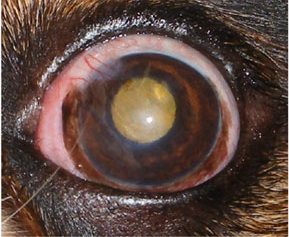
Corneal dystrophy is the development of gray-white opaque lipid deposits of calcium and fats under the surface of both of the dog's corneas. They usually appear in cavaliers between the ages of two and four years.
Corneal dystrophy usually does not affect vision, is not painful, and usually no treatment is necessary.
Corneal dystrophy also is referred to as lipid keratopathy or corneal lipidosis.
RETURN TO TOP
Symptoms
The form of corneal dystrophy (CD) most common among CKCSs
is epithelial/stromal dystrophy, which describe the gray-white opacity
which is visible in the dog's eyes. (See photo below at right below.)
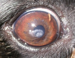 In the cavalier, it
is believed to be caused by hyperlipoproteinemia, which is an inability
to break down lipids or fats, particularly cholesterol and
triglycerides, in the body. This disoder usually is progressive up
to a point but then ceases to grow. and sometimes regresses.
In the cavalier, it
is believed to be caused by hyperlipoproteinemia, which is an inability
to break down lipids or fats, particularly cholesterol and
triglycerides, in the body. This disoder usually is progressive up
to a point but then ceases to grow. and sometimes regresses.
In a 1993 report, Dr. Shiela Crispin found that, especially in the cavalier King Charles spaniel, crystalline stromal dystrophy due to hyperlipoproteinaemmia (raised HDL, LDL, and VLDL) is an ocular manifestation of Cushing's syndrome.
In a September 2018 article, UK investigators describe CD in the cavalier as resembling Schnyder's CD in humans.
RETURN TO TOP
Related Disorders
Corneal dystrophy has been recognized in veterinary research as a manifestation of Cushing's disease in the CKCS. This is not to mean that all cases of corneal dystrophy are symptoms of Cushings, but that any other signs indicating possible Cushings should be looked for when a cavalier is diagnosed with corneal dystophy. In a 1993 report, Dr. Shiela Crispin found that, especially in the cavalier King Charles spaniel, crystalline stromal dystrophy due to hyperlipoproteinaemmia (raised HDL, LDL, and VLDL) is an ocular manifestation of Cushing's syndrome.
RETURN TO TOP
Treatment
Corneal dystrophy usually does not affect vision, is not painful, and usually no treatment is necessary. In an abstract presented to the 2010 WSAVA Congress by Dr. Charlotte Keller, DACVO, DECVO, she suggests that treatment may include the reduction of fat intake. In a June 2015 publication, Dr. Douglas Esson adds that, "Treatment is frequently unnecessary and corticosteroid-based anti-inflammatory medications may exacerbate changes."
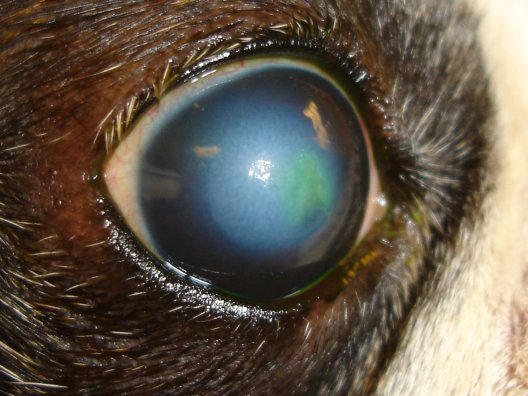
There always are exceptions, however. In some cases, both medical treatment and surgery may be necessary. In a March 2022 article, a cavalier was diagnosed with corneal dystrophy in both eyes and inflammation of the episclera layer of tissues in the left eye. Treatment with neomycin-polymyxin B-dexamethasone ophthalmic suspension and cyclosporine 0.2% ophthalmic ointment did not prevent growth of the crystalline opacities in the left eye. A superficial keratectomy (surgical removal of a portion of the cornea) was performed, which confirmed the presence of "lipid keratopathy" (accumulation of fatty substances -- cholesterol).
Since corneal dystrophy in the CKCS has been found to be a manifestation of Cushing's disease, testing for that disorder also should be performed.
In a March 2015 article, a team of Canadian ophthalmologists noted that:
"Low-fat diets have been recommended anecdotally; however, there are no clinical studies showing that reducing fat in the diet has any significant effect on the density or progression of the lesion."
RETURN TO TOP
Breeders' Responsibilities
 Currently, the Canine Eye Registration Foundation
(CERF) does not deny certification to cavalier King Charles spaniels
which are affected with corneal dystrophy, because the
Genetics
Committee of the ACVO classifies the disorder as a "breeder option" for
CKCSs. See
The Blue Book. However, the Canine Inherited Disorders Database recommends that
all dogs suffering from corneal dystrophy not be bred.
Currently, the Canine Eye Registration Foundation
(CERF) does not deny certification to cavalier King Charles spaniels
which are affected with corneal dystrophy, because the
Genetics
Committee of the ACVO classifies the disorder as a "breeder option" for
CKCSs. See
The Blue Book. However, the Canine Inherited Disorders Database recommends that
all dogs suffering from corneal dystrophy not be bred.
The Cavalier King Charles Spaniel Club, USA recommends that, prior to breeding any Cavalier, the dog have a normal rating or be within CERF "breeder options" from a screening by a board certified veterinary ophthalmologist.
The Canine Health Information Center (CHIC) is a centralized canine health database sponsored by the AKC/Canine Health Foundation (AKC/CHF) and OFA. The CHIC, working with participating parent clubs, provides a resource for breeders and owners of purebred dogs to research and maintain information on the health issues prevalent in specific breeds.
AKC's national breed clubs establish the breed specific testing protocols. Dogs complying with the breed specific testing requirements are issued CHIC numbers. The ACKCSC requires that, to qualify for CHIC certification, cavaliers must have a CERF eye examination, recommending that an initial CERF exam be performed at 8 to 12 weeks, with a follow up exam once the dog reaches 12 months, and annual exams thereafter until age 5 years, and every other year until age 9 years. However, all that is required to qualify for a CHIC certificate is that the breeding stock be examined by a veterinary ophthalmologist. It does not require that the results of the examination show no eye disorders.
RETURN TO TOP
 What You Can Do:
All CKCSs should be examined at least annually by a board certified veterinary ophthalmologist. They are listed on the website of the American College of Veterinary Ophthalmologists (ACVO).
Ocu-GLO Rx is a nutraceutical containing several natural antioxidants in a combination blend formulated specifically for canine eye health. Many veterinary ophthalmologists recommend this product to maintain healthy eyes. Even if your dog has not been diagnosed with a vision disorder, antioxidants contained in Ocu-GLO Rx are considered helpful in keeping dogs' eyes healthy.
RETURN TO TOP
Research News
April 2022:
Cavalier is surgically treated for severe corneal dystrophy.
 In
a
March 2022 article, University of California - Davis veterinary
specialists (Sangwan Park, Lionel Sebbag, Bret A. Moore, M. Isabel
Casanova, Brian C. Leonard, Nicole L. Daley, Kirsten A. Steele, Jennifer
Y. Li, Christopher J. Murphy, Sara M. Thomasy [right])
diagnosed a cavalier King Charles spaniel with corneal dystrophy in both
eyes and inflammation of the episclera layer of tissues in the left eye.
Treatment with neomycin-polymyxin B-dexamethasone ophthalmic suspension
and cyclosporine 0.2% ophthalmic ointment did not prevent growth of the
crystalline opacities in the left eye. A superficial keratectomy
(surgical removal of a portion of the cornea) was performed, which
confirmed the presence of "lipid keratopathy" (accumulation of fatty
substances -- cholesterol).
In
a
March 2022 article, University of California - Davis veterinary
specialists (Sangwan Park, Lionel Sebbag, Bret A. Moore, M. Isabel
Casanova, Brian C. Leonard, Nicole L. Daley, Kirsten A. Steele, Jennifer
Y. Li, Christopher J. Murphy, Sara M. Thomasy [right])
diagnosed a cavalier King Charles spaniel with corneal dystrophy in both
eyes and inflammation of the episclera layer of tissues in the left eye.
Treatment with neomycin-polymyxin B-dexamethasone ophthalmic suspension
and cyclosporine 0.2% ophthalmic ointment did not prevent growth of the
crystalline opacities in the left eye. A superficial keratectomy
(surgical removal of a portion of the cornea) was performed, which
confirmed the presence of "lipid keratopathy" (accumulation of fatty
substances -- cholesterol).
September 2018:
UK geneticists look for gene causing corneal dystrophy in the
cavalier King Charles spaniel.
 In
a
September 2018 abstract, a team of UK's Animal Health Trust gene
investigators (S Maini, SL Ricketts, L Pettitt, JAC Oliver, CS Mellersh
[right]) used whole-gene sequencing of a cavalier King Charles
spaniel affected with corneal dystrophy (CD) to determine if UBIAD1 (and
seven other genes) is a candidate gene for CD in the breed. Mutations of
this gene cause Schnyder's CD in humans, which resembles CD in the CKCS.
They report not finding any evidence that the UBIAD1 gene contains any
variants causing CD in the cavalier. They stated that their analysis is
ongoing and could lead to the identification of genetic factors
underlying this condition.
In
a
September 2018 abstract, a team of UK's Animal Health Trust gene
investigators (S Maini, SL Ricketts, L Pettitt, JAC Oliver, CS Mellersh
[right]) used whole-gene sequencing of a cavalier King Charles
spaniel affected with corneal dystrophy (CD) to determine if UBIAD1 (and
seven other genes) is a candidate gene for CD in the breed. Mutations of
this gene cause Schnyder's CD in humans, which resembles CD in the CKCS.
They report not finding any evidence that the UBIAD1 gene contains any
variants causing CD in the cavalier. They stated that their analysis is
ongoing and could lead to the identification of genetic factors
underlying this condition.
March 2016: Dr. Crispin announces a study of a genetic cause of corneal dystrophy in the CKCS. In a March 2016 article, Dr. Sheila Crispin, of the University of Bristol in the UK, announced that a study is on-going at the Animal Health Trust to determine if a genetic defect involving cholesterol metabolism causes crystalline corneal dystrophy in the cavalier King Charles spaniel.
 June 2015:
US ophthalmologist warns that steroid treatment may worsen
corneal dystrophy. In a June 2015 book on ophthalmic diseases, Dr.
Douglas W. Esson (right) reports that corneal dystrophy occurs mostly in
cavalier King Charles spaniels and three other breeds. He notes that it
is diagnosed based upon clinical findings. Most importantly, he warns:
June 2015:
US ophthalmologist warns that steroid treatment may worsen
corneal dystrophy. In a June 2015 book on ophthalmic diseases, Dr.
Douglas W. Esson (right) reports that corneal dystrophy occurs mostly in
cavalier King Charles spaniels and three other breeds. He notes that it
is diagnosed based upon clinical findings. Most importantly, he warns:
"Treatment is frequently unnecessary and corticosteroid-based anti-inflammatory medications may exacerbate changes."
April 2015: VetCompass
analysis shows frequent diagnoses of corneal diseases in cavaliers.
 In
an
April 2015 report by UK and Australian veterinarians (Jennifer F
Summers, Dan G O'Neill, David B Church, Peter C Thomson, Paul D
McGreevy, David C Brodbelt), the veterinary records of 1,875 cavalier
King Charles spaniels treated between 2009 and 2013 and on the database
of the VetCompass animal health surveillance project, were dissected.
Only 75 of the 1,875 cavaliers had a confirmed KC-registration status.
They found that corneal diseases were diagnosed relatively frequently,
but they could not differentiate between inherited, early forms and
age-related degenerative cataracts.
In
an
April 2015 report by UK and Australian veterinarians (Jennifer F
Summers, Dan G O'Neill, David B Church, Peter C Thomson, Paul D
McGreevy, David C Brodbelt), the veterinary records of 1,875 cavalier
King Charles spaniels treated between 2009 and 2013 and on the database
of the VetCompass animal health surveillance project, were dissected.
Only 75 of the 1,875 cavaliers had a confirmed KC-registration status.
They found that corneal diseases were diagnosed relatively frequently,
but they could not differentiate between inherited, early forms and
age-related degenerative cataracts.
January 2015: UK's Animal Health Trust announces a DNA test for corneal dystrophy in Labradors. The Animal Health Trust and UK's Kennel Club have announced a new DNA test for the genetic mutation causing macular corneal dystrophy (MCD) in Labrador retrievers. MCD is an inherited eye condition which, although painless, can cause severe visual impairment in affected dogs. The mutation for MCD is recessive, which means that only dogs that inherit two copies of the mutation will be affected. Dogs do not typically develop MCD until middle age. Now, affected and carriers Labradors can be identified through one swab test.
RETURN TO TOP
Related Links
Veterinary Resources
Dystrophy, degeneration and infiltration of the canine cornea. S.M. Crispin, K.C. Barnett. J.Small Animal Prac.;Feb. 1983;24(2):63-83. Quote: Three conditions of the cornea in the dog are described; a form of corneal dystrophy (central/paracentral lipid dystrophy, lipidosis, lipoidosis, cholesterolosis), degeneration (fatty and calcareous degeneration) and infiltration (arcus lipoides corneae, anterior embryotoxon, pre-senile arcus). The clinical appearance, together with histopathological and ultrastructural details, are recorded. The age, sex, breed incidence and possible hereditary factors are also included. Reference is made to previous reports in the veterinary literature and the three conditions are compared with similar conditions in man. ... Corneal lipidosis, as has been stated, is not rare in the dog, and is being recognized with increasing frequency. ... Perusal of the breed, age and sex incidence of the cases examined reveals some interesting facts. Four breeds, namely the Rough Collie, Shetland Sheepdog, Cavalier King Charles Spaniel and Alsatian (German Shepherd Dog), account for over two thirds of the total cases seen. This high breed incidence indicates some genetic factor and, referred to before, the term corneal dystrophy in man is reserved for hereditary conditions of the cornea. ... Crispin (1982) has proposed the topographical description of 'diffuse' lipid dystrophy for the type reported as inherited in rlated Airedales and 'annular' lipid dystrophy for the type reported in Siberian Huskies. This communication records central lipid dystrophy as a familial condition in the following breeds: Rough Collie, Shetland Sheepdog, Cavalier King Charles Spaniel and Alsatian (German Shepherd Dog), although no exact mode of inheritance has been demonstrated. In the Rough Collie and Cavalier King Charles Spaniel this type of corneal dystrophy has been seen through three generations.
Crystalline Stromal Dystrophy in the Cavalier King Charles Spaniel. Crispin SM, Proc Am Coll Vet Ophthalmol 17:18, 1986.
Crystalline corneal dystrophy in the dog. Histochemical and ultrastructural study. Crispin SM. Cornea. 1988;7(2):149-61. Quote: "Crystalline stromal dystrophy a condition resembling human Schnyder's crystalline stromal dystrophy, is described in both the Cavalier King Charles Spaniel and Rough Collie breeds of dog. Both clinical cases and controls were normolipoproteinaemic; neither showed evidence of arcus lipoides corneae. Lipid histochemistry indicated the presence of cholesterol, cholesterol ester, and phospholipid in the superficial stroma. Free fatty acids were also detected in some samples, but triglycerides were not detected in any samples. Electron microscopy showed lamellated and nonlamellated vacuoles both within and outside abnormal fibroblasts. Acicular and rhomboidal clefts, some with a characteristic notch, were observed mainly in extracellular locations, although they were also associated with dying fibroblasts. The epithelium, basal lamina, Descemet's membrane, and endothelium were unaffected, apart from an irregularity of the basal lamina in some specimens."
Inherited eye disease in the dog and cat. K. C. Barnett. J. Sm. Anim. Pract. July 1988; doi: 10.1111/j.1748-5827.1988.tb03514.x Quote: Corneal lipidosis is another type of corneal dystrophy which is commonly bilateral, subepithelial, central or paracentral, progressive only to a certain point and the cause of neither visual defect nor irritation of any sort. It occurs particularly in the Shetland sheepdog and the cavalier King Charles spaniel (Fig. l l ) , as well as in other breeds (eg, Siberian husky, rough collie), and more commonly in the bitch than in the dog. It bears similarities to crystalline stromal dystrophy in man.
Ocular manifestations of hyperlipoproteinaemia. S. M. Crispin. J.Small Animal Prac.;Oct. 1993;34(10):500-506. Quote: "The ocular manifestations of hyperlipoproteinaemia include visible lipaemia of ocular blood vessels, corneal opacities, lipaemic aqueous and gross infiltration of the globe itself which is easiest to observe in the peripheral cornea and uveal tract. Both primary and secondary hyperlipoproteinaemia may produce ocular signs and the ocular appearance can be characteristic of the underlying lipoprotein disorder. ... Crystalline stromal dystrophy in a Cavalier King Charles spaniel with secondary hyperlipoproteinaemia associated with Cushing's syndome. ... Crystalline stromal dystrophy [due to] Hyperlipoproteinaemmia (raised HDL, LDL, and VLDL) [is an] ocular manifestation [of the] clinical disorder Cushing's syndrome especially in the Cavalier King Charles Spaniel."
Ocular Disorders Presumed to be Inherited in Purebred Dogs. Genetics Committee, A.C.V.O. 1999.
Control of Canine Genetic Diseases. Padgett, G.A., Howell Book House 1998, pp. 198-199, 239.
Guide to Congenital and Heritable Disorders in Dogs. Dodds WJ, Hall S, Inks K, A.V.A.R., Jan 2004, Section II(65).
Breed Predispositions to Disease in Dogs & Cats. Alex Gough, Alison Thomas. 2004; Blackwell Publ. 44-45.
Visual Morbidity in Thirty-four Families with Schnyder Crystalline Corneal Dystrophy (An American Ophthalmological Society Thesis). Jayne S. Weiss. Trans Am Ophthalmol Soc. 2007 December; 105: 616-648. Quote: "Crystalline stromal dystrophy is the commonest canine corneal lipid deposition and is relatively common in the Cavalier King Charles Spaniel."
Corneal dystrophy. Canine Inherited Disorders Database.
The Eye and Systemic Disease. John Mould. 2008 WSAVA Congress. Quote: "Corneal Lipid ... Most common appearance is paracentral lipid dystrophy: ... Shelties and Cavalier King Charles spaniels."
Ophthalmic Disease in Veterinary Medicine. Charles L. Martin. Manson Publ. 2009; page 475, table 15.1. Quote: "Presumed Inherited Ocular Diseases: Table 15.1: Breed predisposition to eye disease in dogs: Cavalier King Charles Spaniel: ... Corneal dystrophy: epithelial/stromal".
Diagnosis and Treatment of Inherited Canine Corneal Diseases. Kerry L. Ketring. NAVC Conf. June 2009. Quote: After more than 30 years in private practice, I realize the majority of canine ocular diseases have a breed predisposition. This article reflects the most common corneal diseases in the breeds seen in my practice. It is pertinent to keep in mind, however, that breed incidence may reflect just the popularity of the breed. The awareness of breed-specific corneal diseases is a head start in making the correct diagnosis. ... Corneal Dystrophy: ... Any breed may develop a corneal dystrophy but the occurrence is high in the Afghan hound, Akita, boxer, Cavalier King Charles spaniel, cocker spaniel, collie, Dalmatian, golden retriever, Labrador retriever, Lowchen, miniature pinscher, Siberian husky, and Weimaraner.
Non-Ulcerative Corneal Disorders. Charlotte Keller. 2010 WSAVA Congress. Quote: "The White/Yellow/Grey/Blue Cornea: Lipid and mineral deposition in the cornea create a sparkly white, refractile appearance. The deposition of lipid in the cornea has been divided into three clinical types: 1) Crystalline stromal dystrophy--bilateral, axial or paraxial crystalline appearance without inflammation or vascularisation. The deposition of lipid in the cornea without the presence of another disease. Cavalier King Charles Spaniel, Husky Beagle, etc. 2) Lipid keratopathy (corneal lipidosis)--Arcus lipoides corneae--peripheral lesion bilateral ocular manifestation of systemic disease (hypothyroidism, systemic lipid abnormalities). 3) Corneal degeneration--primary or secondary to anterior segment disease (ulcerative keratitis, pannus). Treatment: May include the reduction of fat intake, the control of the underlying systemic diseases and control of ocular disease."
Ocular conditions affecting the brachycephalic breeds. Peter G.C. Bedford. RVC. 2010. Quote: "There are two types of disease which affect the eye of the brachycephalic breeds and both are directly or indirectly related to genetic predisposition. First and by far the commonest are those conditions which are due to be conformation of skull and are related to the exophthalmos which is the common feature of these breeds. Second there are those conditions which have been unwittingly bred into some brachycephalic breeds in the pursuit of desired breed characteristics. In this lecture I will present an overview of all the diseases that the small animal practitioner is likely to encounter in the brachycephalic breeds of pedigree dog. The fourteen breeds I have included for discussion are the Affenpinscher, Boston Terrier, Boxer, Bulldog, Cavalier King Charles and the King Charles Spaniels, (mesaticephalic) French Bulldog, Griffon Bruxellois, Japanese Chin, Lhasa Apso, Pekingese, Pug, Shih Tzu and Tibetan Spaniel. ... Corneal Lipid Dystrophy: The term applies to the characteristic cholesterol and triglyceride deposits in the superficial corneal stroma seen most commonly in the Cavalier King Charles Spaniel. It is clinically benign and seldom affects vision to any noticeable degree. ... Hereditary Cataract: Hereditary cataract is seen in the Boston Terrier and the Cavalier King Charles Spaniel. ... Microphthalmos (MoD): Again the American literature suggests that microphthalmos (MoD) may be inherited in the Cavalier King Charles Spaniel."
Breed Predispositions to Disease in Dogs & Cats (2d Ed.). Alex Gough, Alison Thomas. 2010; Blackwell Publ. 53.
A
cloudy view? David L. Williams. April 2013. Quote: "What is
happening in the cornea of this Cavalier King Charles
Spaniel? This dog has a crystalline stromal lipid dystrophy
- an inherited defect in how the keratocytes - the corneal cells -
deal with lipid, leading to a deposition of lipid crystals in the
centre of the cornea. It looks identical to Schnyder's dystrophy in
people as shown below, but whether the same gene mutation is present
is currently unknown. In people this makes reading and driving at
night difficult, but in the dog the lesion doesn't interfere with
vision (as they rarely read or drive at night!) and the lipid
deposit is unlikely to develop markedly."
Cultivation of corneal epithelial cell sheets on canine amniotic membrane. Eunryel Nam, Ayaka Takahashi, Naoki Fujita, Keiko Tsuzuki, Ryohei Nishimura. Vet.Ophthalmology. July 2013;16(4):263-268. Quote: "Objective: To develop and assess canine corneal epithelial cell sheets cultivated from limbal stem cells on amniotic membrane. Procedures: Canine corneal limbal segments were obtained from six beagle dogs. Cryopreserved denuded amniotic membranes (obtained from Miniature Dachshund and Cavalier King Charles Spaniel breeds) from which the epithelial cells were removed were used as scaffolds. The limbal segments were cultured on these amniotic membranes with 3T3 feeder cells for 2 weeks. The harvested corneal epithelial cell sheets were stained with H&E for histologic analysis. The harvested sheets were analyzed immunohistochemically using a corneal epithelium-specific marker keratin 3(K3) and putative stem cell markers ABCG2, p63, and vimentin. Results: Cultivated cells from the corneal limbal tissues reached confluency in 7-8 days. The cultivated cells adhered to the denuded amniotic membrane and formed a sheet. The cultivated cell sheet was transparent and consisted of five to eight layers. K3 was observed in all layers and ABCG2, p63, and vimentin were notably present in the basal layer of the cultivated canine epithelium by immunofluorescence. Conclusions: Canine corneal epithelial cells were successfully cultivated on the canine amniotic membrane. The cultivated epithelial sheets contained putative stem cells in the basal layer and had a stratified epithelium."
Ocular Disorders presumed to be inherited in purebred dogs. Genetics Committee of the American College of Veterinary Ophthalmologists. Blue Book 6th Ed. October 2013;pp 241-247. "Ocular Disorders Report -- Cavalier King Charles Spaniel" pp 1-6. Quote: "Cavalier King Charles Spaniel: Disorder: E. Corneal dystrophy- epithelial/stromal. Inheritance: Not defined."
The genetics of eye disorders in the dog. Cathryn S. Mellersh. Canine Genetics & Epidemiology. April 2014. Quote: "Inherited forms of eye disease are arguably the best described and best characterized of all inherited diseases in the dog, at both the clinical and molecular level and at the time of writing 29 different mutations have been documented in the scientific literature that are associated with an inherited ocular disorder in the dog. The dog has already played an important role in the identification of genes that are important for ocular development and function as well as emerging therapies for inherited blindness in humans. Similarities in disease phenotype and eye structure and function between dog and man, together with the increasingly sophisticated genetic tools that are available for the dog, mean that the dog is likely to play an ever increasing role in both our understanding of the normal functioning of the eye and in our ability to treat inherited eye disorders. This review summarises the mutations that have been associated with inherited eye disorders in the dog."
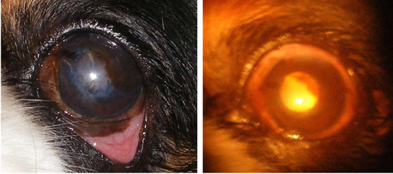
Diagnostic Ophthalmology.
Lynne S. Sandmeyer, Bianca S. Bauer, Bruce H. Grahn. Canadian Vet.
J. March 2015;56(3):301-302.
 Quote:
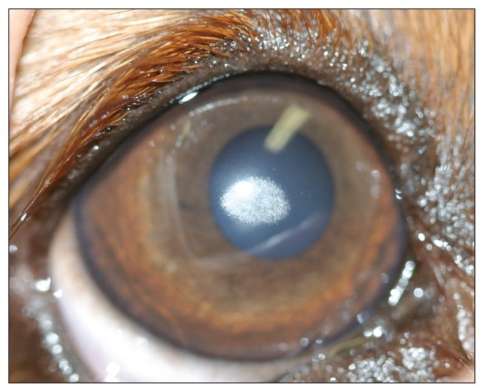
"A 4-year-old spayed female Cavalier King Charles spaniel
dog was examined at the ophthalmology service at the
Western College of Veterinary Medicine for evaluation of white spots
in the corneas. ... A photograph of the right eye at presentation is
provided for your assessment. [See photo at right.]
Discussion: Our
clinical diagnosis was corneal stromal dystrophy. Corneal stromal
dystrophy is a primary, inherited, bilateral opacity of the cornea
which is not associated with ocular inflammation or systemic
disease. Opacities due to stromal dystrophy are depositions of
cholesterol, lipid, and fatty acids within the stroma. Corneal
stromal dystrophy occurs in many dog breeds and has been well
described in the beagle, Siberian husky, Airedale terrier, rough
collie, and Cavalier King Charles spaniel. Mode of
inheritance is known for some breeds, but is variable among breeds.
The main clinical manifestation of corneal stromal dystrophy is a
bilateral, roughly symmetrical, round to oval crystalline opacity in
the axial or paraxial cornea that is not associated with corneal
inflammation, vascularization, or pigmentation. In the
Cavalier King Charles spaniel specifically, opacities
develop between 2 to 4 years of age and consist of fine, striate
crystals closely packed together in the sup-epithelial, axial, or
paraxial cornea. The pathogenesis is unknown but speculated to be a
local abnormality of corneal fibroblasts, possibly with lipid
metabolism and a polygenic mode of inheritance is suspected. The
diagnosis of corneal stromal dystrophy is based on the signalment
and clinical manifestations described. The differential diagnoses
for corneal dystrophy include corneal fibrosis, corneal
degeneration, and lipid keratopathies; all of which are not
inherited or familial, are non-symmetrical, and not necessarily
bilateral. Corneal fibrosis occurs following corneal stromal injury
and is usually a well-demarcated dense gray-white opacity. Corneal
degeneration occurs secondary to pathological conditions of the
cornea, or systemic disease causing lipid, cholesterol, or calcium
deposition. Corneal inflammation, vascularization, and/or melanosis
often accompany the changes. Lipid keratopathy is associated with
systemic lipid abnormalities and is characterized by peripheral
and/or central, crystalline corneal opacities in one or both eyes.
When lipid or mineral deposition is not typical of inherited
dystrophy, serum chemistry and lipid profiles are recommended to
investigate for underlying systemic conditions. In general, corneal
stromal dystrophies do not require or respond to medical therapy.
Topical anti-inflammatory medications may actually exacerbate the
lesion. Lesions may return after keratectomy and therefore, surgery
is not usually recommended. Low-fat diets have been recommended
anecdotally; however, there are no clinical studies showing that
reducing fat in the diet has any significant effect on the density
or progression of the lesion. The prognosis for corneal stromal
dystrophies is excellent. Most opacities are small and do not
progress to involve the whole cornea. They do not tend to cause
visual disturbance and they are usually not associated with ocular
pain."
Quote:
"A 4-year-old spayed female Cavalier King Charles spaniel
dog was examined at the ophthalmology service at the
Western College of Veterinary Medicine for evaluation of white spots
in the corneas. ... A photograph of the right eye at presentation is
provided for your assessment. [See photo at right.]
Discussion: Our
clinical diagnosis was corneal stromal dystrophy. Corneal stromal
dystrophy is a primary, inherited, bilateral opacity of the cornea
which is not associated with ocular inflammation or systemic
disease. Opacities due to stromal dystrophy are depositions of
cholesterol, lipid, and fatty acids within the stroma. Corneal
stromal dystrophy occurs in many dog breeds and has been well
described in the beagle, Siberian husky, Airedale terrier, rough
collie, and Cavalier King Charles spaniel. Mode of
inheritance is known for some breeds, but is variable among breeds.
The main clinical manifestation of corneal stromal dystrophy is a
bilateral, roughly symmetrical, round to oval crystalline opacity in
the axial or paraxial cornea that is not associated with corneal
inflammation, vascularization, or pigmentation. In the
Cavalier King Charles spaniel specifically, opacities
develop between 2 to 4 years of age and consist of fine, striate
crystals closely packed together in the sup-epithelial, axial, or
paraxial cornea. The pathogenesis is unknown but speculated to be a
local abnormality of corneal fibroblasts, possibly with lipid
metabolism and a polygenic mode of inheritance is suspected. The
diagnosis of corneal stromal dystrophy is based on the signalment
and clinical manifestations described. The differential diagnoses
for corneal dystrophy include corneal fibrosis, corneal
degeneration, and lipid keratopathies; all of which are not
inherited or familial, are non-symmetrical, and not necessarily
bilateral. Corneal fibrosis occurs following corneal stromal injury
and is usually a well-demarcated dense gray-white opacity. Corneal
degeneration occurs secondary to pathological conditions of the
cornea, or systemic disease causing lipid, cholesterol, or calcium
deposition. Corneal inflammation, vascularization, and/or melanosis
often accompany the changes. Lipid keratopathy is associated with
systemic lipid abnormalities and is characterized by peripheral
and/or central, crystalline corneal opacities in one or both eyes.
When lipid or mineral deposition is not typical of inherited
dystrophy, serum chemistry and lipid profiles are recommended to
investigate for underlying systemic conditions. In general, corneal
stromal dystrophies do not require or respond to medical therapy.
Topical anti-inflammatory medications may actually exacerbate the
lesion. Lesions may return after keratectomy and therefore, surgery
is not usually recommended. Low-fat diets have been recommended
anecdotally; however, there are no clinical studies showing that
reducing fat in the diet has any significant effect on the density
or progression of the lesion. The prognosis for corneal stromal
dystrophies is excellent. Most opacities are small and do not
progress to involve the whole cornea. They do not tend to cause
visual disturbance and they are usually not associated with ocular
pain."
Prevalence of disorders recorded in Cavalier King Charles Spaniels attending primary-care veterinary practices in England. Jennifer F Summers, Dan G O'Neill, David B Church, Peter C Thomson, Paul D McGreevy, David C Brodbelt. Canine Genetics and Epidemiology. April 2015;2:4. Quote: "This study used large volumes of health data from UK primary-care practices participating in the VetCompass animal health surveillance project to evaluate in detail the disorders diagnosed in a random selection of over 50% of dogs recorded as Cavalier King Charles Spaniels (CKCSs). Confirmation of breed using available microchip and Kennel Club (KC) registration data was attempted. Results: In total, 3624 dogs were recorded as CKCSs within the VetCompass database of which 143 (3.9%) were confirmed as KC-registered via microchip identification linkage of VetCompass to the KC database. ... Microchip data were available in 1692 (46.7%) of the 3624 identified CKCSs. It was possible to crosslink microchip data with KC-registration details in 143 of these dogs; this represented 8.5% of all identified CKCSs with microchip data, and 3.9% of all identified CKCSs. The remaining 3481 dogs were classified as of unknown KC-registration status. The 52% randomly selected sample of all identified CKCSs totalled 1875 dogs: 1800 with unknown and 75 with confirmed KC-registration status. These 1875 dogs were seen at 109 individual clinics during the study period, including 90 (83%) Medivet and 19 (17%) Vets4Pets sites located from north-east to southern England. ...1875 dogs (75 KC registered and 1800 of unknown KC status, 52% of both groups) were randomly sampled for detailed clinical review. Clinical data associated with veterinary care were recorded in 1749 (93.3%) of these dogs. ... Median ages at first and last consultation were 4.0 and 5.25 years, respectively (ranges one month - 17.2 years for both age measures). The most frequent coat colours were Blenheim (44.3%) and tri-colour (30.8%) (Table 1). Of the 1521 dogs with more than one clinical data entry, median time contributed to the study was 1.3 years (range 1 day to 3.6 years). ... Ocular disorders, and particularly corneal diseases, were frequently recorded in study dogs. KCS was particularly frequent, with a proportion of the unspecified corneal disorders (and chronic keratitis cases) possibly also due to undiagnosed KCS. Studies suggest an autosomal recessive mode of inheritance CKCSID in the CKCS. In addition, the typical CKCS skull morphology (with large eyes and shallow eye sockets) may also pre-dispose the breed to corneal damage, exposure keratitis, conjunctival injury and subsequent irritation. Cataracts were recorded relatively frequently in study CKCSs and an inherited basis has been suggested for certain early onset cataracts in the breed. However, the current study could not differentiate between inherited, early forms and age-related degenerative cataracts. DNA screening tests (e.g. CKCSID) and British Veterinary Association (BVA)/KC health schemes (e.g., multifocal retinal dysplasia and hereditary cataract) offer opportunities to reduce population levels of certain inherited conditions through selective breeding. However, the current study indicates that eye disorders remain an important challenge for those concerned with improving the health and welfare of CKCSs. ... Further work This work highlights the value of veterinary practice based breed-specific epidemiological studies to provide targeted and evidence-based health policies. Further studies using electronic patient records in other breeds could highlight their potential disease predispositions."
Corneal Dystrophy. Douglas W. Esson, in Clinical Atlas of Canine and Feline Ophthalmic Disease. John Wiley & Sons, Inc. June 2015;114-115. Quote: "This chapter presents the diagnosis and treatment for corneal dystrophy that mostly occurs in Cavalier King Charles Spaniel, Siberian Husky, Beagle, and Shetland Sheepdog. Corneal dystrophy describes a group of corneal disorders in which cholesterol, phospholipids, and free fatty acids accumulate within the cornea(s). This disorder is bilateral in presentation and is not associated with any preexisting or concurrent corneal or ocular inflammation. Multiple patterns of corneal dystrophy include axial or peripheral, ovoid to circular lesions. Corneal dystrophy is diagnosed based on clinical findings. Treatment is frequently unnecessary and corticosteroid-based anti-inflammatory medications may exacerbate changes. Keratectomy may be indicated in rare cases of secondary ulceration and/or mineralization associated with epithelial discontinuity and discomfort.
Lipids and the eye. Sheila M. Crispin. Vet. J. March 2016. Quote: "Corneal lipid deposition, on occasions associated with hypercholesterolaemia, has been identified in other breeds in the United Kingdom, including the Afghan hound, Cavalier King Charles spaniel, German shepherd dog, Shetland sheepdog and Rough collie. No primary genetic defect has yet been determined in any of these breeds, although research involving Cavalier King Charles spaniels has indicated that a defect of corneal keratocyte metabolism may be the underlying defect in the corneal lipid deposition, termed Crystalline Corneal Dystrophy, or Crystalline Stromal Dystrophy), which resembles human Schnyder Corneal Dystrophy to the extent that it might tentatively be described as Schnyder-like Corneal Dystrophy. The ocular manifestations in the Cavalier King Charles spaniel may be modified by raised circulating lipoproteins, as is the situation in the similar human disorder. ... Whereas Schnyder corneal dystrophy is a rare condition in humans, the canine equivalent, crystalline corneal dystrophy, is not rare in dogs, the only other species in which it has been reported to date. A genetic study of this particular corneal dystrophy in the Cavalier King Charles spaniel, a breed in which the condition is relatively common, is in progress at the Kennel Club Canine Genetics Centre at the Animal Health Trust, Newmarket, UK and focuses on the possibility of a genetically determined metabolic defect involving cholesterol metabolism in the keratocytes of the corneal stroma. There is no inflammatory component and no vascularisation evident and the dystrophy is typified by abnormal accumulation of predominantly lipid, but also detectable acid mucosubstances, within the corneal keratocytes of the anterior stroma, with subsequent keratocyte death and necrosis. Cholesterol crystals associated with keratocyte death are responsible for the characteristic crystalline appearance, but other lipids, notably cholesterol esters, phospholipids and free fatty acids, can be identified in affected corneas. Crystalline corneal dystrophy is bilateral and most obvious in the axial, coolest part of the cornea. Hyperlipoproteinaemia may or may not be present in affected dogs."
Preliminary results of a prospective study of inter- and intra-user
variability of the Royal Veterinary College corneal clarity score
(RVC-CCS) for use in veterinary practice. Rick F. Sanchez,
Charlotte Dawson, Màrian Matas Riera, Natàlia Escanilla.
 Vet.
Ophthalmology. July 2016;19(4):313-318. Quote: Objective: To
introduce a new corneal clarity score for use in small animals and
describe its inter- and intra-user variability. Animals studied:
Twelve dogs [including two cavalier King Charles spaniels] and two
cats with corneal abnormalities and five dogs with healthy corneas.
Materials and Methods: Four examiners scored every patient twice and
never consecutively, focusing on the central cornea. The peripheral
cornea was scored separately. The following scoring system was used
to describe corneal clarity:
Vet.
Ophthalmology. July 2016;19(4):313-318. Quote: Objective: To
introduce a new corneal clarity score for use in small animals and
describe its inter- and intra-user variability. Animals studied:
Twelve dogs [including two cavalier King Charles spaniels] and two
cats with corneal abnormalities and five dogs with healthy corneas.
Materials and Methods: Four examiners scored every patient twice and
never consecutively, focusing on the central cornea. The peripheral
cornea was scored separately. The following scoring system was used
to describe corneal clarity:
-- G0: no fundus reflection is
visible on retroillumination (RI) using a head-mounted indirect
ophthalmoscope.
-- G1: a fundus reflection is visible with RI.
(Images above right are of the same eye taken before and during
indirect ophthalmoscopy in a Cavalier King Charles Spaniel with a
CLCT [corneo-limboconjunctival transposition] OS [conjunctival
pedicle graft laterally]. The axial corneal in this eye was assigned
a G1 as there was a tapetal reflection passing through it with the
indirect ophthalmoscope, but there was not enough clarity to allow
for a 0.1-mm slit beam to travel though it without it being
completely distorted by the time it reached the iris and lens.)
-- G2: a 0.1-mm diameter light beam is visible on the anterior
surface of the iris and/or lens.
 --
G3: gross fundic features are visible when viewed with indirect
ophthalmoscopy (IO) using a head-mounted indirect ophthalmoscope and
a hand-held 30D lens, although fine details are not clear.
(Image at below right is of Cavalier King Charles Spaniel with
keratoconjunctivitis sicca and a CLCT OS. The axial cornea of this
eye was assigned a G3 because indirect ophthalmoscopy was possible.
Gross details of the fundus were visible although the finer details
of the fundus were not.)
--
G3: gross fundic features are visible when viewed with indirect
ophthalmoscopy (IO) using a head-mounted indirect ophthalmoscope and
a hand-held 30D lens, although fine details are not clear.
(Image at below right is of Cavalier King Charles Spaniel with
keratoconjunctivitis sicca and a CLCT OS. The axial cornea of this
eye was assigned a G3 because indirect ophthalmoscopy was possible.
Gross details of the fundus were visible although the finer details
of the fundus were not.)
-- G4: fine details of the fundic
features are clearly visible with IO.
The minimum grades given
were analyzed for inter- and intra-user variability with kappa
analysis. Results: Intra- and interuser variability of the central
corneal clarity ranged from 0.78 to 0.96, showing substantial to
almost perfect reproducibility, and from 0.66 to 0.91, showing
substantial to almost perfect reliability, respectively. Intra- and
interuser variability of the peripheral cornea ranged from 0.83 to
0.95, showing almost perfect agreement, and from 0.53 to 0.91,
showing moderate to almost perfect agreement. Conclusions: The
RVC-CCS is well suited to assess and monitor central corneal clarity
in small animals and to compare outcomes between studies and
different surgeons.
Corneal dystrophy in the cavalier king charles spaniel -- A candidate gene study. S Maini, SL Ricketts, L Pettitt, JAC Oliver, CS Mellersh. Vet. Ophth. September 2018;21(5):E20. Quote: Purpose: To investigate UBIAD1 as a candidate gene for corneal dystrophy (CD) in the Cavalier King Charles Spaniel (CKCS) using whole-genome sequencing (WGS) of an affected dog. CKCS CD resembles Schnyder's CD in humans, which is caused by mutations in the UBIAD1 gene. Methods: WGS of an affected CKCS was compared with 20 WGS of other breeds that did not have documented susceptibility to CD, lipid-related disorders, or hypothyroidism. UBIAD1 and seven additional candidate genes were screened. Subsequent analysis has focused on a global comparison of the CKCS case to 113 WGS of other breeds, and screening of annotated variants that appear only in the case, against human gene sets for corneal dystrophies. Candidate variants have been assayed by Sanger sequencing in a set of 27 additional cases and 12 controls. Results: A scan of the introns and protein coding exons of UBIAD1 did not identify any single nucleotide or complex (structural) variants unique to the CKCS CD case. An analysis of the WGS using currently available human corneal dystrophy gene sets has so far excluded two variants in the SOAT1 and SCAP genes. Conclusions: We did not find any evidence that the UBIAD1 gene harbors causal variants for CKCS CD. The global analysis is ongoing and may be supplemented by a genome-wide association study to facilitate mapping of CD in the CKCS. With the available WGS, this could lead to the identification of genetic factors underlying this condition.
Multimodal ocular imaging of known and novel corneal stromal
disorders in dogs. Sangwan Park, Lionel Sebbag, Bret A.
Moore, M. Isabel Casanova, Brian C. Leonard, Nicole L. Daley,
Kirsten A. Steele, Jennifer Y. Li, Christopher J. Murphy & Sara M.
Thomasy. BMC Vet. Res. March 2022; doi: 10.1186/s12917-022-03214-7.
Quote: Background: Imaging features obtained with Fourier-domain
optical coherence tomography (FD-OCT) and in vivo confocal
microscopy (IVCM) for corneal stromal disorders have been sparsely
reported in dogs. This case report is a compilation of imaging
features for three cases of different stromal disorders of the
canine cornea which have not yet been reported elsewhere. Case
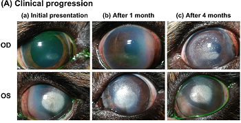 presentation:
... Case 1: A 4-year-old, male-castrated Cavalier King
Charles Spaniel was presented for corneal dystrophy in both
eyes (OU). Ophthalmic examinations revealed a horizontally ovoid,
crystalline opacity in the axial cornea of the right eye (OD) and a
round, dense, white, crystalline opacity in the axial cornea of the
left eye (OS) surrounded by anterior stromal punctate deposits in
the periphery (Fig. 1 Aa). The medial perilimbal cornea was
thickened with tan cellular infiltrates admixed with extensive
stromal vascularization OS, which was clinically consistent with
episcleritis. The STT-1 were 19 mm/min OD and 9 mm/min OS and IOP
were 13 mmHg OU. To improve tear production and episcleritis,
neomycin-polymyxin B-dxamethasone ophthalmic suspension and
cyclosporine 0.2% ophthalmic ointment were prescribed twice daily
OS. The patient was reexamined one month after initial presentation
(Fig. 1 Ab). While the axial corneal opacity OU and the perilimbal
inflammation OS were static, a similar perilimbal keratitis
developed in the nasal cornea OD despite the medical treatment. The
frequency of neomycin-polymyxin B-dexamethasone ophthalmic
suspension was increased to 4-6 times daily OU. Following 3 months
of medical treatment, perilimbal keratitis was markedly reduced OU,
but the axial crystalline opacities had progressed, and a
superficial keratectomy was recommended OS (Fig. 1 Ac). Prior to
surgery, FD-OCT and IVCM were performed. With FD-OCT, dense
hyperreflective dots or lines were visible along the stromal
collagen lamellae and spanned from subepithelium to mid-stroma OU
(Fig. 1B). With IVCM, hyperreflective needle-like lines were visible
in the affected corneas OU (Fig. 1C). Serum biochemistry including
triglycerides, cholesterol, and total T4 concentration were tested;
all were within normal limits. Superficial keratectomy was performed
at 60% stromal depth as determined by the pre-operative images
obtained with FD-OCT. The keratectomy specimen was assessed
histologically, and oil-red O staining demonstrated the presence of
lipid deposition between stromal lamellae, thus confirming the
diagnosis of lipid keratopathy (Fig. 1D). ... Lipid deposition in
case 1 appeared as needle-shaped hyperreflective lines along the
collagen lamellae, which correlated histologically with lipid
clefts. ... Conclusions: In vivo multimodal corneal imaging
facilitated instantaneous microstructural analysis and may be
valuable in the differential diagnosis of corneal stromal disorders
in veterinary clinical practice. The non-specific nature of imaging
findings occurs in some conditions such as mucopolysaccharidosis,
thus in vivo corneal imaging should be complemented with other gold
standard methods of definitive diagnosis.
presentation:
... Case 1: A 4-year-old, male-castrated Cavalier King
Charles Spaniel was presented for corneal dystrophy in both
eyes (OU). Ophthalmic examinations revealed a horizontally ovoid,
crystalline opacity in the axial cornea of the right eye (OD) and a
round, dense, white, crystalline opacity in the axial cornea of the
left eye (OS) surrounded by anterior stromal punctate deposits in
the periphery (Fig. 1 Aa). The medial perilimbal cornea was
thickened with tan cellular infiltrates admixed with extensive
stromal vascularization OS, which was clinically consistent with
episcleritis. The STT-1 were 19 mm/min OD and 9 mm/min OS and IOP
were 13 mmHg OU. To improve tear production and episcleritis,
neomycin-polymyxin B-dxamethasone ophthalmic suspension and
cyclosporine 0.2% ophthalmic ointment were prescribed twice daily
OS. The patient was reexamined one month after initial presentation
(Fig. 1 Ab). While the axial corneal opacity OU and the perilimbal
inflammation OS were static, a similar perilimbal keratitis
developed in the nasal cornea OD despite the medical treatment. The
frequency of neomycin-polymyxin B-dexamethasone ophthalmic
suspension was increased to 4-6 times daily OU. Following 3 months
of medical treatment, perilimbal keratitis was markedly reduced OU,
but the axial crystalline opacities had progressed, and a
superficial keratectomy was recommended OS (Fig. 1 Ac). Prior to
surgery, FD-OCT and IVCM were performed. With FD-OCT, dense
hyperreflective dots or lines were visible along the stromal
collagen lamellae and spanned from subepithelium to mid-stroma OU
(Fig. 1B). With IVCM, hyperreflective needle-like lines were visible
in the affected corneas OU (Fig. 1C). Serum biochemistry including
triglycerides, cholesterol, and total T4 concentration were tested;
all were within normal limits. Superficial keratectomy was performed
at 60% stromal depth as determined by the pre-operative images
obtained with FD-OCT. The keratectomy specimen was assessed
histologically, and oil-red O staining demonstrated the presence of
lipid deposition between stromal lamellae, thus confirming the
diagnosis of lipid keratopathy (Fig. 1D). ... Lipid deposition in
case 1 appeared as needle-shaped hyperreflective lines along the
collagen lamellae, which correlated histologically with lipid
clefts. ... Conclusions: In vivo multimodal corneal imaging
facilitated instantaneous microstructural analysis and may be
valuable in the differential diagnosis of corneal stromal disorders
in veterinary clinical practice. The non-specific nature of imaging
findings occurs in some conditions such as mucopolysaccharidosis,
thus in vivo corneal imaging should be complemented with other gold
standard methods of definitive diagnosis.
Essentials of Veterinary Ophthalmology (4th ed.). Gelatt, Kirk N.; Plummer, Caryn E. Wiley-Blackwell. 2022. Quote: Several forms of corneal dystrophy occuf in specific breeds. These include corneal dystrophy in the ... Cavalier King Charles Spaniel.
The Blue Book. Ocular Disorders Presumed to be inherited in purebred dogs. Genetics Committee of the American College of Veterinary Ophthalmologists. Blue Book 15th Ed. 2023. pp. 297-302.
CONNECT WITH US