
Dry Eye Syndrome and the
Cavalier King Charles Spaniel
- What It Is
- Causes
- Symptoms
- Diagnosis
- Treatment
- Breeders' Responsibilities
- What You Can Do
- Research News
- Related Links
- Veterinary Resources
Many cavalier King Charles spaniels suffer from a painful genetic disorder called dry eye syndrome (keratitis sicca or keratoconjunctivitis sicca -- KCS), according to the American College of Veterinary Ophthalmologists (ACVO). Research studies have shown that cavaliers are more pre-disposed to KCS -- at a relative risk of 11.5% -- than any other breed.
What It Is
 Dry
eye is an inflammation of the cornea and conjunctiva due to an inability to
produce watery tears. It also is referred to as tear film deficiency and aqueous
tear deficiency. It cannot be cured in cavaliers. In the cavalier King Charles
spaniel, the most common cause of dry eye syndrome is an immune-mediated
destruction of the tear glands.
Dry
eye is an inflammation of the cornea and conjunctiva due to an inability to
produce watery tears. It also is referred to as tear film deficiency and aqueous
tear deficiency. It cannot be cured in cavaliers. In the cavalier King Charles
spaniel, the most common cause of dry eye syndrome is an immune-mediated
destruction of the tear glands.
Dry eye prevents the cavalier's eyes from being properly moistened, resulting in chronically dry, burning eyes, and scarring and painful ulceration of the cornea which may lead to decreased vision. The disorder requires frequent medication every day.
Since the cornea does not have blood vessels, tears bring oxygen and nutrients to its cells. Tears ("tear film" or TF) consist of three components -- an outer lipid layer, a middle aqueous layer, and an inner mucus layer. The lipid layer is produced by the meibomian glands; the aqueous layer, which is the main portion of tear film, is produced by orbital lacrimal glands and the nictitans gland; the inner mucus layer is produced by the goblet cells.
Tears lubricate the corneal surface, supply oxygen to the cornea, flush debris, and act as a barrier to microorganisms. An insufficent quantity of tears, or an imbalance within the tears will prevent protecting the cornea from drying and receiving needed nutrients. This is known as evaporative dry eye disease (EDED), a lipid deficiency.
Dry eye can lead to irritation, infection, ulcers, and even blindness.
A rarer but far more severe form of dry eye syndrome in some cavalier King Charles spaniel puppies is a combination of dry eye and a congenital skin condition called "curly coat" or "rough coat" syndrome (ichthyosis keratoconjunctivitis sicca). See Curly Coat for details.
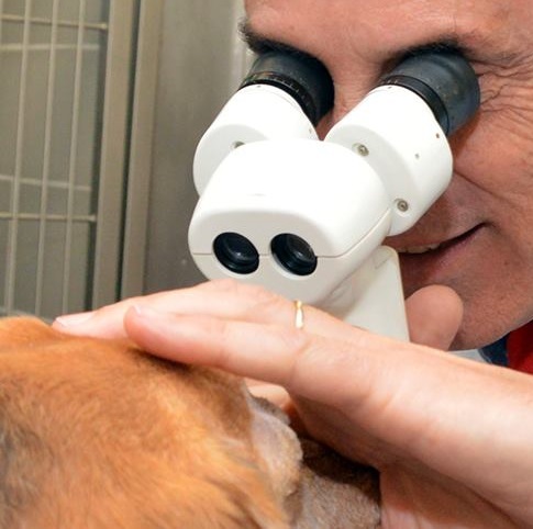
All CKCSs should be examined at least annually by a board certified veterinary ophthalmologist. They are listed on this webpage of the website of the American College of Veterinary Ophthalmologists (ACVO).
RETURN TO TOP
Causes
Causes of KCS in canines are varied. They include congenital, metabolic, infectious, drug induced, neurogenic, radiation, iatrogenic, idiopathic, and immune mediated. Cavaliers tend to have either of two types -- congenital or immune mediated. The congenital version is associated with the curly coat (rough coat) syndrome referred to above and also our our Curly Coat webpage.
The immune mediated version is much more common among CKCSs and is the subject matter of this webpage. It has been found to be the result of immune-mediated inflammation and destruction of the lacrimal glands.
A third significant cause of dry eye in cavaliers is neurogenic keratoconjunctivitis sicca (nKCS) due to the application of certain topcial ear (otitis externa) medications for treating yeast and bacteria infections, specifically those containing terbinafine and/or florfenicol. See this January 2023 article. These topical medications also have been found to cause corneal ulcers in dogs. See our corneal ulcer webpage for more information about that disorder.
RETURN TO TOP
Symptoms
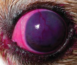
Symptoms of dry eye include:
• chronic redness of the eye
• chronic thick, yellow-green discharge, especially in the morning
• excessive tearing
• excessive blinking
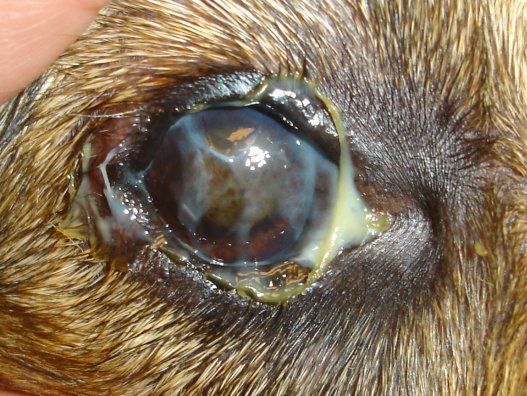
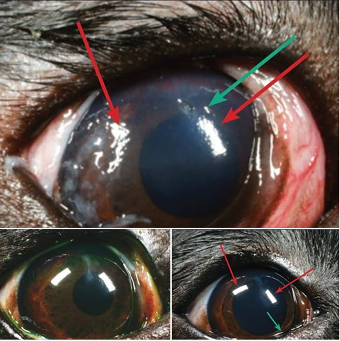
• development of a film over the cornea. (See photo above.)
• abnormal contractions or twitches of the eyelid (blepharospasm)
• squinting, especially when facing the sun or other bright lights (photophobia)
• pigmentation on the corneal surface
• ulcers and abrasions
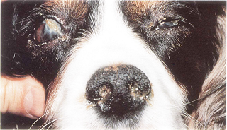
• crusty nostrils (due to lack of fluid to the nasal punctum
RETURN TO TOP
Diagnosis
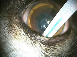
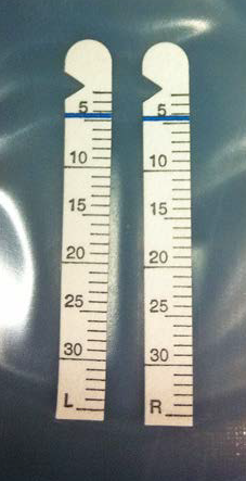 Tear production in the dog's eyes
usually is tested by placing a small strip of
treated paper beneath the lower eyelid. This is called the
Schirmer tear test.
Tear production in the dog's eyes
usually is tested by placing a small strip of
treated paper beneath the lower eyelid. This is called the
Schirmer tear test.
The Schirmer tear test (STT) measures tear production and reflex tear response and is used to diagnose dry eye, as well as other ophthalmic conditions. The STT package contains two sterile test strips, one for each eye. (See photo at right.) The tip of the strip is inserted between the lower eyelid and cornea and left in place for one minute, removed, and read immediately using the scale on the strip.
 A
normal value for dogs is >15 mm wetting/minute. A 10 to 15 mm
wetting/minute result is considered borderline for dry eye, and
treatment should be started if the dog shows signs of
dry eye. Results <10 mm wetting/ minute is
positive for dry eye.
A
normal value for dogs is >15 mm wetting/minute. A 10 to 15 mm
wetting/minute result is considered borderline for dry eye, and
treatment should be started if the dog shows signs of
dry eye. Results <10 mm wetting/ minute is
positive for dry eye.
Evaporative dry eye is tested by tear breakup time (TBUT), in which fluorescein is inserted into the dog's tear film and observed under cobalt blue illumination. The number of seconds between the last blink and the appearance of a dry spot in the tear film is recorded as the TBUT. Times less than ten seconds is regareded as being abnormal.
Another tear measurement device is the ophthalmic surgical sponge, which is made of either cellulose or polyvinyl acetal (PVA). Both the STT and the PVA determine the amount of protein retained after tear extraction. In a March 2018 article, the STT and PVA were compared, and the conclusion was that the Schirmer strips were more reliable than PVA sponges for quantification of total protein content. However, the researchers advised that care should be taken to absorb sufficient volumes of tears with Schirmer strips to minimize the concentrating effect from the absorbent material.
In a
September 2017 article,
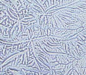 Drs. David Williams
and Heather Hewitt reported observing differing patterns of
Drs. David Williams
and Heather Hewitt reported observing differing patterns of
 dried drops of tears from
control dogs and those affected with dry eye. Patterns from the tears of
normal dogs were "strikingly beautiful" ferning covering the entire face
of the drops (at left). In dogs, including cavaliers, with progressively severe
keratoconjunctivitis sicca (KCS), the dried crystals were smaller and
less fern-like (at right). He found that all eyes with KCS had abnormal ferning
patterns while 39 out of the 50 normal dogs (78%) had so-called 'normal'
ferning patterns. The mean Schirmer tear test type 1 (STT) for dogs
showing 'normal' ferning patterns was 20.6mm/min for the left eye and
21.3mm/min for the right eye. STT values for eyes with 'abnormal'
ferning patterns were 10.9mm/min and 12.4mm/min, these differing from
the normal eyes with STT above 15mm/min significantly. He concluded that
the findings suggest that tear ferning could be a valuable technique for
assessment of the tear film in dogs with KCS.
dried drops of tears from
control dogs and those affected with dry eye. Patterns from the tears of
normal dogs were "strikingly beautiful" ferning covering the entire face
of the drops (at left). In dogs, including cavaliers, with progressively severe
keratoconjunctivitis sicca (KCS), the dried crystals were smaller and
less fern-like (at right). He found that all eyes with KCS had abnormal ferning
patterns while 39 out of the 50 normal dogs (78%) had so-called 'normal'
ferning patterns. The mean Schirmer tear test type 1 (STT) for dogs
showing 'normal' ferning patterns was 20.6mm/min for the left eye and
21.3mm/min for the right eye. STT values for eyes with 'abnormal'
ferning patterns were 10.9mm/min and 12.4mm/min, these differing from
the normal eyes with STT above 15mm/min significantly. He concluded that
the findings suggest that tear ferning could be a valuable technique for
assessment of the tear film in dogs with KCS.
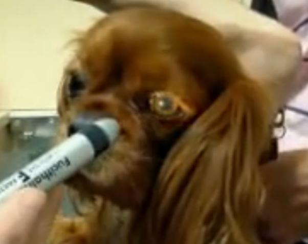
 In
a
December 2017 article, Dr. David L. Williams reported on the success
of using Rose Bengal stain in the eyes of dogs affected with dry eye. Of
the 20 affected dogs in the study, 5 were cavalier King Charles
spaniels. He concluded that there was a reasonable association between
dye staining of the ocular surface and tear production, although clearly
other factors are also important in the genesis of ocular surface damage
in dry eye. (Photo at right is of an 8 year old Cavalier King
Charles spaniel with STT of 4mm/min and staining both of conjunctiva and
cornea together with corneal haze and neovascularisation.)
In
a
December 2017 article, Dr. David L. Williams reported on the success
of using Rose Bengal stain in the eyes of dogs affected with dry eye. Of
the 20 affected dogs in the study, 5 were cavalier King Charles
spaniels. He concluded that there was a reasonable association between
dye staining of the ocular surface and tear production, although clearly
other factors are also important in the genesis of ocular surface damage
in dry eye. (Photo at right is of an 8 year old Cavalier King
Charles spaniel with STT of 4mm/min and staining both of conjunctiva and
cornea together with corneal haze and neovascularisation.)
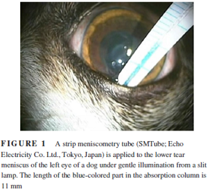
In a
March 2019 article, a team of Japanese veterinary ophthalmologists compared the new strip meniscometry test (SMT) with the
Schirmer tear test (STT) and the phenol red thread test (PRT) to
 evaluate the SMT's relative effectiveness at diagnosing tear-deficient
eyes in dogs. The left eyes of 621 dogs, including 25 cavalier King
Charles spaniels, were evaluated. The SMT is a rapid test, taking only 5
seconds to register results. The researchers report finding that SMT may
be superior to PRT for detecting tear-deficient eyes, and that SMT's
high sensitivity could be useful as a diagnostic tool to rule out normal
eyes within a very short testing time. (In the photo at left here, a
strip meniscometry tube is applied briefly to the lower tear meniscus of
the dog's eye.)
evaluate the SMT's relative effectiveness at diagnosing tear-deficient
eyes in dogs. The left eyes of 621 dogs, including 25 cavalier King
Charles spaniels, were evaluated. The SMT is a rapid test, taking only 5
seconds to register results. The researchers report finding that SMT may
be superior to PRT for detecting tear-deficient eyes, and that SMT's
high sensitivity could be useful as a diagnostic tool to rule out normal
eyes within a very short testing time. (In the photo at left here, a
strip meniscometry tube is applied briefly to the lower tear meniscus of
the dog's eye.)
RETURN TO TOP
Treatment
 Early treatment of dry eye is crucial to preventing destruction of the CKCS's
cornea. Treatment is aimed at increasing tear production, applying artificial
tears, and reducing any bacterial infections, and decreasing inflammation and
scarring of the cornea. The dog's eyes must be kept clean and free of discharge.
The patient may be treated initially with a topical antibiotic or
anti-inflammatory.
Early treatment of dry eye is crucial to preventing destruction of the CKCS's
cornea. Treatment is aimed at increasing tear production, applying artificial
tears, and reducing any bacterial infections, and decreasing inflammation and
scarring of the cornea. The dog's eyes must be kept clean and free of discharge.
The patient may be treated initially with a topical antibiotic or
anti-inflammatory.
RETURN TO TOP
-- topical ointments, etc.
Lacrimostimulant medications such as cyclosporine o.1% and 0.2%, cyclosporine ophthalmic emulsion (Restasis) or ointment (Optimmune) and tacrolimus ophthalmic suspension are commonly prescribed daily for life to increase tear production, and artificial tear solutions must be applied frequently each day to eliminate bacteria, rinse the eyes, and remove discharge. Cyclosporin treats the underlying auto-immune disease and the symptoms, by stimulating the tear glands to resume some tear production, halting the immune destruction of these glands and reducing inflammation of the eyes. Tacrolimus typically is used for cases that do not respond to cyclosporin. See this May 2004 article for more information on lacrimostimulants.
NOTE: In a 2008 study of 25 dogs, including a cavalier, researchers observed that "brachycephalic dogs with a background of chronic keratitis that are treated with nonsteroidal anti-inflammatory drugs [including cyclosporine] are at risk to develop axial corneal SCC [squamous cell carcinoma]. The increase in annual cases of SCC suggests that this phenomenon is a developing problem." See also a 2011 study which concluded, "Chronic inflammatory conditions of the cornea and topical immunosuppressive therapy may be risk factors for developing primary corneal SCC in dogs."
Other frequently prescribed products to help relieve discomfort, itching, and
burning
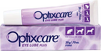 are
Optixcare Eye Lube Plus
(left), sodium hyaluronate (Hy-Optic by Kinetic
Technologies), hyaluronan (I-Drop Vet PLUS
by I-MED Pharma Inc.), and hydroxypropyl methylcellulose and
carboxymethylcellulose sodium (GenTeal).
are
Optixcare Eye Lube Plus
(left), sodium hyaluronate (Hy-Optic by Kinetic
Technologies), hyaluronan (I-Drop Vet PLUS
by I-MED Pharma Inc.), and hydroxypropyl methylcellulose and
carboxymethylcellulose sodium (GenTeal).
Maxitrol (neomycin, polymyxin B, and dexamethasone) is an ointment or drop which may be prescribed for severe cases. It is not intended to be used for long term treatment, as it may have serious side effects, including cataracts and glaucoma.
In a
2013 study, Dr. David L. Williams reports developing a gel
which does not require as many doses per day
 as do the liquid medications. The
gel is a crosslinked hydrogel based upon a modified,
thiolated hyaluronic acid (HA), which he has labelled xCMHA-S.
He stated:
as do the liquid medications. The
gel is a crosslinked hydrogel based upon a modified,
thiolated hyaluronic acid (HA), which he has labelled xCMHA-S.
He stated:
"Further, in a preliminary clinical study of dogs with KCS [including 3 CKCSs], the gel significantly reduced the symptoms associated with KCS within two weeks while only being applied twice daily. The reduction of symptoms combined with the low dosing regimen indicates that this gel may lead to both improved patient health and owner compliance in applying the treatment."
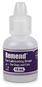
See also Dr. Williams' June 2014 article on this product, which has the brand name Remend by Bayer. He states that it "seems to provide a particularly efficacious tear replacement in each canine KCS patient in which we have tried it".
In a September 2018 article, Taiwan researchers reported that autologous serum (AS) eye drops seemed to be effective and safe for dogs with KCS, and they could improve tear film stability, ocular surface health, and subjective clinical symptoms, especially in dogs younger than 9 years old.
In a May 2020 article, researchers compared the effects of Vizoovet eye drops with GenTeal, to determine the safety and effectiveness of Vizoovet. Vizoovet is a compound of propolis, aloe vera, and chamomile. They reported the result that "squinting, rubbing, ocular discharge, and medication administration scores" by the dogs' owners, all significantly improved throughout the course of the study, and they did not differ significantly between the two groups. The researchers reported no adverse side effects were noted either by the examining clinicians or by the pet owners in either group. They concluded that treatment with Vizoovet was as safe and effective as GenTeal drops at improving clinical signs of dry eye in dogs.
RETURN TO TOP
-- surgery
In severe cases that do not respond to medications, a surgical procedure called a parotid duct transposition (PDT) may be performed in which a duct from a parotid salivary gland is moved from the mouth to the eye. This results in saliva flowing over the eye to keep the eye moist. It is not an ideal treatment for dry eye, because saliva is not the same as tears, and the flow of saliva cannot be as well controlled. The surgery is helpful, however, for those dogs that remain persistently painful and squinty despite trying all forms of medical therapy. In this March 1985 article, Dr. Barnett wrote that:
"The operation of parotid duct transposition is a highly successful surgical procedure in the dog, although it may well produce a wet eye in place of a dry one."
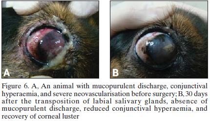 In a
November
2013 article, a team of Brazilian veterinary ophthalmology surgeons
report their success in treating 17 dogs with dry eye by grafting
salivary glands from the dogs' lips to their inner eye lids. The dogs'
dry eye conditions were immune-mediated and unresponsive to medical
treatment.
In a
November
2013 article, a team of Brazilian veterinary ophthalmology surgeons
report their success in treating 17 dogs with dry eye by grafting
salivary glands from the dogs' lips to their inner eye lids. The dogs'
dry eye conditions were immune-mediated and unresponsive to medical
treatment.
They concluded:
"We found a significant clinical improvement in cases of moderate to severe KCS, as well as those which were nonresponsive to medical treatment, as evidenced by clinical examination and statistical tests. Transplantation of labial salivary glands showed that lubrication of the ocular surface by salivary secretion is stable and effective. ... This technique is simple, quick and effective, accessible to any veterinary ophthalmologist surgeon and is of great value for moderate and severe cases of dry keratoconjunctivitis not responsive to medications."
In an April 2021 article, Dr. Chris Dixon at Veterinary Vision in Penrith, Cumbira, UK reported performing a bilateral PDT on a cavalier named Ben, to re-route duct carrying some of his saliva from the mouth onto his eyes. He explained that there are multiple blood vessels and nerves that need to be avoided during the surgery, and that the dissolvable stitch material used is as thin as hair.
RETURN TO TOP
Breeders' Responsibilities
 The Canine Inherited Disorders Database recommends that dogs affected with
dry eye not be bred. Since dry eye is an hereditary disease in cavaliers,
breeders also should never breed any CKCS which has parents or grandparents
which have had dry eye. Dry eye in any littermates of breeding stock should be
taken into consideration. See
The Blue Book.
The Canine Inherited Disorders Database recommends that dogs affected with
dry eye not be bred. Since dry eye is an hereditary disease in cavaliers,
breeders also should never breed any CKCS which has parents or grandparents
which have had dry eye. Dry eye in any littermates of breeding stock should be
taken into consideration. See
The Blue Book.
The Cavalier King Charles Spaniel Club, USA recommends that, prior to breeding any cavalier, the dog have a normal rating from a screening by a board certified veterinary ophthalmologist.
The Canine Health Information Center (CHIC) is a centralized canine health database sponsored by the AKC/Canine Health Foundation (AKC/CHF) and OFA. The CHIC, working with participating parent clubs, provides a resource for breeders and owners of purebred dogs to research and maintain information on the health issues prevalent in specific breeds.
AKC's national breed clubs establish the breed specific testing protocols. Dogs complying with the breed specific testing requirements are issued CHIC numbers. The ACKCSC requires that, to qualify for CHIC certification, cavaliers must have a CERF eye examination, recommending that an initial CERF exam be performed at 8 to 12 weeks, with a follow up exam once the dog reaches 12 months, and annual exams thereafter until age 5 years, and every other year until age 9 years. However, all that is required to qualify for a CHIC certificate is that the breeding stock be examined by a veterinary ophthalmologist. It does not require that the results of the examination show no eye disorders.
Nevertheless, all cavalier breeding stock should be examined by board certified veterinary ophthalmologists at least annually and cleared by the veterinary specialists for dry eye, the closer the examination to the breeding the better.
RETURN TO TOP
 What You Can Do:
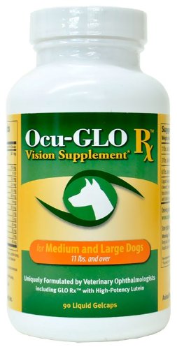
Ocu-GLO Rx is a nutraceutical containing several natural antioxidants in a combination blend formulated specifically for canine eye health. Many veterinary ophthalmologists recommend this product to maintain healthy eyes. Even if your dog has not been diagnosed with a vision disorder, antioxidants contained in Ocu-GLO Rx are considered helpful in keeping dogs' eyes healthy.
RETURN TO TOP
Research News
September 2023:
Topical ear infection medications cause dry eye in a cavalier
and other breeds.
 In
a
January 2023 article, USA and UK ophthalmologiists (Genia R.
Bercovitz [right], Annora M. Gaerig, Emily D. Conway, Jane
Ashley Huey, Mary R. Telle, Renata Stavinohova, Giunio Bruto Cherubini,
Angelo Capasso, Kathern E. Myrna) have found that the topical ear
infection medications containing terbinafine and florfenicol caused 29
dogs, including a cavalier King Charles spaniel, to develop dry eye
(neurogenic keratoconjunctivitis sicca) within one day of first being
applied. Corneal ulcers also developed in 68% of the dogs in this study.
The investigators report that affected dogs had a good prognosis for the
return of normal tear production within a year of ceasing the ear
treatments.
In
a
January 2023 article, USA and UK ophthalmologiists (Genia R.
Bercovitz [right], Annora M. Gaerig, Emily D. Conway, Jane
Ashley Huey, Mary R. Telle, Renata Stavinohova, Giunio Bruto Cherubini,
Angelo Capasso, Kathern E. Myrna) have found that the topical ear
infection medications containing terbinafine and florfenicol caused 29
dogs, including a cavalier King Charles spaniel, to develop dry eye
(neurogenic keratoconjunctivitis sicca) within one day of first being
applied. Corneal ulcers also developed in 68% of the dogs in this study.
The investigators report that affected dogs had a good prognosis for the
return of normal tear production within a year of ceasing the ear
treatments.
June 2021:
Cavaliers rank third in breeds with most dry eye cases during
2013 in a UK study.
 In
a
June 2021 article, UK researchers (D. G. O'Neill [right],
D. C. Brodbelt, A. Keddy, D. B. Church, R. F. Sanchez) reviewed the 2013
VetCompass database of dogs under veterinary care -- 363,898 dogs at 300
veterinary practices in the UK -- to determine the prevalence, incident
risk, and risk factors for keratoconjunctivitis sicca (KCS), commonly
called dry eye. They found 1,456 KCS cases during 2013. Cavalier King
Charles spaniels ranked third in KCS prevalance, behind American cocker
spaniels and West Highland white terriers. Cavaliers also were third in
the "most commmon breed" category, behind the West Highland white
terrier and the English cocker spaniel, and CKCSs were fifth in among
breeds with the highest predispositions, behind American cocker
spaniels, English bulldogs, pugs, and Lhasa apsos. The investigators
concluded by recommending annual quantitative tear tests for cavaliers
and other predisposed breeds. They stated that breed predisposition to
KCS suggests that breeding strategies could aim to reduce extremes of
facial conformation. Finally, the authors recommend adding tear tests to
eye testing for breeding stock.
In
a
June 2021 article, UK researchers (D. G. O'Neill [right],
D. C. Brodbelt, A. Keddy, D. B. Church, R. F. Sanchez) reviewed the 2013
VetCompass database of dogs under veterinary care -- 363,898 dogs at 300
veterinary practices in the UK -- to determine the prevalence, incident
risk, and risk factors for keratoconjunctivitis sicca (KCS), commonly
called dry eye. They found 1,456 KCS cases during 2013. Cavalier King
Charles spaniels ranked third in KCS prevalance, behind American cocker
spaniels and West Highland white terriers. Cavaliers also were third in
the "most commmon breed" category, behind the West Highland white
terrier and the English cocker spaniel, and CKCSs were fifth in among
breeds with the highest predispositions, behind American cocker
spaniels, English bulldogs, pugs, and Lhasa apsos. The investigators
concluded by recommending annual quantitative tear tests for cavaliers
and other predisposed breeds. They stated that breed predisposition to
KCS suggests that breeding strategies could aim to reduce extremes of
facial conformation. Finally, the authors recommend adding tear tests to
eye testing for breeding stock.
May 2020:
Vizoovet eye drops prove safe and effective in treating dry eye
in dogs.
 In
a
May 2020 article, a pair of USA researchers (Dustin Dees [right],
Michael S. Kent) compared the effects of two brands of eye drops --
Vizoovet and GenTeal -- for the treatment of dry eye in dogs to
determine the safety and effectiveness of Vizoovet. Vizoovet is a
compound of propolis, aloe vera, and chamomile. Twenty dogs were
included in the study, with the result that "squinting, rubbing, ocular
discharge, and medication administration scores" by the dogs' owners,
all significantly improved throughout the course of the study, and they
did not differ significantly between the two groups. The researchers
reported no adverse side effects were noted either by the examining
clinicians or by the pet owners in either group. They concluded that
treatment with Vizoovet was as safe and effective as GenTeal drops at
improving clinical signs of dry eye in dogs.
In
a
May 2020 article, a pair of USA researchers (Dustin Dees [right],
Michael S. Kent) compared the effects of two brands of eye drops --
Vizoovet and GenTeal -- for the treatment of dry eye in dogs to
determine the safety and effectiveness of Vizoovet. Vizoovet is a
compound of propolis, aloe vera, and chamomile. Twenty dogs were
included in the study, with the result that "squinting, rubbing, ocular
discharge, and medication administration scores" by the dogs' owners,
all significantly improved throughout the course of the study, and they
did not differ significantly between the two groups. The researchers
reported no adverse side effects were noted either by the examining
clinicians or by the pet owners in either group. They concluded that
treatment with Vizoovet was as safe and effective as GenTeal drops at
improving clinical signs of dry eye in dogs.
March 2019:
Japanese ophthalmologists find strip meniscometry test (SMT) has
high sensitivity for diagnosing dry eye.
 In
a
March 2019 article, a team of Japanese veterinary ophthalmologists
(Keiichi Miyasaka, Yoshiyuki Kazama, Hiroko Iwashita, Shinsuke Wakaiki,
Akihiko Saito) compared the new strip meniscometry test (SMT) with the
Schirmer tear test (STT) and the phenol red thread test (PRT) to
evaluate the SMT's relative effectiveness at diagnosing tear-deficient
eyes in dogs. The left eyes of 621 dogs, including 25 cavalier King
Charles spaniels, were evaluated. The SMT is a rapid test, taking only 5
seconds to register results. The researchers report finding that SMT may
be superior to PRT for detecting tear-deficient eyes, and that SMT's
high sensitivity could be useful as a diagnostic tool to rule out normal
eyes within a very short testing time.
In
a
March 2019 article, a team of Japanese veterinary ophthalmologists
(Keiichi Miyasaka, Yoshiyuki Kazama, Hiroko Iwashita, Shinsuke Wakaiki,
Akihiko Saito) compared the new strip meniscometry test (SMT) with the
Schirmer tear test (STT) and the phenol red thread test (PRT) to
evaluate the SMT's relative effectiveness at diagnosing tear-deficient
eyes in dogs. The left eyes of 621 dogs, including 25 cavalier King
Charles spaniels, were evaluated. The SMT is a rapid test, taking only 5
seconds to register results. The researchers report finding that SMT may
be superior to PRT for detecting tear-deficient eyes, and that SMT's
high sensitivity could be useful as a diagnostic tool to rule out normal
eyes within a very short testing time.
December 2018:
Pre-anesthesia sedatives methadone and acepromazine are found to
decrease tear production in dogs.
 In a
December 2018 article, a team of UK anesthesiologists and
ophthalmologists (Hayley A. Volk [right], Ellie West, Rose Non
Linn-Pearl, Georgina V. Fricker, Ambra Panti, David J. Gould) studied the tear production in the eyes of 30 dogs,
including two cavalier King Charles spaniels, which were to receive
anesthesia for general non-ocular surgeries. Each dog was to receive
customary intra-muscular injections of pre-medication sedatives of both
methadone and acepromazine. Each dog's eyes were Schirmer tear tested
(STT) both before and after injection of the sedatives. The results
showed a reduction in tear production after administration of the two
sedatives. Nine of the dogs, including one of the two cavaliers, had STT
readings following the sedation drop below 15mm/minute. Clinically
normal dogs' STT readings should be greater than 15mm/min. For that
cavalier (dog #26), her STT reading dropped from 21.5mm/min. before
sedation to 11mm/min following sedation. For the other cavalier (dog
#22), her pre-sedation STT reading was 19.5 and her post-sedation STT
reading was borderline at 15.5mm/min. The investigators concluded that:
In a
December 2018 article, a team of UK anesthesiologists and
ophthalmologists (Hayley A. Volk [right], Ellie West, Rose Non
Linn-Pearl, Georgina V. Fricker, Ambra Panti, David J. Gould) studied the tear production in the eyes of 30 dogs,
including two cavalier King Charles spaniels, which were to receive
anesthesia for general non-ocular surgeries. Each dog was to receive
customary intra-muscular injections of pre-medication sedatives of both
methadone and acepromazine. Each dog's eyes were Schirmer tear tested
(STT) both before and after injection of the sedatives. The results
showed a reduction in tear production after administration of the two
sedatives. Nine of the dogs, including one of the two cavaliers, had STT
readings following the sedation drop below 15mm/minute. Clinically
normal dogs' STT readings should be greater than 15mm/min. For that
cavalier (dog #26), her STT reading dropped from 21.5mm/min. before
sedation to 11mm/min following sedation. For the other cavalier (dog
#22), her pre-sedation STT reading was 19.5 and her post-sedation STT
reading was borderline at 15.5mm/min. The investigators concluded that:
"Intramuscular combinations of acepromazine and methadone in dogs may add to the risk of ocular morbidities, such as corneal ulceration, in susceptible individuals."
February 2018:
Dr. Williams finds that dry eye is an immune-mediated T
cell-dominated condition.
 In a
January 2018 article, the leading researcher of dry eye conditions
in dogs, Dr. David Williams (right) and co-researcher Dr. Alice Tighe compared
the dry eye conditions of ten dogs (including one cavalier King Charles
spaniel) suffering from idiopathic keratoconjunctivitis sicca (KCS) with
three dogs with neurogenic KCS (dry eye arising in the nervous system)
and ten dogs with normal tear production. They report finding that T
lymphocyte populations in nictitans glands of dogs with idiopathic KCS
were significantly elevated when compared to T cells in healthy dogs and
those with neurogenic KCS. T cells are recognized as important in the
development of autoimmune disease. They conclude:
In a
January 2018 article, the leading researcher of dry eye conditions
in dogs, Dr. David Williams (right) and co-researcher Dr. Alice Tighe compared
the dry eye conditions of ten dogs (including one cavalier King Charles
spaniel) suffering from idiopathic keratoconjunctivitis sicca (KCS) with
three dogs with neurogenic KCS (dry eye arising in the nervous system)
and ten dogs with normal tear production. They report finding that T
lymphocyte populations in nictitans glands of dogs with idiopathic KCS
were significantly elevated when compared to T cells in healthy dogs and
those with neurogenic KCS. T cells are recognized as important in the
development of autoimmune disease. They conclude:
"We have shown in this immunohistochemical study of the nictitans glands from dogs with idiopathic KCS, neurogenic KCS and normal dogs, that those with iKCS have a significantly greater population of T cells than normal dogs or those with neurogenic KCS. These findings confirm that idiopathic KCS in the dog is an immune-mediated T cell-dominated condition which is highly likely to be autoimmune in nature. Further research to evaluate CD4 and CD8 populations in the tear-producing glands of such canine patients should be undertaken."
December 2017: Dr. David Williams reports on current concepts of dry eye diagnosis and treatment. In a December 2017 article, Dr. David Williams of the Unversity of Cambridge presents a tidy review of the current concepts of diagnosis and treatment of canine keratoconjunctivitis sicca (KCS) -- dry eye. In this review article, he notably points out his recent research on "tear ferning" (comparing fern-like patterns of dried tear drops from dogs with normal tear production and those with severe KCS) and on staining with the vital dye Rose Bengal. He lists cavalier King Charles spaniels among the handful of breeds genetically inclined to suffer from dry eye. He also reviews current treatment options, including a variety of ointments.
December 2017:
Dr. David Williams reports that Rose Bengal staining indicates tear
production level of dry-eye cavaliers.
In
a
December 2017 article, UK ophthalmologist Dr. David L. Williams
reports on the success of using Rose Bengal stain in the eyes of dogs
affected with dry eye (keratoconjunctivitis sicca). Of the 20 affected
dogs in the study, 5 were cavalier King Charles spaniels. He concluded
that there was a reasonable association between dye staining of the
ocular surface and tear production, although clearly other factors are
also important in the genesis of ocular surface damage in dry eye.
(Photo at right is of an 8 year old Cavalier King Charles spaniel
with STT of 4mm/min and staining both of conjunctiva and cornea together
with corneal haze and neovascularisation.)
September 2017:
Dr. David Williams reports patterns in
dried tears differ markedly between dogs with normal eyes and those with
dry eye.
In a September 2017 article, Drs. David Williams
and
Heather
Hewitt report on
discovering differing patterns of dried drops of tears from
control dogs and those affected with dry eye. Patterns from the tears of
normal dogs were "strikingly beautiful" ferning covering the entire face
of the drops (at left). In dogs, including cavaliers, with progressively severe
kerato-conjunctivitis sicca (KCS), the dried crystals were smaller and
less fern-like (at right). He found that all eyes with KCS had abnormal ferning
patterns while 39 out of the 50 normal dogs (78%) had so-called 'normal'
ferning patterns. The mean Schirmer tear test type 1 (STT) for dogs
showing 'normal' ferning patterns was 20.6mm/min for the left eye and
21.3mm/min for the right eye. STT values for eyes with 'abnormal'
ferning patterns were 10.9mm/min and 12.4mm/min, these differing from
the normal eyes with STT above 15mm/min significantly. He concluded that
the findings suggest that tear ferning could be a valuable technique for
assessment of the tear film in dogs with KCS.
June 2016:
Brazilian ophthalmology surgeons transplant lip
salivary glands to dogs' inner eye lids to cure dry eye.
In a
November
2013 article, a team of Brazilian veterinary ophthalmology surgeons
report their success in treating 17 dogs with dry eye by grafting
salivary glands from the dogs' lips to their inner eye lids. The dogs'
dry eye conditions were immune-mediated and unresponsive to medical
treatment.
They concluded:
"We found a significant clinical improvement in cases of moderate to severe KCS, as well as those which were nonresponsive to medical treatment, as evidenced by clinical examination and statistical tests. Transplantation of labial salivary glands showed that lubrication of the ocular surface by salivary secretion is stable and effective. ... This technique is simple, quick and effective, accessible to any veterinary ophthalmologist surgeon and is of great value for moderate and severe cases of dry keratoconjunctivitis not responsive to medications."
August 2015:
UK researchers opine that corneal ulcers in cavaliers may be due to the
breed standard favoring large eyes.
 In a
May 2015 study by
a team of researchers (Rowena M. A. Packer [right], Anke Hendricks, Charlotte C.
Burn) from the UK's Royal Veterinary College, they measured eleven
conformational features demonstrated to be breed-defining (muzzle
length, cranial length, head width, eye width, neck length, neck girth,
chest girth, chest width, body length, height at the withers and height
at the base of tail) of 700 dogs, 31 dogs of which were affected with
corneal ulcers, including three cavalier King Charles spaniels.
They specifically criticized the CKCS breed standard for considering
"large" eyes as a desirable feature, and also noted that the cavalier's
predisposition to dry eye can lead to corneal ulcers. They stated:
In a
May 2015 study by
a team of researchers (Rowena M. A. Packer [right], Anke Hendricks, Charlotte C.
Burn) from the UK's Royal Veterinary College, they measured eleven
conformational features demonstrated to be breed-defining (muzzle
length, cranial length, head width, eye width, neck length, neck girth,
chest girth, chest width, body length, height at the withers and height
at the base of tail) of 700 dogs, 31 dogs of which were affected with
corneal ulcers, including three cavalier King Charles spaniels.
They specifically criticized the CKCS breed standard for considering
"large" eyes as a desirable feature, and also noted that the cavalier's
predisposition to dry eye can lead to corneal ulcers. They stated:
"Several brachycephalic breeds have been identified as being predisposed to dry eye, including the Bulldog, Lhasa Apso, Shih Tzu, Pug, Pekingese, Boston Terrier and Cavalier King Charles Spaniel. Even moderately lowered tear production associated with dry eye may produce clinical signs in brachycephalic dogs, as a larger portion of the globe is exposed. In a UK based study, a higher proportion of brachycephalic dogs that were affected by dry eye were also affected by ulcers, than were non-brachycephalic dogs with dry eye, e.g. 36% of Shih Tzus and 30% of Cavalier King Charles Spaniels versus 17% of dogs in the overall study population."
April 2015:
VetCompass analysis shows frequent diagnoses of dry eye in cavaliers.
 In an
April 2015 report by UK and Australian veterinarians (Jennifer F
Summers, Dan G O'Neill, David B Church, Peter C Thomson, Paul D
McGreevy, David C Brodbelt), the veterinary records of 1,875 cavalier
King Charles spaniels treated between 2009 and 2013 and on the database
of the VetCompass animal health surveillance project, were dissected.
Only 75 of the 1,875 cavaliers had a confirmed KC-registration status.
They found that dry eye [KCS] was particularly frequent, with a
proportion of the unspecified corneal disorders and chronic keratitis
cases possibly also due to undiagnosed KCS.
In an
April 2015 report by UK and Australian veterinarians (Jennifer F
Summers, Dan G O'Neill, David B Church, Peter C Thomson, Paul D
McGreevy, David C Brodbelt), the veterinary records of 1,875 cavalier
King Charles spaniels treated between 2009 and 2013 and on the database
of the VetCompass animal health surveillance project, were dissected.
Only 75 of the 1,875 cavaliers had a confirmed KC-registration status.
They found that dry eye [KCS] was particularly frequent, with a
proportion of the unspecified corneal disorders and chronic keratitis
cases possibly also due to undiagnosed KCS.
 August
2013: Dr.
David L. Williams develops a gel for treating dry eye. In a
2013 study, Dr. David L. Williams (at right)
of UK's Cambridge University, reports developing a gel which does not
require as many doses per day as due the liquid medications. The gel is
a crosslinked hydrogel based on a modified, thiolated hyaluronic acid
(HA), labelled "xCMHA-S". He stated:
August
2013: Dr.
David L. Williams develops a gel for treating dry eye. In a
2013 study, Dr. David L. Williams (at right)
of UK's Cambridge University, reports developing a gel which does not
require as many doses per day as due the liquid medications. The gel is
a crosslinked hydrogel based on a modified, thiolated hyaluronic acid
(HA), labelled "xCMHA-S". He stated:
"Further, in a preliminary clinical study of dogs with KCS [including 3 CKCSs, the gel significantly reduced the symptoms associated with KCS within two weeks while only being applied twice daily. The reduction of symptoms combined with the low dosing regimen indicates that this gel may lead to both improved patient health and owner compliance in applying the treatment."
July 2012: OSU seeks dogs with dry eye for cyclosporine study. Ohio State University's vet school is seeking dogs with dry eye for a study of a new formulation of the topical drug cyclosporine. To qualify for enrollment in this study, dogs must have confirmed diagnosis of dry eye and not be currently treated with a cyclosporine-type drug. All study medication will be provided at no cost; all examination charges following study enrollment will be covered by the study.
Initially, a routine complete ophthalmic examination will need to be performed to determine the patient's eligibility. This includes an evaluation of ocular discharge and comfort, menace and pupillary light responses, penlight examination, slitlamp examination, Schirmer Tear Test (STT), determination of the Tear Break-up time, flourescein stain uptake, determination of intraocular pressure and indirect ophthalmoscopy following dilation of the pupils. If deemed eligible, you are required to fill out a questionaire, and your dog will be randomly assigned to a treatment group (the study drug or 1% cyclosporine). Both medications are to be administered every 12 hours for the duration of the study. The veterinarian will be blinded during the course of the study, i.e. will not know which drug your dog is receiving. A routine STT will need to be performed on Day 7 and 14 which can be performed at OSU or at your local referring veterinarian. A 1 month and 2 month recheck will need to be performed at OSU. During these visits you will need to fill out a questionaire, and a routine complete ophthalmic eximation will be performed on your dog. Contact Dr. David Wilkie, wilkie.1@osu.edu or Dr. Anne Metzler, metzler.134@osu.edu, or call 614-292-3551 for further information. Click here for their webpage. OSU is offering $500.00 to referral veterinarians, so tell your vet about it!
December 2011: UK researchers find dry eye medications have mixed results. In a 2011 UK study of cavaliers suffering from both dry eye and curly coat syndrome, the researchers found that "lacrimostimulant treatment [e.g., cyclosporin] had no statistically significant effect on Schirmer tear test results, although subjectively, this treatment reduced progression of the keratitis [dry eye]."
April 2011: Animal Health Trust Starts DNA Test for Curly Coat in Cavaliers. On April 18, 2011. Animal Health Trust (AHT) begins offering to cavalier breeders its DNA test to detect the mutations causing dry eye/curly coat syndrome, through the AHT's online DNA testing webshop. The DNA test is available world-wide. Read more here.
November
2010: DNA Region for Curly Coat Has Been Found.
 Animal Health Trust (AHT) veterinary geneticist Dr. Tom Lewis
(right)
announced at the UK Cavalier Club's "Cavalier Health Day" on November 20 that
the DNA region for the curly coat syndrome in cavaliers has been located. The
AHT is continuing its research, started by the late Dr. Keith C. Barnett, to
identify the precise mutations of gene(s) causing curly coat syndrome (ichthyosis keratoconjunctivitis sicca).
The Trust's future plan is to offer a DNA test for the mutations to cavalier
breeders.
Animal Health Trust (AHT) veterinary geneticist Dr. Tom Lewis
(right)
announced at the UK Cavalier Club's "Cavalier Health Day" on November 20 that
the DNA region for the curly coat syndrome in cavaliers has been located. The
AHT is continuing its research, started by the late Dr. Keith C. Barnett, to
identify the precise mutations of gene(s) causing curly coat syndrome (ichthyosis keratoconjunctivitis sicca).
The Trust's future plan is to offer a DNA test for the mutations to cavalier
breeders.
March 2009: Dr. Keith C. Barnett died on March 10, 2009. Read his obituary.
 April
2007:
Researchers find cavaliers are more likely to acquire ulcerative
dry eye. Drs. R. F.
Sanchez (England)(right), G. Innocent (Scotland), J.
Mould (England), and F. M. Billson (Australia)
reported in an April 2007
report that the cavalier King Charles spaniels in their study
"showed a more acute disease pattern with a biphasic age distribution at 0 to
less than two years of age, and four to less than six and six to less than eight
years of age, respectively, with more males affected than females and a
significantly higher incidence of ulcerative keratitis in some cases resulting
in corneal perforation."
April
2007:
Researchers find cavaliers are more likely to acquire ulcerative
dry eye. Drs. R. F.
Sanchez (England)(right), G. Innocent (Scotland), J.
Mould (England), and F. M. Billson (Australia)
reported in an April 2007
report that the cavalier King Charles spaniels in their study
"showed a more acute disease pattern with a biphasic age distribution at 0 to
less than two years of age, and four to less than six and six to less than eight
years of age, respectively, with more males affected than females and a
significantly higher incidence of ulcerative keratitis in some cases resulting
in corneal perforation."
March
2007:
UK researchers seek genes causing dry eye and curly coat in
cavaliers. The
 Animal
Health Trust (AHT) in the UK is conducting research to try to establish the
pattern of inheritance of CKCS puppies born with the combination of both dry eye
and curly coat syndrome (ichthyosis keratoconjunctivitis sicca), which appears
to be unique to the cavalier as a breed. According to Dr. Keith C.
Barnett (left), European specialist in
veterinary ophthalmology, who has been studying these conditions for several
years, no cases of the two abnormalities occurring together have been recorded
in any other breed.
Animal
Health Trust (AHT) in the UK is conducting research to try to establish the
pattern of inheritance of CKCS puppies born with the combination of both dry eye
and curly coat syndrome (ichthyosis keratoconjunctivitis sicca), which appears
to be unique to the cavalier as a breed. According to Dr. Keith C.
Barnett (left), European specialist in
veterinary ophthalmology, who has been studying these conditions for several
years, no cases of the two abnormalities occurring together have been recorded
in any other breed.
Dr. Barnett and Dr. Cathryn Mellersh, senior canine geneticist at the AHT, are leading a team of AHT colleagues who are researching the DNA of the puppies. Dr. Mellersh reports that twenty-seven candidate genes have been identified and the tests are currently in progress and final results are pending.
Dr. Barnett requests that breeders who have puppies affected with these combined disorders send blood samples and skin tissue samples from the affected puppies, their siblings, and parents to identify the responsible gene. Contact Dr. Barnett at the AHT if you wish to participate in the research project. He may be reached at Animal Health Trust, Lanwades Park, Kentford, Newmarket, CB8 7UU, United Kingdom; telephone: (+44) (0)8700 502424; email: Keith.Barnett@aht.org.uk Blood samples of 3 to 5 ml should be provided in ETDA anti-coagulant tubes. Alternatively, for very young or old donors, cheek swabs may be used. Samples should be marked for the attention of Dr. K. Barnett and sent to: Sarah Gray, The Animal Health Trust, Lanwades Park, Newmarket Suffolk CB8 7UU UK. Please indicate clearly that the samples are Curly Coat affected or related. Dr. Mellersh may be reached at Animal Health Trust, Lanwades Park, Kentford, Newmarket, Suffolk CB8 7UU, United Kingdom; telephone: (+44) (0)1638 750659 ; email: cathryn.mellersh@aht.org.uk
RETURN TO TOP
Related Links
Veterinary Resources
Keratoconjunctivitis sicca in the dog: a review of two hundred cases. Jane Sansom, K.C. Barnett. J.Sm.Anim.Pract. March 1985; 26(3):121-131. Quote: Two hundred consecutive referred cases of keratoconjunctivitis sicca in the dog were examined over a 9 year period [including 11 cavalier King Charles spaniels]. The clinical signs are described and the cases discussed in sections relating to the aetiology and in particular, the age and sex incidence in the West Highland White Terrier. The suitability of this animal as a model for Sjogrens syndrome in man is discussed. ... Hypothyroidism: The association between KCS and hypothyroidism has already been suggested (Peruccio, 1982) and a further two cases in this series supported this theory although the hypothyroidism had not previously been suspected. Nine other cases were sampled but radioimmune assay of serum samples for thyroxine (T4) revealed normal levels. Both dogs were male, a 5-year-old Cavalier King Charles Spaniel and a 7-yearold Shetland Sheepdog. Both showed clinical signs of hypothyroidism in that they were obese and lethargic with a dry coat and soft, pendulous testicles. Blood samples revealed hypercholesterolaemia and low T4 values and both dogs showed a marked improvement in condition when treated with thyroid fed by mouth. The Cavalier King Charles had an extremely painful left eye with blepharospasm and photophobia, marked conjunctival congestion and thick mucopurulent discharge, an oedematous cornea with vascular fringe and a central deep ulcer. The Schirmer tear test reading was zero. The other eye had a slightly tacky discharge with a Schirmer reading of 8 mm of wetting per minute.
A new perspective on canine keratoconjunctivitis sicca. Treatment with ophthalmic cyclosporine. Kaswan RL and Salisbury MA. Vet Clin North Am (Small Anim Pract). 1990;20:583-613. Quote: "Canine breeds predisposed to keratoconjunctivitis sicca: Cavalier King Charles spaniel ... relative risk 11.5%."
Ocular Disorders Presumed to be Inherited in Purebred Dogs. ACVO 1999.
Control of Canine Genetic Diseases. Padgett, G.A., Howell Book House 1998, pp. 198-199, 240.
Dry eye and curly coat in the Cavalier King Charles Spaniel. Barnett, KC, Veterinary Ophthalmology 6 (4), 343-350, Dec. 2003.
Guide to Congenital and Heritable Disorders in Dogs. Dodds WJ, Hall S, Inks K, A.V.A.R., Jan 2004, Section II(179).
Lacrimostimulants and lacrimomimetics. Bruce H. Grahn, Eric S. Storey. Vet. Clin. Sm. Anim. May 2004;34(3):739-753. Quote: A thorough understanding of tear film physiology and the clinical manifestations of tear film abnormalities enables the veterinarian to diagnose and treat quantitative (decreased aqueous layer) and qualitative (decreased mucin or lipid layers) tear film abnormalities accurately and to monitor the responses to lacrimo-stimulatory and lacrimomimetic therapy. This article reviews the embryology, anatomy, and physiology of the lacrimal glands; glands of the nictitating membrane; goblet cells; and tarsal glands as well as the pathophysiology of tear film deficiencies. We also review lacrimostimulants, including cyclosporine, tacrolimus, sirolimus, pilocarpine, and lacrimomimetics (tear film replacements).
Breed Predispositions to Disease in Dogs & Cats. Alex Gough, Alison Thomas. 2004; Blackwell Publ. 44-45.
Ophthalmic Disease in Veterinary Medicine. Martin C.L. Manson Publ. 2005.
Congenital keratoconjunctivitis sicca and ichthyosiform dermatosis in the cavalier King Charles spaniel. K. C. Barnett. J.Sm.Anim.Prac. 2006 Sep;47(9):524-8. Quote: "Objectives: To record a previously unreported congenital and hereditary condition affecting the eyes and skin in the cavalier King Charles spaniel. ... In the cavalier King Charles spaniel, the coat abnormality was noted at birth by the breeders as a 'curly coat', with deterioration of the skin signs as the animal became adult."
Canine keratoconjunctivitis sicca: disease trends in a review of 229 cases. R. F Sanchez, G Innocent, J Mould, F. M Billson. J.Sm.Anim.Pract.; April 2007;48(4): 211-217. Quote: "There were 44 breeds in the study, with four breeds, English cocker spaniels [45 dogs], cavalier King Charles spaniels [44 dogs], West Highland white terriers [37 dogs] and shih-tzus, making up 58 per cent of the cases. ... In contrast, cavalier King Charles spaniels and shih-tzus showed a more acute disease pattern with a biphasic age distribution at 0 to less than two years of age, and four to less than six and six to less than eight years of age, respectively, with more males affected than females and a significantly higher incidence of ulcerative keratitis in some cases resulting in corneal perforation. ... In the USA, predisposed breeds include cavalier King Charles spaniels (CKCS), English bulldogs, Lhasa apsos, shih-tzus, West Highland white terriers (WHWT), pugs, bloodhounds, American cocker spaniels, Pekingeses, Boston terriers, miniature schnauzers and Samoyeds (Kaswan and Salisbury 1990)."
Dry Eye in Veterinary Ophthalmology. Cameron Whittaker, Robin G. Stanley. 32d WSAVA Conf. August 2007. Quote: "What Causes KCS? The vast majority of KCS cases are caused by the body's own immune system i.e., an autoimmune disease directed against the lacrimal gland. Work done in the early 1980s showed that there was a strong mono-nuclear cell infiltrate of lymphocytes and plasma cells into the lacrimal gland suggesting an autoimmune basis. There also seems to be a strong breed predilection for dry eye. Breeds predisposed include ... Cavalier King Charles Spaniels."
Immunopathogenesis of Keratoconjunctivitis Sicca in the Dog. David L. Williams. Vet Clin Small Anim. March 2008. 38(2):251-268. Quote: Keratoconjunctivitis sicca (KCS), more commonly known as dry eye, is an inflammatory condition of the ocular surface caused by a pathologic reduction in the aqueous component of the tear film. It is seen commonly in the dog and defined as a Schirmer tear test with a reading of less than 10 mm in one minute. While KCS may be caused by neurological disease or drug toxicity, most cases are immune-mediated. Whereas the immunological basis of autoimmune KCS has been extensively investigated in humans and experimental rodent models, little research has been undertaken in the dog. It is hoped that this review spurs further research into the etiopathogenic factors in canine KCS. ... Canine breeds predisposed to keratoconjunctivitis sicca: Cavalier King Charles Spaniel, relative risk 11.5% [highest risk level of all breeds of dogs].
Corneal squamous cell carcinoma in dogs with a history of chronic keratitis. R. R. Dubielzig, C. S. Schobert and J. Dreyfus. Vet Ophth; 2008;11(6):413-429. Quote: "Purpose: Corneal squamous cell carcinoma (SCC) is a rare tumor in dogs. The COPLOW has seen a recent increase in primary SCC in the axial cornea. We report here on 25 cases. Methods: Twenty-five cases of primary axial corneal SCC were selected from the COPLOW collection which includes more that 6000 neoplastic specimens. ... Results: The number of canine corneal SCC has risen in the past several years from 1 case per year from 1998 to 2004, jumping to 6 cases in 2005, 8 cases in 2006, and 7 cases in 2007. Brachycephalic breeds are overrepresented. The breed distribution included 8 Pugs, 5 Bulldog, 2 Boxers, 2 Greyhound, 2 Shi Tzu, 2 Border Collie, 2 Pekinese, 1 Bassett, 1 Chow, 1 Cocker, and 1 Cavalier King Charles Spaniel. No correlation to sex was found. Out of the 25 cases, 21 showed signs of chronic keratitis prior to developing SCC. In the remaining 4 cases the prior corneal history was unknown. Within the group of 25, 10 cases had been treated with cyclosporine alone, 4 with tacrolimus alone, 5 with both cyclosporine and tacrolimus, and 6 treated with other drugs or unknown. Follow-up information was obtained from 23 cases with a follow-up interval of between 5 days and 31 months (mean: 7.9 months). Three dogs had died for reasons unrelated to the ocular disease. One dog had recurrent disease extending deeply into the cornea. Conclusions: Brachycephalic dogs with a background of chronic keratitis that are treated with nonsteroidal antiinflammatory drugs are at risk to develop axial corneal SCC. The increase in annual cases of SCC suggests that this phenomenon is a developing problem."
Congenital keratoconjunctivitis sicca and ichthyosiform dermatosis (ckcsid) in the Cavalier King Charles Spaniel (CKCS) dog: a candidate gene study. C. Hartley, K. C. Barnett, C. S. Mellersh, L. Pettitt and O. P. Forman. Vet Ophthal (2009) 12(6):379-385. Quote: "Purpose: To identify causative mutation(s) for CKCSID in CKCS dogs using a candidate gene approach. Methods: DNA samples from 21 cases/parents were collected. Canine candidate genes (CCGs) for similar inherited human diseases were chosen. Twenty-eight candidate genes were identified by searching the Pubmed database (http://www.ncbi.nlm.nih.gov/sites/entrez/query.fcgi). Canine orthologs of human candidate genes were identified using the Ensembl orthologue prediction facility (http://www.ensembl.org/index.html). Two microsatellites flanking each candidate gene were selected and primers to amplify each microsatellite were designed using the Whitehead Institute primer design website (http://frodo.wi.mit.edu/cgibin/primer3/primer3_www.cgi). The microsatellites associated with all 28 CCGs were genotyped on a panel of 21 DNA samples from CKCS dogs (13 affected, 8 carriers). Genotyping data was analysed to identify markers homozygous in affected dogs and heterozygous in carriers (homozygosity mapping). Results: None of the microsatellites associated with 25 of the CCGs displayed an association with CKCSID in the 21 DNA samples tested. Three CCGs associated microsatellites were monomorphic across all samples tested. Conclusion: Twenty five CCGs were excluded as cause of CKCSID. Three CCGs could not be excluded from involvement in the inheritance of CKCSID."
Ophthalmic Disease in Veterinary Medicine. Charles L. Martin. Manson Publ. 2009; page 475, table 15.1. Quote: Presumed Inherited Ocular Diseases: Table 15.1: Breed predisposition to eye disease in dogs: Cavalier King Charles Spaniel: ... Exposure keratopathy/macroblepharon.
Breed Predispositions to Disease in Dogs & Cats (2d Ed.). Alex Gough, Alison Thomas. 2010; Blackwell Publ. 51, 54.
New DNA tests for Cavalier King Charles spaniels. Vet Rec 2011 168(14):370. "NEW DNA tests to detect the mutations causing congenital keratoconjunctivitis sicca and ichthyosiform dermatosis (dry eye and curly coat syndrome) and episodic falling in Cavalier King Charles spaniels will be available from the Animal Health Trust (AHT) later this month. Episodic falling is a neurological condition induced by exercise, excitement or frustration. The dog's muscle tone increases and the animal is unable to relax its muscles, becomes rigid and falls over. Dry eye and curly coat syndrome results in an affected dog producing no tears, so its eyes become sore. The skin becomes flaky and dry, particularly around the feet, which can make standing and walking difficult and painful. The syndrome appears to be unique to Cavalier King Charles spaniels and most dogs diagnosed with it are euthanased. Researchers at the Kennel Club Genetics Centre at the AHT have now identified the mutations responsible for the two conditions. This has allowed the development of the new DNA tests, which will be available from the AHT from April 18. Cathryn Mellersh, head of canine genetics at the AHT, said: To date there has been no long-term effective treatment for either dry eye and curly coat syndrome or episodic falling so the development of the DNA tests is an important breakthrough for breeders and owners of Cavalier King Charles spaniels. As with all inherited disease, it's important that breeders are armed with the facts and that they still continue to use carrier dogs in their breeding programmes. Breeding a carrier with a non-carrier will not produce affected puppies; however, breeding just clear dogs with other clear dogs could reduce the gene pool within the breed and this could lead to other health problems in the future."
Superficial corneal squamous cell carcinoma occurring in dogs with chronic keratitis. Jennifer Dreyfus, Charles S. Schobert, Richard R. Dubielzig. Vet Ophth; May 2011;14(3):161-168. Quote: "Objective: Canine corneal squamous cell carcinoma (SCC) is a rare tumor, with only eight cases previously published in the veterinary literature. The Comparative Ocular Pathology Lab of Wisconsin (COPLOW) has diagnosed 26 spontaneously occurring cases, 23 in the past 4 years [three of which were cavalier King Charles spaniels]. This retrospective study describes age and breed prevalence, concurrent therapy, biologic behavior, tumor size and character, and 6-month survival rates after diagnosis. Results: A search of the COPLOW database identified 26 corneal SCC cases diagnosed from 1978 to 2008. There is a strong breed predilection (77%) in brachycephalic breeds, particularly those prone to keratoconjunctivitis sicca. The mean age was 9.6 years (range 6-14.5 years). Follow-up information >6 months was available for 15 of 26 cases. Recurrence occurred in the same eye in nine cases, seven of which were incompletely excised at the time of first keratectomy. No cases were known to have tumor growth in the contralateral eye and no cases of distant metastases are known. Where drug history is known, 16 of 21 dogs had a history of treatment with topical immunosuppressive therapy (cyclosporine or tacrolimus) at the time of diagnosis. Conclusion: Chronic inflammatory conditions of the cornea and topical immunosuppressive therapy may be risk factors for developing primary corneal SCC in dogs. SCC should be considered in any differential diagnosis of corneal proliferative lesions. Superficial keratectomy with complete excision is recommended, and the metastatic potential appears to be low."
Genetic Connection: A Guide to Health Problems in Purebred Dogs, Second Edition. Lowell Ackerman. July 2011; AAHA Press; pg 168. Quote: "Table 11.5 -- Breeds Most Commonly Affected with Keratoconjunctivitis Sicca (KCS): ... Cavalier King Charles spaniel...."
Keratoconjunctivitis associated with eosinophils in dogs: A retrospective study of 35 cases (2004-2009). G. de Geyera, I. Raymond-Letronb. Pratique Medicale et Chirurgicale de l'Animal de Compagnie. doi:10.1016/j.anicom.2011.09.002. Quote: "The objective of this study is to present the clinical and histopathologic features of dogs with keratoconjunctivitis selected based on eosinophils detected in corneal histopathology. Thirty-five cases were reviewed focusing on breed, history, ophthalmic lesions, results of cytology and intradermal allergy testing for 19 allergens, and response to treatment which included keratectomy, topical antibiotics, and corticosteroids in variable conjunction with cyclosporine. Results are: patients included 18 males and 17 females, 9 months to 12 years of age (mean 6.8 years). Among the 34 pure bred dogs were seven Boxers, five French Bulldogs and four Labrador Retrievers. History was that of uni- or bilateral chronic or recurrent corneal ulcers or chronic keratitis. Lesions most commonly were located in the temporal cornea with vessels extending from the limbus centrally to the mid-periphery to form a dense meshwork of thin vessels with an associated superficial stromal infiltrate and a superficial ulcer and associated corneal edema. Conjunctival inflammation and follicular hyperplasia of the bulbar surface of the third eyelid were a consistent finding. Ocular surface cytology showed a predominance of neutrophils and lymphocytes and infrequently eosinophils. Intradermal allergy testing showed a positive reaction to injected aeroallergens in 23 of 26 tested dogs with house dust mite the most common positive allergen. Corneal histopathology showed a hyperplasic epithelium, a lacking basal membrane in the area of corneal defect, an epitheliostromal clivage, a hyalinized acellular zone on the superficial stroma, and corneal infiltrate with neutrophils, monocytes and variable eosinophils. Treatment was effective in all dogs with complete resolution of the ulcers; variable recurrence was successfully managed by topical corticosteroids. In conclusion, this study indicates that eosinophils may participate to the corneal infiltrate of dogs with keratitis associated or not with chronic or recurrent ulcer. Hypotheses include an allergy."
Congenital keratoconjunctivitis sicca and ichthyosiform dermatosis in 25 Cavalier King Charles spaniel dogs - part I: clinical signs, histopathology, and inheritance. Claudia Hartley, David Donaldson, Ken C. Smith, William Henley, Tom W. Lewis, Sarah Blott, Cathryn Mellersh, Keith C. Barnett. Vet.Opht. 29Dec2011. Quote: "The clinical presentation and progression (over 9 months to 13 years) of congenital keratoconjunctivitis sicca and ichthyosiform dermatosis (CKCSID) in the Cavalier King Charles spaniel dog are described for six new cases and six previously described cases. Cases presented with a congenitally abnormal (rough/curly) coat and signs of KCS from eyelid opening. Persistent scale along the dorsal spine and flanks with a harsh frizzy and alopecic coat was evident in the first few months of life. Ventral abdominal skin was hyperpigmented and hyperkeratinized in adulthood. Footpads were hyperkeratinized from young adulthood with nail growth abnormalities and intermittent sloughing. Long-term follow-up of cases (13/25) is described. Immuno-modulatory/lacrimostimulant treatment had no statistically significant effect on Schirmer tear test results, although subjectively, this treatment reduced progression of the keratitis. Histopathological analysis of samples (skin/footpads/ lacrimal glands/salivary glands) for three new cases was consistent with an ichthyosiform dermatosis, with no pathology of the salivary or lacrimal glands identified histologically. Pedigree analysis suggests the syndrome is inherited by an autosomal recessive mode."
Keratoconjunctivitis Sicca in the dog. Sarah Cooper. UK Companion Animal. Oct. 2012; 17(8):37-42.. Quote: "Dry eye or keratoconjunctivitis sicca is a reduction in the aqueous component of the pre-ocular tear film causing inflammation of the ocular surface. It is an important condition in dogs and diagnosis with a Schirmer Tear Test at an early stage can aid in successful treatment."
Ocular Disorders Presumed to be inherited in purebred dogs. Genetics Committee of the American College of Veterinary Ophthalmologists. Blue Book 6th Ed. 2013. pp. 241-247. Quote: "Cavalier King Charles Spaniel: Disorder: B. Keratoconjunctivitis Sicca (Dry Eye). Inheritance: Not defined."
A Crosslinked HA-Based Hydrogel Ameliorates Dry Eye Symptoms in Dogs. David L. Williams, Brenda K. Mann. Int.J. of Biomaterials. 2013. Quote: "Keratoconjunctivitis sicca, commonly referred to as dry eye or KCS, can affect both humans and dogs. ... With immune-mediated KCS in dogs, there is a predisposition for specific breeds having a higher prevalence. These breeds include English Bulldogs, West Highland White Terriers, Cavalier King Charles Spaniels, American and English Cocker Spaniels, and Pugs, with the prevalence reaching as high as 20% in these breeds. ... The standard of care in treating KCS typically includes daily administration of eye drops to either stimulate tear production or to hydrate and lubricate the corneal surface. Lubricating eye drops are often applied four to six times daily for the life of the patient. In order to reduce this dosing regimen yet still provides sufficient hydration and lubrication, we have developed a crosslinked hydrogel based on a modified, thiolated hyaluronic acid (HA), xCMHA-S. This xCMHA-S gel was found to have different viscosity and rheologic behavior than solutions of noncrosslinked HA. The gel was also able to increase tear breakup time in rabbits, indicating a stabilization of the tear film. Further, in a preliminary clinical study of dogs with KCS [including 3 CKCSs], the gel significantly reduced the symptoms associated with KCS within two weeks while only being applied twice daily. The reduction of symptoms combined with the low dosing regimen indicates that this gel may lead to both improved patient health and owner compliance in applying the treatment."
Labial salivary glands transplantation in the treatment of dry eye in
dogs by autograft. Leticia Sera Castanho, Hamilton Moreira,
Carmen Austrália Paredes Marcondes Ribas, Antônio Felipe Paulino de
Figueiredo Wouk, Manuella Sampaio, Tatiana Giordano. Revista Brasileira
de Oftalmologia. November 2013.
Quote: Objective: To evaluate the
clinical effects of lips salivary gland secretion as ocular lubricant
for dry eye relief in mild cases, severe and refractory to medical
treatment, through the transposition technique of salivary glands
autograft to the conjunctival fornix. Methods: Seventeen dogs exhibiting
autoimmune dry eye with no satisfactory response to clinical treatment
were selected. Lacrimal Schirmer Test and Tear Film break-up time (BUT)
preoperative tests were performed to estimate the quantity and the
quality of produced tear. Animals were submitted to complete ophthalmic
exams routine preoperative, each 15 days for two months and then each 30
days for more two months after surgery, totalizing six returns. Photos
were taken before and after surgical procedure for photo archive.
Photoshop software was utilized for corneal neovascular evaluation.
Results: Mucopurulent secretion, conjunctival hyperemia and
blepharospasm diminished in all cases, as well as occurred stabilization
of pre existent damages with important reduction of corneal
neovascularization. The transposition resulted on break-up time tests
improvement but no significant changes on Schirmer tests. Conclusion:
This technique is simple, quick and effective, accessible to any
veterinary ophthalmologist surgeon and is of great value for moderate
and severe cases of dry keratoconjunctivitis not responsive to
medications.
Efficacy of a crosslinked hyaluronic acid-based hydrogel as a tear film supplement: a masked controlled study. Williams DL, Mann BK. PLoS One. June 2014;9(6):e99766. Quote: Keratoconjunctivitis sicca (KCS), or dry eye, is a significant medical problem in both humans and dogs. Treating KCS often requires the daily application of more than one type of eye drop in order to both stimulate tear prodcution and provide a tear supplement to increase hydration and lubrication. A previous study demonstrated the potential for a crosslinked hyaluronic acid-based hydrogel (xCMHA-S) to reduce the clinical signs associated with KCS in dogs while using a reduced dosing regimen of only twice-daily administration. The present study extended those results by comparing the use of the xCMHA-S to a standard HA-containing tear supplement in a masked, randomized clinical study in dogs with a clinical diagnosis of KCS [including two cavalier King Charles spaniels]. The xCMHA-S was found to significantly improve ocular surface health (conjunctival hyperaemia, ocular irritation, and ocular discharge) to a greater degree than the alternative tear supplement (P = 0.0003). Further, owners reported the xCMHA-S treatment as being more highly effective than the alternative tear supplement (P = 0.0024). These results further demonstrate the efficacy of the xCMHA-S in reducing the clinical signs associated with KCS, thereby improving patient health and owner happiness.
Diagnosis & Treatment of Keratoconjunctivitis Sicca in Dogs. Lori J. Best, Diane V.H. Hendrix, Daniel A. Ward. Today's Veterinary Practice. July 2014;16-22. Quote: "Many breeds are predisposed to primary KCS, including, but not limited to, the American cocker spaniel, cavalier King Charles spaniel, West Highland white terrier, and brachycephalic breeds (eg, English bulldog)."
 Advances
in treating ocular issues. Christine Heinrich. Vet. Times.
November 24, 2014:8-12. Quote: "Canine dry eye is a painful and blinding
disease, which, unfortunately, is not usually successfully managed with
only the use of tear replacements - even if the client has the time and
inclination to comply with complex treatment schedules. In the past, it
must have been disheartening - even with frequent applications of tear
replacements - for colleagues to watch patients with severe dry eye
continuing to suffer from excessive amounts of tenacious ocular
discharge, progressive corneal pigmentation, vision loss and, at times,
devastating corneal ulceration. ... Having started my career in
ophthalmology as an intern in 1995, I've never had to manage dry eye
patients without the benefit of this drug. The ability of Optimmune to
restore tear production and resolve chronic keratitis and pigmentation
can be astonishing and, in many patients, normal ocular surface health
can be restored with ongoing use of the drug (Figures 5a to 5c [of a
cavalier King Charles spaniel at right. Top: 5a: ).
Initially, most vets would have formulated their own ciclosporin eye
drops in oil, until Optimmune was launched in the mid-1990s as the
licensed drug to treat immune-mediated canine dry eye. It is often
criticised as a relatively expensive drug that is required life-long to
maintain tear production in dry eye patients. The use of alternative,
less costly tear replacements might, therefore, be tempting to the vet
and owner; however, even in initially mild cases of dry eye, this is, in
my view, a false economy, as without the use of immune-modulators, the
destruction of the tear glands will progress, eventually resulting in a
blind eye at risk of corneal ulceration. Finally, eyes with advanced KCS
and Schirmer tear tests (STT) of 0mm to 2mm of wetting have a
much-reduced chance to respond positively to the use of Optimmune than
those diagnosed and treated earlier in the course of the disease, when
STT readings still exceed 2mm of wetting. Careful client education is,
therefore, crucial in ensuring compliance with the ongoing use of the
drug and efficient dry eye control. To date, it remains the mainstay in
the management of immunemediated canine KCS and only few refractory
patients require the use of more concentrated formulations of
ciclosporin or of more potent topical immunomodulators, such as
tacrolimus. However, veterinary licensed preparations of the latter are
not yet available and their use has to follow the cascade system."
Advances
in treating ocular issues. Christine Heinrich. Vet. Times.
November 24, 2014:8-12. Quote: "Canine dry eye is a painful and blinding
disease, which, unfortunately, is not usually successfully managed with
only the use of tear replacements - even if the client has the time and
inclination to comply with complex treatment schedules. In the past, it
must have been disheartening - even with frequent applications of tear
replacements - for colleagues to watch patients with severe dry eye
continuing to suffer from excessive amounts of tenacious ocular
discharge, progressive corneal pigmentation, vision loss and, at times,
devastating corneal ulceration. ... Having started my career in
ophthalmology as an intern in 1995, I've never had to manage dry eye
patients without the benefit of this drug. The ability of Optimmune to
restore tear production and resolve chronic keratitis and pigmentation
can be astonishing and, in many patients, normal ocular surface health
can be restored with ongoing use of the drug (Figures 5a to 5c [of a
cavalier King Charles spaniel at right. Top: 5a: ).
Initially, most vets would have formulated their own ciclosporin eye
drops in oil, until Optimmune was launched in the mid-1990s as the
licensed drug to treat immune-mediated canine dry eye. It is often
criticised as a relatively expensive drug that is required life-long to
maintain tear production in dry eye patients. The use of alternative,
less costly tear replacements might, therefore, be tempting to the vet
and owner; however, even in initially mild cases of dry eye, this is, in
my view, a false economy, as without the use of immune-modulators, the
destruction of the tear glands will progress, eventually resulting in a
blind eye at risk of corneal ulceration. Finally, eyes with advanced KCS
and Schirmer tear tests (STT) of 0mm to 2mm of wetting have a
much-reduced chance to respond positively to the use of Optimmune than
those diagnosed and treated earlier in the course of the disease, when
STT readings still exceed 2mm of wetting. Careful client education is,
therefore, crucial in ensuring compliance with the ongoing use of the
drug and efficient dry eye control. To date, it remains the mainstay in
the management of immunemediated canine KCS and only few refractory
patients require the use of more concentrated formulations of
ciclosporin or of more potent topical immunomodulators, such as
tacrolimus. However, veterinary licensed preparations of the latter are
not yet available and their use has to follow the cascade system."
Prevalence of disorders recorded in Cavalier King Charles Spaniels attending primary-care veterinary practices in England. Jennifer F Summers, Dan G O'Neill, David B Church, Peter C Thomson, Paul D McGreevy, David C Brodbelt. Canine Genetics and Epidemiology. April 2015;2:4. Quote: "This study used large volumes of health data from UK primary-care practices participating in the VetCompass animal health surveillance project to evaluate in detail the disorders diagnosed in a random selection of over 50% of dogs recorded as Cavalier King Charles Spaniels (CKCSs). Confirmation of breed using available microchip and Kennel Club (KC) registration data was attempted. Results: In total, 3624 dogs were recorded as CKCSs within the VetCompass database of which 143 (3.9%) were confirmed as KC-registered via microchip identification linkage of VetCompass to the KC database. ... Microchip data were available in 1692 (46.7%) of the 3624 identified CKCSs. It was possible to crosslink microchip data with KC-registration details in 143 of these dogs; this represented 8.5% of all identified CKCSs with microchip data, and 3.9% of all identified CKCSs. The remaining 3481 dogs were classified as of unknown KC-registration status. The 52% randomly selected sample of all identified CKCSs totalled 1875 dogs: 1800 with unknown and 75 with confirmed KC-registration status. These 1875 dogs were seen at 109 individual clinics during the study period, including 90 (83%) Medivet and 19 (17%) Vets4Pets sites located from north-east to southern England. ...1875 dogs (75 KC registered and 1800 of unknown KC status, 52% of both groups) were randomly sampled for detailed clinical review. Clinical data associated with veterinary care were recorded in 1749 (93.3%) of these dogs. ... Median ages at first and last consultation were 4.0 and 5.25 years, respectively (ranges one month - 17.2 years for both age measures). The most frequent coat colours were Blenheim (44.3%) and tri-colour (30.8%) (Table 1). Of the 1521 dogs with more than one clinical data entry, median time contributed to the study was 1.3 years (range 1 day to 3.6 years). ... Ocular disorders, and particularly corneal diseases, were frequently recorded in study dogs. KCS [dry eye] was particularly frequent, with a proportion of the unspecified corneal disorders (and chronic keratitis cases) possibly also due to undiagnosed KCS. Studies suggest an autosomal recessive mode of inheritance CKCSID in the CKCS. In addition, the typical CKCS skull morphology (with large eyes and shallow eye sockets) may also pre-dispose the breed to corneal damage, exposure keratitis, conjunctival injury and subsequent irritation. Cataracts were recorded relatively frequently in study CKCSs and an inherited basis has been suggested for certain early onset cataracts in the breed. However, the current study could not differentiate between inherited, early forms and age-related degenerative cataracts. DNA screening tests (e.g. CKCSID) and British Veterinary Association (BVA)/KC health schemes (e.g., multifocal retinal dysplasia and hereditary cataract) offer opportunities to reduce population levels of certain inherited conditions through selective breeding. However, the current study indicates that eye disorders remain an important challenge for those concerned with improving the health and welfare of CKCSs. ... Further work This work highlights the value of veterinary practice based breed-specific epidemiological studies to provide targeted and evidence-based health policies. Further studies using electronic patient records in other breeds could highlight their potential disease predispositions."
Impact of Facial Conformation on Canine Health: Corneal
Ulceration. Rowena M. A. Packer, Anke Hendricks,
Charlotte C. Burn. PlosOne. May 2015. Quote: "Concern has arisen
in recent years that selection for extreme facial morphology in
the domestic dog may be leading to an increased frequency of eye
disorders. Corneal ulcers are a common and painful eye problem
in domestic dogs that can lead to scarring and/or perforation of
the cornea, potentially causing blindness. Exaggerated
juvenile-like craniofacial conformations and wide eyes have been
suspected as risk factors for corneal ulceration. ... Several
brachycephalic breeds have been identified as being predisposed
to dry eye, including the Bulldog, Lhasa Apso, Shih Tzu, Pug,
Pekingese, Boston Terrier and Cavalier King Charles
Spaniel. Even moderately lowered tear production
associated with dry eye may produce clinical signs in
brachycephalic dogs, as a larger portion of the globe is
exposed. In a UK based study, a higher proportion of
brachycephalic dogs that were affected by ulcers, than were
non-brachycephalic dogs with dry eye, e.g. 36% of Shih Tzus and
30% of Cavalier King Charles Spaniels versus
17% of dogs in the overall study population. ... This study
aimed to quantify the relationship between corneal ulceration
risk and conformational factors including relative eyelid
aperture width, brachycephalic (short-muzzled) skull shape, the
presence of a nasal fold (wrinkle), and exposed eye-white. A 14
month cross-sectional study of dogs entering a large UK based
small animal referral hospital for both corneal ulcers and
unrelated disorders was carried out. Dogs were classed as
affected if they were diagnosed with a corneal ulcer using
fluorescein dye while at the hospital (whether referred for this
disorder or not), or if a previous diagnosis of corneal ulcer(s)
was documented in the dogs' histories. Of 700 dogs recruited, measured and clinically examined, 31 were affected by corneal
ulcers. Most cases were male (71%), small breed dogs (mean± SE
weight: 11.4±1.1 kg), the most common being the Pug (n = 12
affected), the Shih Tzu (n = 4), the Bulldog and the
Cavalier King Charles Spaniel (n = 3). ... Morphometric
data were collected for each dog using previously defined
measuring protocols, measuring 11 conformational features
that were demonstrated to be breed-defining: muzzle length,
cranial length, head width, eye width, neck length, neck girth,
chest girth, chest width, body length, height at the withers and
height at the base of tail (all in cm). ... A further
morphometric predictor of interest for corneal ulcers was
craniofacial ratio, (CFR): the muzzle length divided by the
cranial length, which quantifies the degree of brachycephaly,
was used to differentiate skull morphologies. [Photos at right:
"This Cavalier King Charles
 Spaniel
has a craniofacial ratio of 0.27 (muzzle length 28mm / cranial
length 102mm), and a relative palpebral fissure width value of
33.3% ((palpebral fissure width 34mm / cranial length 102mm)
*100"] ...
[B]rachycephalic dogs (craniofacial ratio <0.5) were twenty
times more likely to be affected than non-brachycephalic dogs. A
10% increase in relative eyelid aperture width more than tripled
the ulcer risk. Exposed eye-white was associated with a nearly
three times increased risk. The results demonstrate that
artificially selecting for these facial characteristics greatly
heightens the risk of corneal ulcers, and such selection should
thus be discouraged to improve canine welfare."
Spaniel
has a craniofacial ratio of 0.27 (muzzle length 28mm / cranial
length 102mm), and a relative palpebral fissure width value of
33.3% ((palpebral fissure width 34mm / cranial length 102mm)
*100"] ...
[B]rachycephalic dogs (craniofacial ratio <0.5) were twenty
times more likely to be affected than non-brachycephalic dogs. A
10% increase in relative eyelid aperture width more than tripled
the ulcer risk. Exposed eye-white was associated with a nearly
three times increased risk. The results demonstrate that
artificially selecting for these facial characteristics greatly
heightens the risk of corneal ulcers, and such selection should
thus be discouraged to improve canine welfare."
Immune-mediated keratoconjunctivitis sicca in dogs: current perspectives on management. Pier Luigi Dodi. Vet Med: Research & Repts. October 2015;6:341-7. Quote: Keratoconjunctivitis sicca (KCS) is a frequent canine ophthalmic disease, resulting from the deficiency of one or more elements in the precorneal tear film. There are different known causes of KCS in dogs, including congenital, metabolic, infectious, drug induced, neurogenic, radiation, iatrogenic, idiopathic, and immune mediated, though the last one is the most prevalent form in dogs. ... in the Yorkshire Terrier, Bedlington Terrier, English Cocker Spaniel, and Cavalier King Charles Spaniels. For this, last breed KCS is associated with ichthyosiform dermatosis (dry eye curly coat syndrome). ... Immune mediated: ... Breed prevalence of immunomediated KCS in clinical research made in UK and in the USA is Cavalier King Charles Spaniels, English Bulldogs ... Initially, clinical signs of KCS include blepharospasm caused by ocular pain, mucoid to mucopurulent ocular discharge, and conjunctival hyperemia; secondary bacterial infection may also occur, with chronicity, corneal epithelial hyperplasia, pigmentation, neovascularization, and corneal ulceration. The diagnosis of KCS is based on the presence of consistent clinical signs and measurement of decreased aqueous tear production using the Schirmer tear test. Therapy is based on administering the following topical drugs: ocular lubricant, mucolytics, antibiotics, corticosteroids, pilocarpine, and immunomodulators. These last drugs (eg, cyclosporine, pimecrolimus, and tacrolimus) have immunosuppressive activity and stimulate tear production. Furthermore, the nerve growth factor is a new subject matter of the research. Although these therapies are advantageous, stimulation of natural tear production seems to provide the highest recovery in clinical signs and prevention of vision loss.
Canine Keratoconjunctivitis Sicca. Alison Clode. Clinician's Brief. December 2015;81-85. Quote: "Predisposed breeds for presumptive immune-mediated KCS include: Cavalier King Charles spaniel ... ."
Preliminary results of a prospective study of inter- and intra-user
variability of the Royal Veterinary College corneal clarity score
(RVC-CCS) for use in veterinary practice. Rick F. Sanchez,
Charlotte Dawson, Màrian Matas Riera, Natàlia Escanilla. Vet.
Ophthalmology. July 2016;19(4):313-318. Quote: Objective: To introduce a
new corneal clarity score for use in small animals and describe its
inter- and intra-user variability. Animals studied: Twelve dogs
[including two cavalier King Charles spaniels] and two cats with corneal
abnormalities and five dogs with healthy corneas. Materials and Methods:
Four examiners scored every patient twice and never consecutively,
focusing on the central cornea. The peripheral cornea was scored
separately. The following scoring system was used to describe corneal
clarity:
-- G0: no fundus reflection is visible on retroillumination
(RI) using a head-mounted indirect ophthalmoscope.
-- G1: a fundus
reflection is visible with RI.
-- G2: a 0.1-mm diameter light beam is
visible on the anterior surface of the iris and/or lens.
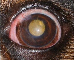 --
G3: gross fundic features are visible when viewed with indirect
ophthalmoscopy (IO) using a head-mounted indirect ophthalmoscope and a
hand-held 30D lens, although fine details are not clear. (Image at
below right is of Cavalier King Charles Spaniel with
keratoconjunctivitis sicca and a CLCT OS. The axial cornea of this eye
was assigned a G3 because indirect ophthalmoscopy was possible. Gross
details of the fundus were visible although the finer details of the
fundus were not.)
--
G3: gross fundic features are visible when viewed with indirect
ophthalmoscopy (IO) using a head-mounted indirect ophthalmoscope and a
hand-held 30D lens, although fine details are not clear. (Image at
below right is of Cavalier King Charles Spaniel with
keratoconjunctivitis sicca and a CLCT OS. The axial cornea of this eye
was assigned a G3 because indirect ophthalmoscopy was possible. Gross
details of the fundus were visible although the finer details of the
fundus were not.)
-- G4: fine details of the fundic features are
clearly visible with IO.
The minimum grades given were analyzed for
inter- and intra-user variability with kappa analysis. Results: Intra-
and interuser variability of the central corneal clarity ranged from
0.78 to 0.96, showing substantial to almost perfect reproducibility, and
from 0.66 to 0.91, showing substantial to almost perfect reliability,
respectively. Intra- and interuser variability of the peripheral cornea
ranged from 0.83 to 0.95, showing almost perfect agreement, and from
0.53 to 0.91, showing moderate to almost perfect agreement. Conclusions:
The RVC-CCS is well suited to assess and monitor central corneal clarity
in small animals and to compare outcomes between studies and different
surgeons.
Dry Eye in Dogs: When Good Glands Go Bad. Shelby Reinstein. Vet. Team Brief. February 2017. Quote: Dry eye, or keratoconjunctivitis sicca (KCS), is a common condition in dogs characterized by decreased tear production that most often results from idiopathic lacrimal gland inflammation with secondary glandular atrophy. Neurogenic KCS is caused by loss of parasympathetic innervation to the lacrimal gland and is less common than immune-mediated KCS. Neurogenic KCS occurs secondary to chronic otitis, peripheral neuropathies, idiopathic disease, and primary neurologic disease. Decreased tear production results in corneal and conjunctival cellular hypoxia, debris accumulation, and bacterial overgrowth, causing inflammation of the ocular surface. Clinical signs include conjunctival hyperemia, squinting, and thick, sticky discharge. ... KCS should be suspected in all patients with clinical signs, especially in those with breed predisposition, including Cavalier King Charles spaniel, cocker spaniel, English bulldog, pug, shih tzu, West Highland white terrier, and Lhasa apso.
Tear fluid collection in dogs and cats using ophthalmic sponges. Sebbag L, Harrington DM, Mochel JP. Vet. Ophthalmol. August 2017. doi: 10.1111/vop.12502. Quote: Objective: To compare the use of two ophthalmic sponges for tear collection in dogs and cats. Animals Studied: Ten healthy dogs and 10 healthy cats. [None were cavalier King Charles spaniels.] Procedures: A strip (4×10 mm) of either cellulose or polyvinyl acetal (PVA) sponge was inserted into the ventral fornix of each eye for either 15, 30, or 60 s. The wetted strip was placed into a 0.2-mL tube that was first punctured at its bottom. Tears were eluted through the drainage hole into a 1.5-mL tube via centrifugation. Tear volume absorbed (VA) and tear volume recovered (VR) were calculated as the difference of the post- and precollection weight of the 0.2-mL tube and 1.5-mL tube, respectively. Recovery ratio (RR) was determined as the ratio between VR and VA. Results: Ophthalmic sponges were well tolerated by all subjects. In dogs and cats, median (95% range) VA, VR, and RR were as follows: 44 μL (11-106 μL) and 16 μL (2-43 μL); 27 μL (1-84 μL) and 6 μL (0-29 μL); 64% (7-91%) and 35% (0-86%), respectively. PVA sponges achieved significantly greater VR in cats and RR in both species. All parameters were significantly greater with a collection time of 60 vs. 30 and 15 s. Body weight was associated with VA and VR in dogs but not cats. Conclusions: Polyvinyl acetal is better than cellulose for tear collection given its superior recovery. Ophthalmic sponges could facilitate routine analysis of tear fluid in dogs and cats, although further studies are needed to evaluate the quality of tears obtained with this method.
Tear ferning in normal dogs and dogs with keratoconjunctivitis sicca. David Williams, Heather Hewitt. Open Vet. J. September 2017;7(3). Quote: This study evaluates tear ferning as an ancillary technique for the evaluation of the canine tear film in normal eyes and eyes affected by keratoconjunctivitis sicca (KCS). Thirty dogs with KCS [including two cavalier King Charles spaniels] and 50 control dogs with normal tear film were evaluated with a full ophthalmoscopic examination and a Schirmer tear test type 1 (STT) determined before tear samples were obtained from the medial canthus with a microhaematocrit capillary tube. 10ul of tear was placed on a microscope slide and the time to first formation of a fern of crystallised tear solute was determined. The appearance of the ferning pattern was graded and correlated with the STT value. All eyes with KCS had abnormal ferning patterns while 39 out of the 50 normal dogs (78%) had so-called 'normal' ferning patterns. The mean STT for dogs showing 'normal' ferning patterns was 20.6mm/min for the left eye and 21.3mm/min for the right eye. STT values for eyes with 'abnormal' ferning patterns were 10.9mm/min and 12.4mm/min, these differing from the normal eyes with STT above 15mm/min significantly. These findings suggest that tear ferning could be a valuable technique for assessment of the tear film in dogs with KCS.
Measurement of tear osmolarity in the canine eye: a new diagnostic tool for canine keratoconjunctivitis sicca? Williams DL, Buckingham A. Research & Reviews: J. Vet. Sci. December 2017;3(2):8-12. Quote: In this study the osmometer was compared with the Schirmer Tear Test I (STT) in 100 dogs from the patient population at the Queen's Veterinary School hospital, University of Cambridge and a UK animal rehoming facility. 153 eyes were ophthalmoscopically normal and 47 had varying degrees of keratoconjunctivitis sicca (KCS) as determined by STT values below 15mm/min. [including 2 cavalier King Charles spaniels]. ... Corneal ulceration is a frequent sequela, particularly in exophthalmic breeds, such as the Pug or Cavalier King Charles Spaniel, where an imperfect lid closure may exacerbate the problem. ... Animals underwent a full ophthalmic examination and then were sampled using the TearLab device to measure tear osmolarity followed by a standard Schirmer tear test I. Tear osmolality in eyes with normal tear production was compared to that in eyes with KCS using the Student's T test. Correlation between STT and osmolarity data was investigated using the Pearson rank correlation coefficient. Tear osmolality was significantly higher in eyes with KCS (350 ± 27 mOsm) than in normal eyes (339 ± 23 mOsm) this difference significant at p<0.02. As high tear omolarity is considered to be a key factor in the pathogenesis of ocular surface damage in eyes with aqueous tear film deficiency in human patients, measuring tear film osmolarity in dogs with suspected KCS may be an important diagnostic step in these animals.
Ocular surface Rose Bengal staining in normal dogs and dogs with
Keratoconjunctivitis Sicca: Preliminary findings. Williams DL,
Griffiths A. Insights in Vet. Sci. December 2017;1:42-46. Quote: Dry eye
or keratoconjunctivitis sicca, is commonly seen in the dog. Veterinary
ophthalmologists diagnose this aqueous tear deficiency using the
Schirmer tear test (STT), but this measures tear production and does not
indicate ocular surface pathology.
The
vital dye Rose Bengal is commonly used in the diagnosis of dry eye in
human patients but until now has not been reported in veterinary
patients. Here we corelate the degree of Rose Bengal staining with the
STT value and find a reasonable association between dye staining of the
ocular surface and tear production, although clearly other factors are
also important in the genesis of ocular surface damage in dry eye. ...
The KCS group comprised 20 adult dogs with 36 eyes with STT<15mm/min))
all diagnosed with to uni-ocular or bilateral KCS. Only eyes with KCS
were included in the study. The breeds were West Highland White Terriers
(6), Cavalier King Charles Spaniels (4), English Cocker
Spaniel (3), American Bulldog (2), Lhasa Apso (2), Pug (1), Mastiff (1)
and crossbreed (1). ... Whatever the exact mechanism, the stain
evaluates ocular surface pathology rather than merely tear production as
does the STT. For that reason the Rose Bengal stain should be a valuable
addition to the diagnostic tests used in canine dry eye.
(Photo at right is of an 8 year old Cavalier King Charles spaniel with
STT of 4mm/min and staining both of conjunctiva and cornea together with
corneal haze and neovascularisation.)
Canine Keratoconjunctivitis Sicca: Current Concepts in Diagnosis and Treatment. David Williams. J. Clin. Ophthalmol. & Optom. December 2017;1(1):105. Quote: Keratoconjunctivitis sicca or dry eye is a commonly recognised condition of canine patients in veterinary practice. The majority of cases are seen in a number of pedigree dog breeds from West Highland White terriers, English and American Cocker Spaniels, Cavalier King Charles spaniels, Lhasa Apsos and Shuh Tzus among a number of other breeds. In these animals an immune-mediated destruction of the lacrimal gland appears to be taking place and topical cyclosporine has, for many years, been a valuable treatment option in these animals. Other causes of dry eye are neurological dysfunction and drug-related lacrimotoxicity. In these cases topical cyclosporine is not effective and treatment with tear replacement drops is required. Another option is parotid duct transposition which can give a lasting improvement in ocular surface pathology. Diagnosis of canine dry eye is primarily by use of the Schirmer tear test, in which tear strip wetting of 15-20mm/min is normal and a wetting of less than 10mm/min is characteristic of dry eye. ... Our research has shown an average STT of one thousand dogs with normal eyes of 18.6mm/min but different breeds have significantly different STT values. Interesting a number of breeds classically recognised as predisposed to KCS, namely the West Highland white terrier, the American and English Cocker Spaniel, the Lhasa Apo, Shih Tzu and Cavalier King Charles Spaniel have significantly lower STT results. ... Other diagnostic modalities such as staining with the vital dye Rose Bengal, determination of tear ferning or measurement of tear film breakup time or tear film osmolarity are used to varying degrees by those investigating the canine tear film but in clinical practice the Schirmer tear test remains the key diagnostic test. Understanding the pathogenesis and treatment of canine keratoconjunctivitis sicca is important for the animals thus affected, but also in the manner it provides a useful spontaneous model for dry eye in human patients. Other models include rodents with inherited predisposition to dry eye and those in which a continual airflow dries the ocular surface, but the spontaneous canine model has the advantage of an eye much more similar in size to the human eye than is the mouse or rat globe and an outbred species living in the same environment as the human patients.
Immunohistochemical evaluation of lymphocyte populations in the nictitans glands of normal dogs and dogs with keratoconjunctivitis sicca. David L. Williams, Alice Tighe. Open Vet. J. January 2018;8(1):47-52. Quote: Idiopathic canine keratoconjunctivitis sicca (iKCS) is a common condition of the canine eye involving a deficiency in aqueous tear production which is commonly held to have an immune-mediated, as most probably an autoimmune aetiopathogenesis. Yet to date no direct evaluation has been made of the inflammatory cell populations in the lacrimal tissue of dogs with iKCS. Here we sought to quantify T and B lymphocyte populations in the lacrimal tissue of the nictitans glands of dogs with iKCS those with neurological KCS (nKCS)and also in dogs with tear production within the recognized normal levels and no ocular surface signs of KCS. Nictitans glands were obtained from 10 healthy dogs with no signs or history consistent with KCS at post-mortem or after enucleation. Nictitans glands were also obtained at parotid duct transposition surgery from ten dogs [including one cavalier King Charles spaniel] with idiopathic KCS and three with neurogenic KCS. Histological sections form the lacrimal tissue were processed immunohistochemically with primary monoclonal antibodies recognizing the T lymphocyte CD3 antigen and the B lymphocyte CD79a antigen. Cell numbers were counted in 10 randomly sampled representative high-power fields in five sections. Statistical significance of differences in cell numbers was determined using analysis of variance with significance achieved at p=0.05. Nictitans glands from dogs with iKCS showed elevated numbers of T and B lymphocytes compared with those from dogs with normal tear production. The increase in the T cell population was highly statistically significant (p=0.0025) while the increase in B cells, while statistically significant was less pronouncedly so (p=0.049). T and B lymphocyte numbers were not significantly elevated in nictitans glands from dogs with neurogenic KCS compared with those in dogs with normal tear production. The elevation in the T cell population seen in dogs with idiopathic KCS strongly supports the widely held assumption that this disease is an immune-mediated and probably autoimmune. The lack of increase in T cell populations in dogs with nKCS strongly suggests that the changes in iKCS are causing the tear deficiency and not resulting from it.
Effect of tear collection on lacrimal total protein content in dogs and cats: a comparison between Schirmer strips and ophthalmic sponges. Lionel Sebbag, Emily M. McDowell, Patrick M. Hepner, Jonathan P. Mochel. BMC Vet. Res. March 2018;14:61. Quote: Background: Quantification of lacrimal total protein content (TPC) is an important tool for clinical scientists to understand disease pathogenesis, identify potential biomarkers and assess response to therapy, among other applications. However, TPC is not only affected by disease state but also by the method used for tear collection. Thus, the purpose of this study is to determine the impact on TPC of two methods of tear collection in dogs and cats: Schirmer strips and polyvinyl acetal (PVA) sponges. Methods: (i) In vivo - Ten healthy dogs and 10 healthy cats were examined. [None were cavalier King Charles spaniels.] Each animal underwent two sessions, separated by 10 min, in which a Schirmer strip was placed in one randomly selected eye until the 20-mm mark was reached, while a strip of PVA sponge was placed in the other eye for 1 min. (ii) In vitro - Schirmer strips and PVA sponges were spiked with various volumes of four bovine serum albumin solutions (0.5, 4, 10, and 20 mg/mL). In both experiments, the wetted absorbent materials were centrifuged for 1 min, and the TPC was quantified on the extracted fluid using Direct Detect™ infrared spectroscopy. Results: Lacrimal TPC in dogs and cats ranged from 5.2 to 14.6 mg/mL and from 6.2 to 20.6 mg/mL, respectively. In cats, TPC was significantly lower with Schirmer strips vs. PVA sponges (P<0.001). In dogs, the volume absorbed by PVA sponges was negatively correlated with TPC (r= −0.48, P=0.033). The inter-session coefficient of variation was significantly lower with Schirmer strips vs. PVA sponges in both species (P≤0.010). In vitro, both absorbent materials resulted in a 'concentrating effect' of the TPC obtained post-centrifugation, which was most pronounced when the volume absorbed was low, especially for Schirmer strips. Conclusion: Schirmer strips provide a repeatable method to quantify lacrimal TPC in dogs and cats. Schirmer strips are more reliable than PVA sponges (i.e. lower inter-session CV%) for quantification of TPC in canine and feline tears. However, care should be taken to absorb sufficient volumes of tears with Schirmer strips to minimize the concentrating effect from the absorbent material.
Effects of autologous serum eye drops for treatment of keratoconjunctivitis sicca in dogs. Han-Syuan Lin, Shiun-Long Lin, Feng-Jen Chang. Taiwan Vet. J. September 2018;44(3):111-117. Quote: In humans, autologous serum (AS) eye drops has been applied for the treatment of refractory keratoconjunctivitis sicca (KCS) for several decades. However, there are few researches to investigate the AS eye drops in dogs with KCS. The objective of this study was to evaluate the effects of AS eye drops on treatment of KCS in dogs. Eighteen eyes of ten client-owned dogs with refractory KCS were used in this study. Schirmer tear test (STT), tear film breakup time (TBUT), fluorescein (FL) staining score, and Rose Bengal (RB) staining score were used to measure the status of cornea prospectively at baseline and 1-3 months after treatment. Additionally, the results were further stratified by their STT value, sex, and age. The results indicated that the mean TBUT, FL staining score, and RB staining score were significantly improved after treatment except STT. In 18 eyes, 77.8% eyes had decreased mucopurulent ocular discharge and 38.9% eyes got wet. Besides, both TBUT and RB staining score were significantly improved in a subgroup of dogs with age less than 9 years old. As far as we know, this study is the first trial to determine the efficacy and safety of 20% AS eye drops for cKCS. In conclusion, AS eye drops seemed to be effective and safe for dogs with KCS, and it could improve tear film stability, ocular surface health, and subjective clinical symptoms, especially in dogs younger than 9 years old.
Effect of methadone and acepromazine premedication on tear production in dogs. Hayley A Volk, Ellie West, Rose Non Linn-Pearl, Georgina V Fricker, Ambra Panti, David J Gould. Vet. Rec. open. December 2018;5:e000298. Quote: Objectives: To evaluate the combined effect of intramuscular acepromazine and methadone on tear production in dogs undergoing general anaesthesia for elective, non-ocular procedures. Design: Prospective, non-randomised, pre-post treatment study. Setting: Patients were recruited from a referral practice in the UK. Methods: Thirty client-owned dogs [including two cavalier King Charles spaniels] were enrolled in this study and received a combined intramuscular premedication of methadone (0.3 mg/kg) and acepromazine (0.02 mg/kg) before general anaesthesia for elective, non-ocular procedures. Full ophthalmic examination was performed and tear production was quantified using the Schirmer tear test-1 (STT-1). On the day of general anaesthesia, an STT-1 was performed before (STT-1a) and after (STT-1b) intramuscular premedication with methadone/acepromazine. Results: Using a general linear model, a significant effect on STT-1 results was found for premedication with methadone/acepromazine (P=0.013), but not eye laterality (P=0.527). Following premedication, there was a significant reduction observed in the mean STT-1 readings of left and right eyes between STT-1a (20.4±2.8 mm/min) and STT-1b (16.9±4.1 mm/min; P<0.001). Significantly more dogs had an STT-1 reading less than 15 mm/min in one or both eyes after premedication (30 per cent; 9/30 dogs) [including one of the CKCSs] compared with before premedication (6.7 per cent; 2/30 dogs; P=0.042). Conclusions: An intramuscular premedication of methadone and acepromazine results in a decrease in tear production in dogs before elective general anaesthesia. This may contribute to the risk of ocular morbidities, such as corneal ulceration, particularly in patients with lower baseline tear production.
A novel strip meniscometry method for measuring aqueous tear volume in dogs: Clinical correlations with the Schirmer tear and phenol red thread tests. Keiichi Miyasaka, Yoshiyuki Kazama, Hiroko Iwashita, Shinsuke Wakaiki, Akihiko Saito. Vet. Ophthal. March 2019; doi: 10.1111/vop.12664. Quote: Objectives: The strip meniscometry test (SMT) is a novel method for quantitative measurement of tear volume with only five seconds. We aimed to evaluate clinical correlations of SMT with the gold standard Schirmer tear test (STT) and phenol red thread test (PRT) in dogs, including normal and tear-deficient eyes. Animals studied: Left eyes from 621 outpatient dogs with and without ocular disorders were evaluated [including 25 cavalier King Charles spaniels]. Procedures: Each subject underwent SMT, PRT, and STT without topical anesthesia in the described order with five-minute intervals. The total population was divided into four groups by classifying tear deficiency severity based on STT results: "severe" (0-5 mm/min), "moderate" (6-10 mm/min), "subclinical" (11-14 mm/min), and "normal" (15 or more mm/min). Results: The strongest correlation coefficient was found between SMT-STT (0.676), followed by PRT-STT (0.637) and SMT-PRT (0.600) pairs. Mean(SD) scores of SMT, PRT, and STT in total population were 9.47 (4.08) mm/5 s, 33.30 (8.52) mm/15 s, and 16.47 (7.01) mm/min. Significant differences were found among STTclassified groups, both using SMT and PRT results. Receiver operating characteristic (ROC) curves revealed that SMT better agreed with STT than PRT; agreement increased with increasing STT severity. A cutoff for SMT was identified at 10 mm/5 s to discriminate normal eyes from tear-deficient eyes, yielding high sensitivities and acceptable specificities. Conclusions: SMT could be superior to PRT for discriminating tear-deficient eyes. The high sensitivity of SMT could be useful as an initial diagnostic tool to rule out normal eyes with the short testing time.
A retrospective evaluation of systemic and/or topical pilocarpine treatment for canine neurogenic dry eye: 11 cases. Michaela L. Wegg. Vet. Ophthal. December 2019;doi: 10.1111/vop.12731. Quote: Objective: To assess the response to topical and/or systemic pilocarpine in dogs with neurogenic dry eye. Method: Medical records of dogs diagnosed with dry eye between 2015 and 2018 were reviewed [including one cavalier King Charles spaniel]. Cases were excluded if STT values were decreased bilaterally, if dogs were lost to follow-up, or if surgical measures (parotid duct transposition) were undertaken within thirty days of presentation. Dogs were on treatment with topical pilocarpine (0.1%, every 6 hours) and/or oral pilocarpine (starting dose 2%, one drop per 10 kg every twelve hours). Results: Eleven cases were included in the study, seven females and four males [1 cavalier male] with mean age of 10 years. Seven cases had xeromycteria, two cases had facial nerve paralysis, and one case had Horner's syndrome. Seven cases (63.6%) had successful outcome following pilocarpine treatment, return to normal STT (15-25mm/minute), in an average of 24 ± 5.1 days. Of these cases, five had both systemic and topical treatment, one had just topical treatment, and one had just systemic treatment. The average time to normal tear production on treatment with topical pilocarpine ± systemic was 23 days (range 9-48 days). The number of systemic drops until a positive response varied between individuals from 0.8drops/10kg to 7drops/10kg. Conclusion: Pilocarpine treatment (topical ± systemic) is an effective therapy for unilateral dry eye disease in cases suspected to be neurogenic in origin. Most cases responded within 30 days. Side effects included topical irritation to the ophthalmic solution and systemic effects from oral pilocarpine, such as diarrhea and regurgitation.
Efficacy of adjunctive therapy using Vizoovet in improving clinical signs of keratoconjunctivitis sicca in dogs: A pilot study. D. Dustin Dees, Michael S. Kent. Vet. Ophthal. May 2020; doi: 10.1111/vop.12763. Quote: Objective: To assess the clinical safety and efficacy of adjunctive therapy using Vizoovet to ameliorate clinical signs of keratoconjunctivitis sicca (KCS) in dogs. Animals studied: Twenty client‐owned dogs. Procedures: Canine patients diagnosed with KCS were enrolled in this prospective study. Patients were randomly selected to receive either Vizoovet or GenTeal drops twice daily in addition to twice daily tacrolimus 0.03% solution. Data were collected from only one eye of each patient and included STT-1, IOP, TFBUT, and results of objective clinical scoring performed by pet owners. Statistical significance was set at P ≤ .05. Results: In all, 20 dogs (20 eyes) were enrolled in this prospective randomized study. Females (n = 12; 60%) outnumbered males (n = 8; 40%) and all dogs were spayed/neutered. Mean age of all dogs was 10.6 ± 3.79 years. In both treatment groups, the improvement in STT-1 values over the course of the study was significant. When comparing the STT-1 improvements between groups, no significance was found. In both groups, the improvement in TFBUT was significant. When comparing the TFBUT improvements between groups, no significance was found. Squinting, rubbing, ocular discharge, and medication administration scores all significantly improved throughout the course of the study; however, they did not differ significantly between groups. Throughout the study, no adverse side effects were noted clinically or by the pet owner in either group. Conclusions and Clinical Relevance: Adjunctive treatment with Vizoovet was as safe and effective as GenTeal drops at improving clinical signs of dry eye in dogs.
Keratoconjunctivitis sicca in dogs under primary veterinary care in the UK: an epidemiological study. D. G. O'Neill, D. C. Brodbelt, A. Keddy, D. B. Church, R. F. Sanchez. J. Sm. Anim. Pract. June 2021; doi: 10.1111/jsap.13382. Quote: Objectives: To estimate the frequency and breed-related risk factors for keratoconjunctivitis sicca (KCS) in dogs under UK primary veterinary care. Methods: Analysis of cohort electronic patient record data through the VetCompass Programme. Risk factor analysis used multivariable logistic regression. Results: There were 1456 KCS cases overall from 363,898 dogs [prevalence 0.40%, 95% confidence interval (CI) 0.38-0.42] and 430 incident cases during 2013 (1-year incidence risk 0.12%, 95% CI 0.11-0.13). ... The breeds with the highest KCS prevalence were American cocker spaniel, West Highland white terrier, cavalier king Charles spaniel, Lhasa apso, English bulldog, English bull terrier and English cocker spaniel. ... The most common breeds among the KCS cases were West Highland white terrier (n = 212, 14.56%), English cocker spaniel (195, 13.39%), cavalier king Charles spaniel (158, 10.85%), shih-tzu (118, 8.10%) and Lhasa apso (113, 7.76%). ... The breeds with the highest predispositions (i.e. aORs) in the present primary-care study were American cocker spaniel, English bulldog, pug, Lhasa apso, cavalier king Charles spaniel, English bull terrier, English cocker spaniel, basset hound, West Highland white terrier, shih-tzu and king Charles spaniel. ... Brachycephalic dogs had 3.63 (95% CI 3.24-4.07) times odds compared to mesocephalics. Spaniels had 3.03 (95% CI 2.69-3.40) times odds compared to non-spaniels. Dogs weighing at or above the mean bodyweight for breed/sex had 1.25 (95% CI 1.12-1.39) times odds compared to body weights below. Advancing age was strongly associated with increased odds. Clinical significance: Quantitative tear tests are recommended within yearly health examinations for breeds with evidence of predisposition to KCS and might also be considered in the future within eye testing for breeding in predisposed breeds. Breed predisposition to KCS suggests that breeding strategies could aim to reduce extremes of facial conformation. ... The authors make recommendations to reduce the incidence and clinical impact of KCS by including quantitative tear tests in eye testing as part of the annual physical examination of all dogs in the list of predisposed breeds, especially as they approach advanced age, and to consider adding it within eye testing for breeding animals as we attempt to gain a more complete understanding of the condition and how it affects these breeds.
Long-lasting otic medications may be a rare cause of neurogenic keratoconjunctivitis sicca in dogs. Genia R. Bercovitz, Annora M. Gaerig, Emily D. Conway, Jane Ashley Huey, Mary R. Telle, Renata Stavinohova, Giunio Bruto Cherubini, Angelo Capasso, Kathern E. Myrna. JAVMA. January 2023; doi: 10.2460/javma.22.07.0301. Quote: Objective: To characterize the clinical course and long-term prognosis of a suspected novel cause of neurogenic keratoconjunctivitis sicca (nKCS) secondary to florfenicol, terbinafine hydrochloride, mometasone furoate (Claro and Neptra) or florfenicol, terbinafine, betamethasone acetate (Osurnia). Animals: 29 client-owned dogs [including one cavalier King Charles spaniel]. Procedures: Online survey and word-of-mouth recruitment were conducted to identify dogs that developed clinical signs of nKCS after application of otitis externa medication containing terbinafine and florfenicol. A retrospective analysis of medical records of dogs meeting inclusion criteria was then conducted. Included dogs had onset of clinical signs of nKCS within 1 day after application of otitis externa medications containing terbinafine and florfenicol and had documentation of low Schirmer tear test value (< 15 mm/min) of affected eyes. Results: 29 dogs with medical records available for review met the inclusion criteria [including one cavalier King Charles spaniel]. Documented return of clinically normal tear production was identified in 24 of 29 dogs, with a median time from application of ear medication to documented return of clinically normal tear production of 86 days (range, 19 to 482 days). A corneal ulcer was diagnosed in 68% (20/29). Multivariable Cox regression analysis showed being referred to an ophthalmologist (P = .03) and having a deep ulcer (P = .02) were associated with a longer time to documentation of Schirmer tear test > 15 mm/min. Clinical Relevance: Dogs that developed nKCS within 1 day after application of otitis externa medications containing terbinafine and florfenicol had a good prognosis for return of normal tear production within 1 year.
The Blue Book. Ocular Disorders Presumed to be inherited in purebred dogs. Genetics Committee of the American College of Veterinary Ophthalmologists. Blue Book 15th Ed. 2023. pp. 297-302.
CONNECT WITH US