
Facial Nerve Paralysis in the
Cavalier King Charles Spaniel
 What It Is
Idiopathic facial nerve paralysis or Bell's palsy or facial palsy is reportedly common in the cavalier King Charles spaniel. It has been identified in several veterinary journal artickws, including this June 2005 article, this July 2011 article, and this March 2012 article, among otheres.
What It Is
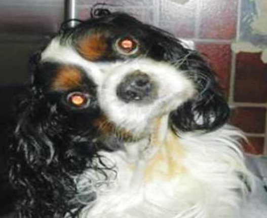
Idiopahtic facial nerve paralysis is the most common form of facial nerve dysfunction (FND). It is described as a sudden onset of either unilateral or bilateral facial nerve paralysis with no other abnormal signs -- no evidence of infection, reduced thryroid function, or injury. Signs may include drooping or inability to blink eye lids or move lips, ears, or other muscles of the face. (See the chart of signs below.) Onset of the signs usually are sudden and do not worsen over time. The signs typically occur in dogs aged 2 years or older.
The cause of facial nerve paralysis is said to be
idiopathic facial neuropathy in 75% of the cases, which
 essentially means the cause has not been determined. It may
also be diagnosed as Horner's Syndrome. In 40% to
70% of the cases, it has been combined with
vestibular syndrome. The cause of that combination likewise is
unknown. In fact, that combination condition has been named "facial and
vestibular neuropathy of unknown origin" (FVNUO). Seriously(!).
In a November 2016 abstract, French researchers studied 69 dogs with facial nerve paralysis. The report does not provide a count of cavalier King Charles spaniels included in the study, but the researchers concluded that the CKCS and the French bulldog should be added to the list of predisposed breeds to the disorder. Additionally, they found:
"Idiopathic facial paralysis was diagnosed in 48% of dogs. Vestibular signs were the most common additional clinical signs and were observed in 36% of dogs with idiopathic facial paralysis. Peripheral nervous system disease was diagnosed in 19% of dogs, and central nervous system disease occurred in 30% of dogs. ... Improved diagnostic methods enabled the diagnosis of a higher percentage of inflammatory/infectious diseases, which were absent in the central nervous system aetiologies of a previous similar study, and revealed metabolic (hypothyroidism), inflammatory and neoplastic aetiologies for peripheral nervous system disease."
In a February 2018 article, a team of French veterinary researchers reported finding primary secretory otitis media (PSOM) as the cause of two cavaliers drooling from their lips and with facial paralysis. One of the two cavaliers also was diagnosed with Chiari-like malformation and syringomyelia (CM/SM). The clinicians performed myringotomies and flushed the tympanic bullae of both CKCSs and then treated with corticosteroids. The dogs' clinical signs improved for several months. They concluded:
"These two cases highlight the importance of otitis media in the differential diagnosis of facial paralysis and focus on the originality of primary secretory otitis media in the Cavalier King Charles Spaniels."
For more information, see Dr. Clare Rusbridge's YouTube video on "Idiopathic facial nerve paralysis (Bell's Palsy) in the dog".
RETURN TO TOP
Symptoms
In Dr. Clare Rusbridge's YouTube video on "Idiopathic facial nerve paralysis (Bell's Palsy) in the dog", she offers this chart of indications of facial nerve paralysis:

In a December 2019 article by Univeristy of Sydney, Australia researchers, they reviewed the records of 122 dogs presented at their hospital from 2001 to 2016 which had symptoms of facial nerve paralysis (FNP). FNP can be a symptom of any of a variety of disorders, including primary secretory otitis media (PSOM), direct injury, hypothyroidism, and drug sensitivity. Idiopathic FNP (IFNP) applies to FNP symptoms due to unknown causes. It is not considered to be effectively treatable. Of the 36 purebred dogs in the entire study, 15 (42%) were cavaliers, the highest number of purebreds by far. Of the 15 purebreds diagnosed with IFNP, cavaliers numbered 9 (60%). The authors concluded:
"This study found differences in the incidence of FNP causes compared with the only previous study, which are presumed to be a result of geographic and temporal differences. Breeds and age groups are significant risk factors correlated to acquisition of FNP and IFNP. Vestibular dysfunction, hypersalivation and keratoconjunctivitis sicca were common findings in dogs with IFNP. Dogs with IFNP have guarded prognosis for recovery, and often require life-long eye lubrication. There is no evidence to date that treatment alters the outcome in dogs with IFNP."
In a July 2026 article, Spanish researchers studied 74 dogs diagnosed with facial nerve dysplasia (FND), including 3 cavaliers (4.1%), to determine the extend of ocular manifestations in such dogs. They included loss or reduction of blink reflex, impaired eyelid closure, and decreased tear production predispose the cornea to desiccation, ulceration, infection, and perforation. Ulcerative keratitis was the most common ocular finding, detected in 52 eyes (52/84; 61.9%). Corneal ulceration was statistically associated with a higher incidence of uveitis.
For more information, see Dr. Clare Rusbridge's YouTube video on "Idiopathic facial nerve paralysis (Bell's Palsy) in the dog".
RETURN TO TOP
Diagnosis
In a May 2021 article, in which magnetic resonance imaging (MRI) was performed on 19 dogs, including 2 cavaliers (11%), which were diagnosed with unilateral idiopathic facial neuropathy, the clinicians were able to observe in the MRI scans, characteristics of idiopathic facial neuropathy which included ipsilateral atrophy and digastric muscle difference, enabling them to predict the likelihood of the return to normal facial nerve function over time.
For more information, see Dr. Clare Rusbridge's YouTube video on "Idiopathic facial nerve paralysis (Bell's Palsy) in the dog".
RETURN TO TOP
Treatment
Treatment options include (1) lubricating the eyes, as the dog may not be able to fully close one or both of its eye lids, resulting in dry eye, and (2) oral care, because the dog may have difficulty chewing, and drooling may cause a skin condition. In humans, early (within 72 hours of onset) short term treatment with a high dose of corticosteroids has been shown to provide rapid and more complete return of facial nerve function.
Recovery may be within weeks, but in the most cases the abnormality is permanent. There is an increased risk of exposure corneal ulcers in dogs with protruding eyes, such as cavaliers. Facial nerve paralysis in the CKCS may also be associated with primary secretory otitis media (PSOM), masticatory muscle myositis (MMM), hypothyroidism, and/or vestibular syndrome.
In an April 2017 article, a cavalier suffered facial nerve paralysis which caused sleep apnea and an inability to blink her eyes. Specialists at the Animal Health Trust performed surgery on her eyelids to help protect her eyes while awaiting nerve function to slowly return. The author states:
"Fortunately most dogs cope very well with such nerve paralysis, which can be permanent, as long as we can keep their eyes healthy."
In an October 2017 abstract, Dr. Dustin Dees of Austin, Texas reports successfully treating a cavalier suffering from idiopathic facial nerve paralysis by using continuous wave laser treatments at four acupuncture sites corresponding to a branch of the facial nerve. The method is called "photobiomodulation". Treatments were performed twice a week for five weeks. The results were successful. The dog's blinking ability substantially improved, and corneal dryness resolved with marked improvement of the dog's Schirmer testing.
For more information, see Dr. Clare Rusbridge's YouTube video on "Idiopathic facial nerve paralysis (Bell's Palsy) in the dog".
RETURN TO TOP
Research News
December 2019:
Australian 16-year veterinary school study of facial nerve
paralysis finds cavaliers at the top of the list.
 In
a
December 2019 article by Univeristy of Sydney, Australia researchers
(MK Chan, J-ALML Toribio, JM Podadera, Georgina Child [right])
they reviewed the records of 122 dogs presented at their hospital from
2001 to 2016 which had symptoms of facial nerve paralysis (FNP). FNP can
be a symptom of any of a variety of disorders, including primary
secretory otitis media (PSOM), direct injury, hypothyroidism, and drug
sensitivity. Idiopathic FNP (IFNP) applies to FNP symptoms due to
unknown causes. It is not considered to be effectively treatable. Of the
36 purebred dogs in the entire study, 15 (42%) were cavalier King
Charles spaniels, the highest number of purebreds by far. Of the 15
purebreds diagnosed with IFNP, cavaliers numbered 9 (60%). The authors
concluded:
"This study found differences in the incidence of FNP causes compared with the only previous study, which are presumed to be a result of geographic and temporal differences. Breeds and age groups are significant risk factors correlated to acquisition of FNP and IFNP. Vestibular dysfunction, hypersalivation and keratoconjunctivitis sicca were common findings in dogs with IFNP. Dogs with IFNP have guarded prognosis for recovery, and often require life-long eye lubrication. There is no evidence to date that treatment alters the outcome in dogs with IFNP."
October
2017:
Cavalier with facial nerve paralysis is successfully treated
with laser treatment at acupuncture points.
 In an
October 2017 abstract, Dr. Dustin Dees (right) of Austin,
Texas reports successfully treating a cavalier King Charles spaniel
suffering from idiopathic facial nerve paralysis by using continuous
wave laser treatments at four acupuncture sites corresponding to a
branch of the facial nerve. The method is called "photobiomodulation".
Treatments were performed twice a week for five weeks. The results were
successful. The dog's blinking ability substantially improved, and
corneal dryness resolved with marked improvement of the dog's Schirmer
testing.
November 2016: French study adds cavaliers to list of breeds predisposed to facial nerve paralysis. In a November 2016 abstract, French researchers studied 69 dogs with facial nerve paralysis. The report does not provide a count of cavalier King Charles spaniels included in the study, but the researchers concluded that the CKCS and the French bulldog should be added to the list of predisposed breeds to the disorder. Additionally, they found:
"Idiopathic facial paralysis was diagnosed in 48% of dogs. Vestibular signs were the most common additional clinical signs and were observed in 36% of dogs with idiopathic facial paralysis. Peripheral nervous system disease was diagnosed in 19% of dogs, and central nervous system disease occurred in 30% of dogs. ... Improved diagnostic methods enabled the diagnosis of a higher percentage of inflammatory/infectious diseases, which were absent in the central nervous system aetiologies of a previous similar study, and revealed metabolic (hypothyroidism), inflammatory and neoplastic aetiologies for peripheral nervous system disease."
RETURN TO TOP
RETURN TO TOP
Veterinary Resources
Results of magnetic resonance imaging in dogs with vestibular disorders: 85 cases (1996-1999). Laurent S. Garosi, Ruth Dennis, Jacques Penderis, Christopher R. Lamb, Mike P. Targett, Rodolfo Cappello, Agnes J. Delauche. J Am Vet Med Assoc February 2001;218:385-391.Quote: Objective: To determine results of magnetic resonance (MR) imaging in dogs with vestibular disorders (VD) and correlate results of MR imaging with clinical findings. Design: Retrospective study. Animals: 85 dogs. Procedure: Information on signalment, clinical signs, and presumptive lesion location was obtained from the medical records, and MR images were reviewed. Results: 27 dogs had peripheral VD, 37 had central VD, and 21 had paradoxical VD. Of the 27 dogs with peripheral VD, 11 (41%) had MR imaging abnormalities involving the ipsilateral tympanic bulla compatible with otitis media (6 also had abnormalities involving the petrous portion of the ipsilateral temporal bone compatible with otitis interna), 7 (26%) had MR imaging abnormalities compatible with middle ear neoplasia, 2 (7%) had an ipsilateral cerebellopontine angle lesion, and 7 (26%) did not have MR imaging abnormalities. ... Two of these dogs had concurrent facial paralysis, and 1 had concurrent Horner's syndrome. ... All dogs with central and paradoxical VD had abnormalities evident on MR images. Of the 37 dogs with central VD, 13 (35%) had an extra-axial lesion, 6 (16%) had an intra-axial lesion, and 18 (49%) had multiple intra-axial lesions. In 23 (62%) dogs with central VD, lesions on MR images corresponded with location suspected on the basis of clinical signs. Of the 21 dogs with paradoxical VD, 12 (57%) had an extra-axial lesion, 5 (24%) had an intra-axial lesion, and 4 (19%) had multiple intra-axial lesions. Location of lesions on MR images agreed with location suspected on the basis of clinical signs in 19 (90%) dogs. ... However, some of these dogs had concurrent facial paralysis or Horner's syndrome, which are not usually observed in dogs with idiopathic peripheral VD.4 This suggests that these dogs may have had an underlying middle or inner ear abnormality and emphasizes the lack of sensitivity of MR imaging techniques used to date. Conclusions and Clinical Relevance: Results suggest that MR imaging may be helpful in the diagnosis and treatment of VD in dogs.
Neurological diseases of the Cavalier King Charles spaniel. Rusbridge, C. J. Small Anim. Prac., June 2005, 46(6): 265-272. Quote: "Facial nerve paralysis (Rusbridge and others 2000) and deafness (Skerritt and Skerritt 2001) have also been associated with the condition. However idiopathic facial palsy is common in the CKCS and so is hearing impairment (Munro and Cox 1997)."
Bell's palsy with concomitant idiopathic cranial nerve polyneuropathy in seven dogs. L. Motta, U. Michal Altay, G. C. Skerritt. J.Small Animal Practice. July 2011;5297):397. Quote: "Recently, we encountered seven dogs (two boxers, one Labrador retriever, three cavalier King Charles spaniels [43%] and one Hungarian vizsla; median age 6.7 years; 5:2 male:female ratio) that were referred for investigation of acute onset of unilateral facial nerve paralysis."
Neurological Diseases in the Cavalier King Charles Spaniel: Chiari, Episodic Falling, and More. Jacques Penderis. March 2012. Quote: "Idiopathic facial nerve paralysis: this is the most common cause of facial nerve paralysis in dogs (75% of cases in one study) and is common in the CKCS. There is an acute onset of unilateral or bilateral facial nerve paralysis with no other abnormalities evident. In some cases this may be combined with idiopathic vestibular syndrome. In some cases there will be recovery in weeks, but in the majority of cases the abnormality is permanent. The deficits are largely cosmetic, although there is an increased risk of exposure corneal ulcers in dogs with protruding eyes. In other dogs little intervention is required, besides keeping the lips clean to prevent moist dermatitis in dogs with excessive drooling and drooping lips."
Facial and vestibular neuropathy of unknown origin in 16 dogs. A. Jeandel, J. L. Thibaud, S. Blot. J. Sm. Anim. Pract. January 2016. Quote: "Objectives: The aim of this study was to describe the signalment, clinical presentation, diagnostic findings and long-term follow-up in dogs with concomitant facial and vestibular neuropathy of unknown origin [FVNUO]. Methods: Appropriate cases were located through medical record searches. Inclusion criteria comprised dogs that had: clinical signs of facial paralysis with concomitant peripheral vestibular syndrome, thyroid function tests, no abnormalities on magnetic resonance imaging of the brain and tympanic bullae, and cerebrospinal fluid analysis. Results: Sixteen dogs met the inclusion criteria [including 2 cavalier King Charles spaniels]. Facial paralysis had acute onset (<24 hours) in all dogs, thyroid function was within normal limits. ... None of the 16 dogs received any treatment except artificial tears. Follow-up was incomplete for three dogs. In eight dogs (62%), vestibular signs resolved [including one of the cavalier King Charles spaniels]. The median time to complete PVS [peripheral vestibular syndrome] resolution was 55 days (range, 3 to 365). A return to normal facial movement was reported in five dogs (39%) [including one of the cavalier King Charles spaniels]. The median time to FP [facial paralysis] resolution was 45 days (range, 7 to 730). Complete resolution of both PVS and FP was observed in four dogs (31%) [including one of the cavalier King Charles spaniels]. The nine other dogs exhibited long-term sequelae: five dogs (38%) displayed a persistent or intermittent head tilt; two dogs (15%) did not recover facial motor function after 18 and 24 months; ipsilateral hemifacial contracture developed in six other dogs (46%) [including one of the cavalier King Charles spaniels]. The median time between the initial and last follow-up examination was 18 months (range, 6 to 42). ... Clinical Significance: Facial and vestibular neuropathy of unknown origin shares similarities with idiopathic facial paralysis. The prognosis for return of normal facial and vestibular function is guarded and there may be relapse after recovery."
Facial Nerve Paralysis in Dogs: A Retrospective Study of 69 Cases. C. Ricco, L. Giraud, L. Cauzinille. J. Vet. Int. Med. November 2016;30(6):1953. Quote: Facial paralysis is readily recognised in small-animal veterinary practice because of its manifestation of facial asymmetry; the idiopathic form has been previously reported to be present in 75% of dogs. The purpose of this study in dogs was to classify and determine the origin of facial nerve dysfunction using enhanced diagnostic procedures, including magnetic resonance imaging (MRI). The medical records of 69 dogs admitted for facial paralysis were reviewed. Neurological examination confirmed facial nerve abnormalities, which were all investigated with MRI. Idiopathic facial paralysis was diagnosed in 48% of dogs. Vestibular signs were the most common additional clinical signs and were observed in 36% of dogs with idiopathic facial paralysis. Peripheral nervous system disease was diagnosed in 19% of dogs, and central nervous system disease occurred in 30% of dogs. Two new predisposed breeds are added, the French bulldog and the Cavalier King Charles spaniel. Improved diagnostic methods enabled the diagnosis of a higher percentage of inflammatory/infectious diseases, which were absent in the central nervous system aetiologies of a previous similar study, and revealed metabolic (hypothyroidism), inflammatory and neoplastic aetiologies for peripheral nervous system disease.
J-Lo's Story. James Warland. Animal Health Trust. April 2017. Quote: J-Lo, a seven year old Cavalier King Charles Spaniel, suffers from several different health problems, including severe sleep apnoea, syringomyelia and facial nerve paralysis, which causes her problems with her eyes. J-Lo's sleep apnoea means she often wakes up suddenly because she can't breathe. ... J-Lo has been referred to the Animal Health Trust (AHT), near Newmarket, several times and has used almost the full range of the AHT's state-of-the-art equipment, including MRI and CT scans, fluoroscopy and endoscopy to help the vets of many different specialties at the AHT investigate her different health problems. ... James Warland, one of the vets who is treating J-Lo at the AHT, said: "J-Lo's sleep apnoea has got so extreme that she has had to be hospitalised for monitoring and more tests to work out the best way to treat her. ... The main problem with J-Lo's eyes is that the facial nerve paralysis prevents her from blinking. She has had a temporary surgery on her eyelids to help protect her eyes while we hope the nerve function will slowly return. Fortunately most dogs cope very well with such nerve paralysis, which can be permanent, as long as we can keep their eyes healthy. ... We look forward to seeing J-Lo for a check-up again soon."
Photobiomodulation for treatment of idiopathic facial nerve paralysis in a dog. Dustin D. Dees. Vet. Ophthal. October 2017. DOI: 10.1111/vop.12519; E-13. Quote: Purpose: To describe a novel method for treatment of facial nerve paralysis in a dog. Methods: Photobiomodulation (Low-level laser therapy) was performed on a 10-year-old neutered male Cavalier King Charles Spaniel with right-sided idiopathic facial nerve paralysis. Informed owner consent was obtained prior to treatment. The laser unit (GradyVet P- 3000; Class 3b; 810 nm) utilized a precision single-diode trigger point probe (500 mW) with the dose calculated by accompanying software. A total of four sites were treated corresponding to the acupuncture points Gall Bladder-1, 2, 3 and Triple Heater-17. These points correlated with the anatomic position of the auriculopalpebral branch of the facial nerve. Laser settings were continuous wave, 500 mW, 32 s at each site. Treatments were performed twice weekly for five weeks. Results: Initial ophthalmic examination showed complete absence of palpebral reflex, moderate upper eyelid ptosis, mild neurotrophic keratitis, and Schirmer tear test-1 (STT-1) value of 17 mm/min OD. After ten treatments, the upper eyelid ptosis and axial corneal dryness resolved. STT-1 value improved to 21 mm/min. Blinking ability was improved allowing for 50% coverage of the cornea. Patient follow up at 3 and 6 months showed stability in blinking ability. Conclusion: In this case, photobiomodulation was successful in improvement of blinking ability and STT-1 values in a dog with unilateral facial nerve paralysis.
Facial paralysis associated with primary secretory otitis media in Cavalier King Charles Spaniels: Study of 2 cases. M. Debuigne, A. Drut, N. Soetart, D. Guillemaille, T. Brement, M. Fusellier, D. Fanuel, V. Bruet. Revue Veterinaire Clinique. February 2018. DOI: 10.1016/j.anicom.2018.01.002 Quote: Primary secretory otitis media is a disease due to an accumulation of mucus in the tympanic bulla, mostly described in the Cavalier King Charles Spaniel. Clinical signs are numerous and can be subtle; facial paralysis is possible but signs of pruritus or pain are most commonly reported. A 4-year-old male and a 7-year-old spayed female Cavalier King Charles have been presented for lip drooping. Neurologic exam showed a generalized decrease in facial muscle motricity. Because of the history of these animals, a primary otitis media was firstly suspected. Imaging of tympanic bullae showed a unilateral or bilateral otitis media. Video-otoscopic examinations and myringotomy revealed acellular mucus in the concerned tympanic bullae. The myringotomy and the flus h of tympanic bullae, associated with corticosteroids, improved the clinical signs for several months. These two cases highlight the importance of otitis media in the differential diagnosis of facial paralysis and focus on the originality of primary secretory otitis media in the Cavalier King Charles Spaniels.
Incidence, cause, outcome and possible risk factors associated with facial nerve paralysis in dogs in a Sydney population (2001-2016): a retrospective study. MK Chan, J‐ALML Toribio, JM Podadera, G Child. Australian Vet. J. December 2019;doi: 10.1111/avj.12906. Quote: Objective: This study aims to assess the incidence and causes of facial nerve paralysis (FNP) in dogs in the Sydney region. Its outcome and possible risk factors are investigated to prognosticate and aid design of diagnostic and treatment plans. Design: Retrospective case study. Methods: Client-owned dogs presented to the University Veterinary Teaching Hospital, Sydney (UVTHS), between 2001 and 2016 with FNP were included (n = 122). ... Thirty-six pure breed dogs were included in this study. When comparing the odds of FNP in each breed with that for crossbreed dogs, Cavalier King Charles Spaniels (n = 15) and Cocker Spaniels (n = 5) were statistically over-represented. ... The incidence of each cause of FNP was investigated. A reference population of noncases seen at the UVTHS during the same time period was used to study the association between idiopathic facial nerve paralysis (IFNP) and gender, age and breed. Results: IFNP (29.5%) was the most common diagnosis. Male dogs had increased odds of IFNP compared with female dogs. Age was a significant risk factor for both the occurrence of FNP and IFNP. There was higher occurrence of IFNP among middle-aged dogs (5-13 years) and reduced risk in juvenile dogs (less than 2 years). Cavalier King Charles Spaniels were over-represented for FNP [15 out of 36 purebreds] and IFNP [9 out of 15 purebreds]. For IFNP, 6 of 16 dogs with known follow-up showed definitive resolution within 3 years of diagnosis. Concurrent vestibular signs were common in dogs with middle/inner ear abnormality and intracranial disease; and were also seen in 50% of dogs with IFNP. Conclusion: The results of this study demonstrate statistical predilections in age, gender and breed for IFNP. Guarded prognosis for recovery should be given to dogs diagnosed with IFNP and supportive management instigated.
Magnetic resonance imaging of the caudal portion of the digastric muscle in canine idiopathic facial neuropathy. Ombeline McGregor, Mark J. Plested, Elsa Beltran. Vet. Radiol. & Ultrasound. May 2021; doi: 10.1111/vru.12974. Quote: Idiopathic is the most common etiology for acute onset of facial neuropathy in dogs with limited number of studies describing MRI characteristics. A retrospective, observational study was performed using archived records, aiming to describe the MRI features of the caudal portion of the digastric muscle in dogs diagnosed with idiopathic facial neuropathy and to determine correlation with prognosis. Client-owned dogs presented to a referral hospital between 2009 and 2019, diagnosed with unilateral idiopathic facial neuropathy and having undergone MRI, with images including the caudal portion of the digastric muscle, were included (n = 19) [including 2 cavalier King Charles spaniels (11%)]. MRI appearance of the affected muscle, including degree of muscle atrophy, signal intensity, enhancement post-contrast, and enhancement characteristics of the affected facial nerve, was described and compared to the contralateral, clinically unaffected caudal portion of the digastric muscle. Correlation between MRI appearance and outcome at 1-month and 3-months following onset of clinical signs was investigated. The majority of patients demonstrated some degree of muscle atrophy (n = 17, 89%), hyperintensity in T2W (n = 17, 89%), and pre-contrast T1W (n = 15, 79%) images, as well as contrast enhancement of the affected muscle (n = 14, 74%) and affected facial nerve (n = 9, 47%). There was no statistically significant correlation between atrophy or enhancement of the affected caudal portion of the digastric muscle nor between enhancement of the affected facial nerve and outcome. Hyperintensity both in T2W images and pre-contrast T1W images was significantly correlated with a worse prognosis. Ensuring inclusion and evaluation of this muscle in MRI may therefore be indicated in canine idiopathic facial neuropathy.
Ocular Manifestations in Dogs With Facial Nerve Dysfunction. L. Gaztelu, M. Leiva, T. Pena, J. Ríos. Vet. Ophth. July 2026; doi: 10.1111/vop.70219. Quote: Objective: To characterize the spectrum and frequency of ocular abnormalities associated with facial nerve dysfunction (FND) in dogs and to assess clinical outcomes, with specific comparison between brachycephalic (BC) and non-brachycephalic (NBC) breeds. Methods: Medical records of dogs with FND evaluated between 2009 and 2023 at the Ophthalmology Service of the Hospital Clínic Veterinari, Universitat Autonoma de Barcelona, were retrospectively reviewed. Results: Seventy-four dogs (84 eyes) were included, with French Bulldog being the most frequently affected breed (20; 27.0%). ... French Bulldog was the most frequently affected breed (20; 27.0%), followed by mixed-breed (10; 13.5%), Golden Retriever (5; 6.6%), Boxer (5; 6.6%), Maltese (4; 5.4%), Cavalier King Charles Spaniel (3; 4.1%), West Highland White Terrier (3; 4.1%), Pug (2; 2.7%), and Rottweiler (2; 2.7%). ... Facial nerve paralysis affected 63.1% of eyes, whereas paresis affected 36.9%. The main etiologies were idiopathic (14.9%), iatrogenic (13.5%), and otitis media/interna (12.2%). Corneal sensitivity and Schirmer tear test I were significantly reduced in affected eyes, without significant differences between skull types. Ulcerative keratitis occurred in 61.9% of eyes, being more frequent and severe in BC dogs (p = 0.007). Facial function recovered in 47.6% of dogs. Recovery was unaffected by age or skull type, differed significantly by sex (p = 0.043), and tended to be higher in dogs with paresis (p = 0.075). Worse ocular outcomes were linked to lower esthesiometry values (p = 0.023) and to the presence of anterior uveitis (p = 0.037). At the end of the study, 14 eyes (17.6%) were enucleated. Conclusions: FND carries a guarded prognosis in dogs, with fewer than half achieving functional recovery. Ocular surface disorders, particularly neurogenic keratoconjunctivitis sicca and neuroparalytic ulcerative keratitis, were frequent and often led to poor outcomes, especially in BC dogs. Early ophthalmic management is crucial to preserve vision.
CONNECT WITH US