
Hydrocephalus and Ventriculomegaly
in the Cavalier King Charles
Spaniel

Cavalier King Charles spaniels are predisposed to disorders in the brain related to the circulation of their cerebrospinal fluid (CSF), which include enlargement of their ventricular system (ventriculomegaly) and the more severe version, hydrocephalus.
Other breeds at higher risk to develop hydrocephalus include the Boston terrier, English bulldog, Pekingese, Chihuahua, pomeranian, pug, cairn terrier, toy poodle, and Yorkshire terrier. Brachycephalic breeds in general are most susceptible to developing ventriculomegaly and/or hydrocephalus. In general, hydrocephalus is the most common malformation of the central nervous system in dogs.
What It Is
Ventriculomegaly and hydrocephalus are disorders resulting from abnormal circulation of cerebrospinal fluid (CSF) through the venrticular system in the dog's brain.
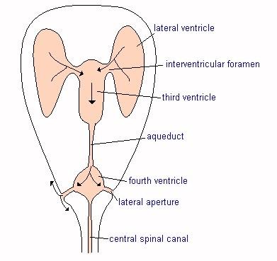
Ventricular System
The brain's ventricular system consists of four cavities, all of which
are connected with each other and the central canal of the spinal cord.
The four ventricles are known as the two lateral ventricles (also called
the first and
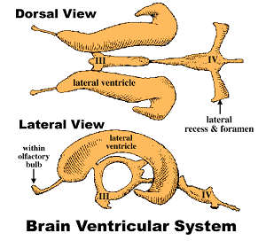 second ventricles), the third ventricle, and the fourth
ventricle. The ventricles are the source of CSF and are the brain's
repository of CSF.
second ventricles), the third ventricle, and the fourth
ventricle. The ventricles are the source of CSF and are the brain's
repository of CSF.
A lateral ventricle is in each of the two cerebral hemispheres. The third ventricle (III in diagram at right) is in the diencephalon. The fourth ventricle (IV) is in both the metencephalon (the cerebellum and pons) and the myelencephalon (the medulla oblongata). The third and fourth ventricles are connected by a mesencephalic aqueduct located in the mesencephalon (the midbrain). CSF flows from the fourth ventricle through two exits, a lateral recess and foramen.
In brachycephalic dogs, the lateral cerebral ventricles can be relatively large compared to mesaticephalic dogs. In general, a rule-of-thumb being that, the smaller the dog, the larger the ventricles.
RETURN TO TOP
Cerebrospinal Fluid (CSF)
CSF is a mostly clear, transcellular fluid produced from blood
plasma, which continuously circulates through
 the
brain and down and back up the spinal cord, to cushion the brain and the
spinal cord and the central nervous system (CNS), regulate changes in
intra-cranial pressure, and also supply them with nutrients, and rid
them of waste products. CSF is a constant secretion from tissues in the
ventricles, and fresh CSF replaces older CSF which is absorbed through
the lymphatics in the base of the skull and also into the bloodstream
(called "venous sinus drainage"), so that the same volume of CSF is
maintained at all times.
the
brain and down and back up the spinal cord, to cushion the brain and the
spinal cord and the central nervous system (CNS), regulate changes in
intra-cranial pressure, and also supply them with nutrients, and rid
them of waste products. CSF is a constant secretion from tissues in the
ventricles, and fresh CSF replaces older CSF which is absorbed through
the lymphatics in the base of the skull and also into the bloodstream
(called "venous sinus drainage"), so that the same volume of CSF is
maintained at all times.
Breathing controls the movement of CSF through the ventricular system. Upon deep inhalation and them exhalation, the CSF flows in one direction or the opposite. Apnea (the temporary cessation of breathing) particularly involuntarily while the dog is asleep, Airway disorders in general, such as inhalation resistance, intermittent reductions in oxygen flow, and other disorders common to brachycephalic breeds, will disrupt the flow of CSF.
RETURN TO TOP
Ventriculomegaly
 Ventriculomegaly is the medical term for oversized or enlarged
ventricles (also, "ventricular distension" or "distention"). Ventriculomegaly results
when excessive amounts of CSF are produced, either by increased
production, or obstruction of its flow, or the failure of waste-product
CSF to be absorbed into the lymphatics and the bloodstream.
Ventriculomegaly is the medical term for oversized or enlarged
ventricles (also, "ventricular distension" or "distention"). Ventriculomegaly results
when excessive amounts of CSF are produced, either by increased
production, or obstruction of its flow, or the failure of waste-product
CSF to be absorbed into the lymphatics and the bloodstream.
Enlarged ventricles in brachycephalic dogs are associated with the loss of cerebral white matter. It is believed that increased CSF pressure and distension of the ventricular system cause the stretching of blood vessels and of white matter fibers, causing reduced blood flow as well as damage to the white matter.
In the cavalier King Charles spaniel, the primary cause of ventriculomegaly is attributed to obstruction of the CSF's pathway. Two roadblocks to CSF flow in the CKCS have been suggested: (1) overcrowding of the caudal cranial fossa (CCF); and (2) reduced absorption of waste-product CSF. The CCF is the inner portion of the skull which houses the brainstem and cerebellum. The CCF of the cavalier is abnormally smaller in size than in other breeds of its size, and this condition is attributed to the cavailer being a brachycephalic breed with a brain oversized for its skull and/or a skull undersized for its brain.
The result of the obstruction of CSF flow through the brain is excessive production of CSF and forced enlargement of the ventricles, hence: ventriculomegaly. Ventriculomegaly is enlargement of the ventricles without any clinical signs (asymptomatic), such as visible enlargement of the skull, as in hydrocephalus.
However, researchers have found direct relationships between enlarged ventricles and both Chiari-like malformation (CM) and syringomyelia (SM) in the CKCS. Cavaliers with CM/SM have been found to have reduced volume CCF sinuses. Venous sinuses are a network of channels in the brain, which receive blood from the brains veins and also receive waste CSF and empty blood into the jugular vein.
In a June 2013 report, UK and German neurology researchers Joe Fenn, Martin J. Schmidt, Harriet Simpson, Colin J. Driver, and Holger A. Volk, having compared 22 cavaliers with SM and 12 without SM, found that in CKCSs with SM the percentage of space taken by venous sinuses in the brain is significantly lower than the volume occupied by the brain's parenchyma. The report concludes that: "These results support a role for reduced venous drainage and parenchymal 'overcrowding' of the CCF [caudal cranial fossa] in the pathophysiology of SM."
In an October 2016 abstract, German researchers compared the perfusion of blood in the periventricular white matter of 23 cavalier King Charles spaniels with ventriculomegaly compared to control dogs consisting of 10 healthy Beagles. They found that cerebral blood flow and volume were significantly lower in the cavaliers. They concluded that the dogs with ventriculomegaly may have a form of normal pressure hydrocephalus (NPH).
In a July 2022 article, Italian researchers reviewed the clinical records of 43 cavaliers diagnosed with CM, to to calculate the sizes of their lateral ventricles and determine if there is any the association between ventriculomegaly and clinical signs, or ventricular asymmetry. Ventriculomegaly was identified in 70% of dogs. They concluded that "the prevalence of ventriculomegaly in Cavalier King Charles Spaniels is high".
Below is a comparison between a canine brain with normal lateral cerebral ventricles (A) and one with enlarged lateral ventricles (B). From a May 2015 study led by Dr. Martin J. Schmidt.
In a November 2023 article, Dr. Schmidt and cohorts proposed a standardized grading system of ventricular distension, ranging from Grade 0 (normal lateral ventricles) to Grade 1 (minimally) to Grade 2 (mildly) to Grade 3 (moderately) to Grade 4 (severely) to Grade 5 (end stage).
RETURN TO TOP
Hydrocephalus
Hydrocephalus is related to ventriculomegaly in that it is defined as an active stretching (distention or distension) of the brain's ventricular system, due to inadequate flow of CSF from the point of its production within the ventricles to its point of elimination (absorption by the lymphatics and/or venous sinuses). Cavaliers with Chiari-like malformation and syringomyelia (CM/SM) have reduced volume venous sinuses.
Hydrocephalus is classified as either "obstructive", if there is blockage of flow in the CSF channels, or "communicating", due to imbalances in CSF production or elimination. Most cases are obstructive.
Hydrocephalus also is distinguished from ventriculomegaly in that the hydrocephalic cavalier suffers clinical symptoms, such as neurological abnormalities, enlarged forebrain skulls, head and neck pain, difficulties in walking and judging distances, among a variety of other odd behaviors. Hydrocephalus creates increased pressure within the cranium and may lead to degeneration of brain tissue.
While obstruction of CSF flow is the main cause of ventriculomegaly
and hydrocephalus, contributing factors may include reduced movement of
CSF caused by other conditions, such as sleeping disorders, particularly
sleep apnea (the temporary cessation of breathing while asleep) and
other inabilities to inhale and exhale deeply. These are hallmarks of
brachycephalic airway obstruction syndrome (BAOS), which is
characterized by upper
 respiratory tract abnormalities resulting in
significant upper airway obstruction.
respiratory tract abnormalities resulting in
significant upper airway obstruction.
In some cases of cavaliers having hydrocephalus, the growth plates in the forehead of the skull are prevented from fusing together as the puppy matures, leaving a soft spot between the eyes or above them, called an open bregmatic fontanel (fontanelle) also called the molera. (See photo at right.)
Some researchers have described ventriculomegaly in cavaliers as a form of "normal pressure hydrocephalus" (NPH). In an October 2016 abstract, German researchers compared the perfusion of blood in the periventricular white matter of 23 cavalier King Charles spaniels with ventriculomegaly compared to control dogs consisting of 10 healthy Beagles. They found that cerebral blood flow and volume were significantly lower in the cavaliers. They concluded that the dogs with ventriculomegaly may have a form of normal pressure hydrocephalus (NPH).
In an August 2017 article, Dr. Schmidt and his team studied the ventricle system of 42 cavaliers -- 32 CKSCs with ventriculomegaly and 10 control CKCSs. They used "dynamic susceptibility contrast perfusion" magnetic resonance imaging (DSC-PMRI), which allows them to quantify the volume of blood passing through the brain tissue. They found that cerebral blood flow (CBF) is reduced in the periventricular white matter of CKCSs with ventriculomegaly, which makes some increase of intraventricular pressure likely. They stated that these findings make it plausible that ventriculomegaly may be a form of internal hydrocephalus. They observed that when intraventricular pressure is increased, a higher cerebrospinal fluid (CSF) volume is forced from the ventricles at a higher velocity.
RETURN TO TOP
Symptoms
B y definition, cavaliers which have
ventriculomegaly do not display
any outward symptoms. Signs that a dog may suffer from hydrocephalus
can be variable, including:
y definition, cavaliers which have
ventriculomegaly do not display
any outward symptoms. Signs that a dog may suffer from hydrocephalus
can be variable, including:
• Domed or apple-shaped skull
• Wide set eyes
• Open fontanel (molera)
• Lack of cordination of legs
• Kicking out front legs
• Weak hind legs
• Restless or erratic behaviors
• Compulsive circling*
• Diffiulty eating and drinking
• Difficulty training
• Mental dullness
• Blindness
• Seizures
* See Dr. Clare Rusbridge's YouTube video on the specific causes of circling.
RETURN TO TOP
Diagnosis
An initial diagnosis of hydrocephalus is a visual one, based upon the clinical signs, such as the facial symptoms, particularly the domed skull and possibly open fontanel. Ventriculomegaly cannot be diagnosed in that manner, because it shows no outward signs.
Ultrasound imaging of the brain can be used to diagnose an open fontanel and through it to show enlargement of the brain's lateral ventricles. However, hydrocephalus is not necessarily based upon the size of the ventricles. If the height of the lateral ventricle is greater than 25% of the height of the brain, that would indicate significant ventriculomegaly. If ventricle height is greater than 65% of the brain, then that would indicate that hydrocephalus is likely.
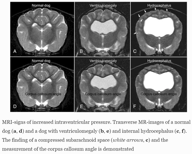
More advanced imaging, computed tomography (CT) and especially magnetic resonance imaging (MRI), is necessary to fully determine the extent of the ventriculomegaly or hydrocephalus, to identify obstructive lesions, and to rule out other disorders. MRI is more sensitive than CT in diagnosing these disorders. However, CT is useful for follow-up of previously diagnosed patients. (See below, the MRI images comparing normal dogs with dogs diagnosed with ventriculomegaly and hydrocephalus. From Laubner 2015.)
RETURN TO TOP
Treatment
Treatment options include medications and/or surgery.
Medication
The main goals of medicine are (a) to reduce CSF production and (b)
to treat symptoms such as seizures. Medical treatment is used for milder cases or when surgery is not an
option, and for short-term care of severe cases before surgery can be
scheduled. However, medical management is not curative of hydrocephalus.
Nevertheless, patients with mild or moderate symptoms may be managed on
a long-term basis with
 prescription
drugs.
prescription
drugs.
Medicines prescribed to decrease production of CSF and provide temporary relief of symptoms include:
• Acetazolamide (Diamox), a carbonic anhydrase inhibitor that decreases CSF production (10 mg/kg PO 3 times a day). Dr. Rusbridge provides a detailed discussion of acetazolamide on her YouTube channel at this link.
• Furosemide, a loop diuretic which deceases CSF formation, for emergency use only (0.5-2.0 mg/kg PO once or twice a day). Dr. Rusbridge provides a detailed discussion of furosemide on her YouTube channel at this link.
• Omeprazole, which may reduce CSF production in some dogs (0.5-1.0 mg/kg PO once a day). See this October 2019 article. Dr. Rusbridge also provides a detailed discussion of omeprazole on her YouTube channel at this link.
• Cimetidine (Tagamet, Zitac), which is a histamine H2-receptor antagonist -- an antihistamine. Histamine contributes to inflammation and causes smooth muscles to constrict. Cimetidine is diffused into the cerebrospinal fluid and reportedly may contribute to reducing the flow of CSF. Cimetidine is a potent inhibitor of several families of cytochrome P450 enzymes and can also inhibit transporter pumps and decrease the renal excretion of some drugs, including clearance of many drugs, such as theophylline, lidocaine, midazolam, and propranolol. See this June 2013 report. Dr. Rusbridge provides a detailed discussion of cimetidine on her YouTube channel at this link.
Another H2-blocker occasionally prescribed is famotidine (Pepcid AC). Dr. Rusbridge provides a detailed discussion of famotidine on her YouTube channel at this link.
• Mannitol is an intravenously injected diuretic which treats swelling of the brain in dogs and thereby lowers intra-cranial pressure and expands blood vessel openings in the brain. It is recommended for emergency use only (0.5-1.0 g/kg IV over 15-20 minutes). Mannitol is the alcohol form of the sugar, mannose, and it is an osmotic diuretic and a mild renal vasodilator.
• Isosorbide (hydonaol) is an oral diuretic which has been found to reduce CSF pressure, but only temporarily, and it has adverse side effects which preclude long-term usage.
• Glucocorticoids, such as dexamethasone, methylprednisolone sodium succinate, or prednisone (0.25-0.5 mg/kg PO twice a day), are prescribed to reduce CSF production and attempt to reduce inflammation and eliminate the clinical signs, and may continue to be administered daily or every other day, at low dosages, for the rest of the patient's life. Long term use has consequences, including hyperadrenocorticism. Dr. Rusbridge provides a detailed discussion of corticosteroids on her YouTube channel at this link.
Seizures may be treated with phenobarbitone, an anti-epileptic medication.
For long-term relief of symptoms, there is no good evidence that any drug is helpful. However, there is some evidence that topiramate (Topamax), another anticonvulsant medication, and high doses of omeprazole may be useful. But their adverse effects would limit that usefulness. Dr. Rusbridge provides a detailed discussion of topiramate on her YouTube channel at this link, and at this link.
RETURN TO TOP
Surgery
Surgical shunting is the "definitive" remedy for relieving pressure on the brain caused by excess CSF which is causing the dog severe symptoms, particularly neurological deficits. Its goals are to halt progression of the hydrocephalus as well as relieve the dog of the symptoms of the disorder. It usually is recommended by neurologists as soon as possible in order to avoid the likelihood of more severe symptoms as the hydrocephalus progresses over time.
Ventriculoperitoneal shunt (VPS)
A "ventriculoperitoneal shunt" (VPS) is a flexible
tube, called a catheter, on end of which is placed into one of the
lateral ventricles of the dog's brain, and the other end placed much
farther down the dog's body, to
 drain
excess CSF away from the brain, where it can be absorbed by the body.
The purpose of shunting is to reduce pressure on the brain caused by the
excess CSF.
drain
excess CSF away from the brain, where it can be absorbed by the body.
The purpose of shunting is to reduce pressure on the brain caused by the
excess CSF.
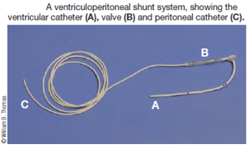
A shunt (right) consists of three basic parts: (1) a ventricular inflow catheter tubing, which drains the CSF from the lateral ventricle; (2) a one-way pressure cutoff valve which regulates the intracranial pressure by controlling the amount of fluid which flows through the tubing; and (3) an outflow (distal) catheter at the other end of the tubing, which is located lower in the dog's body, such as in the abdomen, to be absorbed into the bloodstream and passed through the urinary system. The catheters and tubing usually are made of radiopaque silicone.
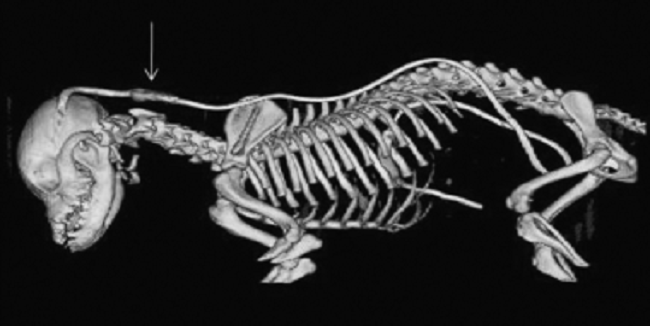
A computed tomography (CT) view showing
location of ventriculoperitoneal shunt placement, from its insertion
into the skull and down into the abdomen. Arrow points to the one-way
pressure cut-off valve.
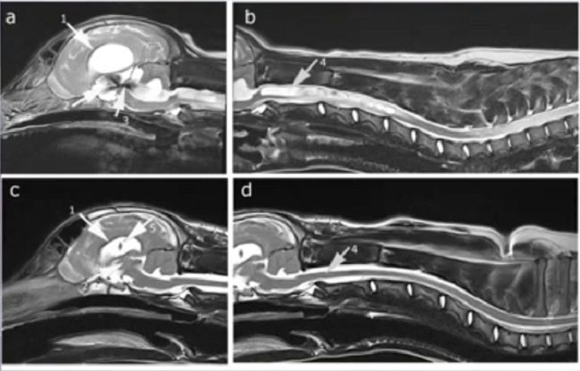
VP shunts have become a standard method of treating hydrocephalus in dogs. At the 2023 meeting of the British Veterinary Neurology Society, Dr. Ana Fernandez Cid reported on the successful insertion of a VPS for management of CM/SM in a cavalier. The series of MRI images below, courtesy of Dr. Rusbridge, show in (a) the arrow pointing to the enlarged ventricles, and in (b) a massive syrinx and developing pre-syrinx, prior to VP shunt surgery. In the post-operative MRI a month later, image (c) shows the two arrows pointing to the reduced size ventricles and the shunt within them. In (d), the arrow points to the completely collapsed syrinx and an almost normal spinal cord.
There are risks of compications and failure of VP shunts (e.g., see this April 2011 article, and this July 2024 article). Proper placement of the ventricular catheter reduces the risk of catheter obstruction. Shunt disconnection and overshunting continue to be serious complications after VPS.
RETURN TO TOP
Ventriculo-atrial shunt
Ventriculo-atrial shunts are intended to divert excess CSF into the dog's internal jugular vein and then into the right atrium of the heart. This shunting method provides permanent intravenous pressure that resists CSF outflow if intraventricular pressure (IVP) falls below intravenous pressure, thereby avoiding overshunting without the need for a shunt valve.
RETURN TO TOP
Ventriculosinusal shunt
Ventriculosinusal shunts provide vascular ventricular drainages to the sagittal-, or transverse sinus. Ae major disadvantage of this technique is the risk of venous thrombosis and sepsis.
RETURN TO TOP
Other shunts
Other shungint options include ventriculopleural shunt, ventriculo-gallbladder shunt, ventriculoureteral shunt, drainage into the thoracic duct, or spinal epidural space, and lumboperitoneal shunt (LPS).
RETURN TO TOP
Dedication
 This article is dedicated to a most charming Blenheim cavalier King Charles
spaniel named Oblio (right), a puppy mill dog who was rescued by a family
who knew of his health issues and gave him the best of care (he also was diagnosed with
Chiari-like malformation and syringomyelia and mitral valve
disease and another brain disorder called lissencephaly, which is
very rare, but Oblio had it). He has lived with the usual symptoms of hydrocephalus: a
dome-shaped head, open fontanel, wide-set eyes, slow growth, partial
blindness, compulsive circling, head tilting, occasional
disorientation, erratic behavior, lack of coordination, seizures,
and difficulty eating without help. Oblio survived the hydrocephalus
but passed away in 2022 at the age of 10 years, from advanced congestive heart failure due to
his MVD. He was a happy boy who lived a full life, and his family
has documented much of it on his Facebook page,
Oblio's Point.
This article is dedicated to a most charming Blenheim cavalier King Charles
spaniel named Oblio (right), a puppy mill dog who was rescued by a family
who knew of his health issues and gave him the best of care (he also was diagnosed with
Chiari-like malformation and syringomyelia and mitral valve
disease and another brain disorder called lissencephaly, which is
very rare, but Oblio had it). He has lived with the usual symptoms of hydrocephalus: a
dome-shaped head, open fontanel, wide-set eyes, slow growth, partial
blindness, compulsive circling, head tilting, occasional
disorientation, erratic behavior, lack of coordination, seizures,
and difficulty eating without help. Oblio survived the hydrocephalus
but passed away in 2022 at the age of 10 years, from advanced congestive heart failure due to
his MVD. He was a happy boy who lived a full life, and his family
has documented much of it on his Facebook page,
Oblio's Point.
RETURN TO TOP
Research News
July 2022:
Italian study of 43 cavaliers with Chiari-like malformation
showed no relationship with enlarged lateral ventricles.
 In a
July 2022 article, Italian researchers (Federica Tirrito [right], Francesca
Cozzi, Martina Bonaldi, Stefania Corazzo, Barbara Contiero, Rocco
Lombardo) reviewed the clinical records of 43 cavalier King Charles
spaniels diagnosed with Chiari-like malformation (CM), to to calculate
the sizes of their lateral ventricles and determine if there is any the
association between ventriculomegaly (dilated -- enlarged -- lateral
ventricles) and clinical signs, ventricular asymmetry, grade of CM
syringomyelia (SM) and degree of medullary kinking. Twenty-eight (65%)
dogs were male and 15 (35%) were female. The most common initial
clinical signs were scratching and neck pain. Ventriculomegaly was
identified in 70% of dogs, Chiari-like malformation grade 2 [(CM2);
cerebellum impacted or herniated through the foramen magnum] was
observed in 77% of cases, ventricular asymmetry in 54% and SM in 80% of the
cavaliers. The median medullary kinking index was 37.77%, and 28% of the
dogs had epileptic seizures. They report finding:
In a
July 2022 article, Italian researchers (Federica Tirrito [right], Francesca
Cozzi, Martina Bonaldi, Stefania Corazzo, Barbara Contiero, Rocco
Lombardo) reviewed the clinical records of 43 cavalier King Charles
spaniels diagnosed with Chiari-like malformation (CM), to to calculate
the sizes of their lateral ventricles and determine if there is any the
association between ventriculomegaly (dilated -- enlarged -- lateral
ventricles) and clinical signs, ventricular asymmetry, grade of CM
syringomyelia (SM) and degree of medullary kinking. Twenty-eight (65%)
dogs were male and 15 (35%) were female. The most common initial
clinical signs were scratching and neck pain. Ventriculomegaly was
identified in 70% of dogs, Chiari-like malformation grade 2 [(CM2);
cerebellum impacted or herniated through the foramen magnum] was
observed in 77% of cases, ventricular asymmetry in 54% and SM in 80% of the
cavaliers. The median medullary kinking index was 37.77%, and 28% of the
dogs had epileptic seizures. They report finding:
• No significant association between the grade of SM and class of ventriculomegaly.
• No significant association between dimension of lateral ventricles and any other conditions.
• No significant association between medullary kinking index and class of ventriculomegaly.
• No significant association between primary secretory otitis media (PSOM) and any clinical signs.
They concluded that "the prevalence of ventriculomegaly in Cavalier King Charles Spaniels is high but this finding does not seem related to the severity of clinical signs, presence of Chiari-like malformation, syringomyelia and craniocervical junction abnormalities such as medullary kinking." (See a September 2021 article on an abstract of the same study, below.)
 March 2022:
Lateral ventriculomegaly is found in 23 of 30 cavaliers, in a UK
MRI study.
In
a March
2022 article, UK researchers Carina Rotter (right),
Danielle Whittaker, and Clare Rusbridge reviewed the medical records of
39 cavalier King Charles spaniels diagnosed with myocluonus, a syndrome
displaying symptoms of spasmodic jerky contractions of groups of
muscles, mainly of the head and forelimbs when the dog is standing or
sitting. In this study, 23 of 30 were diagnosed by MRI to have lateral
ventriculomegaly, which was mild in 9/23 dogs, moderate in 11 and marked
in 3 dogs. One dog had hydrocephalus which was managed with placement of
a ventriculo-peritoneal shunt.
March 2022:
Lateral ventriculomegaly is found in 23 of 30 cavaliers, in a UK
MRI study.
In
a March
2022 article, UK researchers Carina Rotter (right),
Danielle Whittaker, and Clare Rusbridge reviewed the medical records of
39 cavalier King Charles spaniels diagnosed with myocluonus, a syndrome
displaying symptoms of spasmodic jerky contractions of groups of
muscles, mainly of the head and forelimbs when the dog is standing or
sitting. In this study, 23 of 30 were diagnosed by MRI to have lateral
ventriculomegaly, which was mild in 9/23 dogs, moderate in 11 and marked
in 3 dogs. One dog had hydrocephalus which was managed with placement of
a ventriculo-peritoneal shunt.
September 2021:
No relationship between severity of CM/SM and ventriculomegaly is
found in 43 cavaliers in Italian study.
In a
September 2021 presentation
at the 2021 ESVN-ECVN Symposium, a team of Italian veterinary
neurological researchers (Federica Tirrito [right], F. Cozzi,
S. Corazzo, B. Contiero, R. Lombardo) examined the MRI scans of 43
cavalier King Charles spaniels diagnosed with Chiari-like malformation
(CM) to determine if
ventriculomegaly is associated with CM and syringomyelia (SM), based upon symptoms,
ventricular shape, the grades of CM and SM, kinking of the medulla, and
epileptic seizures. They report finding that the common initial clinical
signs were scratching and neck pain. In addition:
• Ventriculomegaly was identified in 70% of the dogs;
• CM grade 2 was observed in 77% of cases;
• ventricular asymmetry was identified in 54% of the dogs;
• SM was identified in 80% of the dogs;
• median medullary kinking index was 37.77%;
• 28% of the dogs presented epileptic seizures.
They found no significant association between dimension the of the lateral ventricles and either any symptoms or MRI findings, and no significant association was identified between ventriculomegaly and epilepsy. They concluded that the prevalence of ventriculomegaly in CM-affected cavaliers is high but does not appear to be related to the severity of clinical signs, to CM/SM, to medullary kinking index, and/or to epileptic seizures. (See a July 2022 article on the full article of the same study, above.)
RETURN TO TOP
RETURN TO TOP
Veterinary Resources
Neurological signs and results of magnetic resonance imaging in 40 cavalier King Charles spaniels with Chiari type 1-like malformations. Lu D, Lamb CR, Pfeiffer DU, Targett MP. Vet Rec. Aug 2003;153(9):260-3. Quote: In human beings a Chiari type 1 malformation is a developmental condition characterised by cerebellar herniation and syringohydromyelia. Abnormalities compatible with such a malformation were identified by magnetic resonance imaging in 39 cavalier King Charles spaniels with neurological signs and in one neurologically normal cavalier King Charles spaniel that was examined postmortem. The dogs with these abnormalities had a wide variety of neurological signs, but there was no apparent correlation between the neurological signs and the severity of cerebellar herniation, syringohydromyelia or hydrocephalus.
Identification of Arachnoid Cysts in the Quadrigeminal Cistern Using Ultrasonography. Miyoko Saito, Natasha J. Olby, Kathy Spaulding. Vet. Radiol. & Ultrasound. May 2005; doi: 10.1111/j.1740-8261.2001.tb00966.x. Quote: The ultrasonographic findings in three small breed dogs [2 cavalier King Charles spaniels] with intracranial arachnoid cysts are described. Ultrasound images were obtained via the foramen magnum, temporal window and persistent bregmatic fontanelle (when possible). In transverse, dorsal and sagittal transcranial ultrasound images there was marked dilation of the lateral ventricles and a well-defined, oval to triangular-shaped an-echoic area between the caudal aspect of the occipital lobes, dorsal to the midbrain, and rostral to the cerebellum. These findings were consistent with a diagnosis of concurrent hydrocephalus and an arachnoid cyst within the quadrigeminal cistern.
Treatment of Hydrocephalus with Ventriculoperitoneal Shunting in Twelve Dogs. Nadia Shihab, Emma Davies, Patrick J. Kenny, Shenja Loderstedt, Holger A. Volk. Vet. Surg. April 2011; doi:10.1111/j.1532-950X.2011.00832.x. Quote: Objective: To report use of ventriculoperitoneal shunt in dogs for management of hydrocephalus for which no cause could be identified. Study Design: Case series. Animals: Dogs with hydrocephalus (n=12) [including 1 cavalier King Charles spaniel]. Methods: Medical records (June 2003-June 2009) were reviewed to determine preoperative clinical findings, initial postoperative, and long-term outcome. Additional follow-up information was obtained from owners and referring veterinarians. Results: All dogs had signs of forebrain dysfunction, 7 had vestibular signs and 3 had signs of spinal pain. Postoperative complications included pain (n=4), undershunting because of shunt kinking (n=1) and seizures (n=1). Initial improvement occurred in all dogs and was sustained in 9 dogs, 1 of which required revision surgery. ... Thus, 3 of 12 dogs had died or were euthanatized because of conditions relating to the hydrocephalus or shunt failure. Outcome and Complications: A failure rate of 25% in this group of 12 dogs, similar to that reported for ventriculoatrial shunting in dogs. ... Undershunting because of shunt kinking occurred in 1 dog and was associated neurologic deterioration, but revision surgery resulted in improvement of clinical signs. It is probable that tight loops of redundant catheter, placed to allow for future dog growth, in close proximity to the valve contributed to formation of the kink. ... Overshunting can also lead to the development of subdural hematomas. ... Conclusions: Sustained clinical improvement can be achieved in hydrocephalus with no active underlying cause by use of ventriculoperitoneal shunting.
Volume reduction of the jugular foramina in Cavalier King Charles Spaniels with syringomyelia. Martin Jürgen Schmidt, Nele Ondreka, Maren Sauerbrey, Holger Andreas Volk, Christoph Rummel, Martin Kramer. BMC Vet. Res. September 2012; doi: 10.1186/1746-6148-8-158. Quote: Background: Understanding the pathogenesis of the chiari-like malformation in the Cavalier King Charles Spaniel (CKCS) is incomplete, and current hypotheses do not fully explain the development of syringomyelia (SM) in the spinal cords of affected dogs. This study investigates an unconventional pathogenetic theory for the development of cerebrospinal fluid (CSF) pressure waves in the subarachnoid space in CKCS with SM, by analogy with human diseases. In children with achondroplasia the shortening of the skull base can lead to a narrowing of the jugular foramina (JF) between the cranial base synchondroses. This in turn has been reported to cause a congestion of the major venous outflow tracts of the skull and consequently to an increase in the intracranial pressure (ICP). Amongst brachycephalic dog breeds the CKCS has been identified as having an extremely short and wide braincase. A stenosis of the JF and a consequential vascular compromise in this opening could contribute to venous hypertension, raising ICP and causing CSF jets in the spinal subarachnoid space of the CKCS. In this study, JF volumes in CKCSs with and without SM were compared to assess a possible role of this pathologic mechanism in the development of SM in this breed. Results: Computed tomography (CT) scans of 40 CKCSs > 4 years of age were used to create three-dimensional (3D) models of the skull and the JF. Weight matched groups (7-10 kg) of 20 CKCSs with SM and 20 CKCSs without SM were compared. CKCSs without SM presented significantly larger JF -volumes (median left JF: 0.0633 cm3; median right JF: 0.0703 cm3) when compared with CKCSs with SM (median left JF: 0.0382 cm3; median right JF: 0.0434 cm3). There was no significant difference between the left and right JF within each group. Bland-Altman analysis revealed excellent reproducibility of all volume measurements. Conclusion: A stenosis of the JF and consecutive venous congestion may explain the aetiology of CSF pressure waves in the subarachnoid space, independent of cerebellar herniation, as an additional pathogenetic factor for the development of SM in this breed.
The association between Chiari-like malformation, ventriculomegaly and seizures in cavalier King Charles spaniels. C.J. Driver, K. Chandler, G. Walmsley, N. Shihab, H.A. Volk. Vet.J. February 2013;195(2):235-237; doi: 10.1016/j.tvjl.2012.05.014 . Quote: Cavalier King Charles spaniels (CKCSs) with Chiari-like malformation (CM) and associated seizures are frequently diagnosed with idiopathic epilepsy. There could be an association between ventriculomegaly (V) or caudal fossa overcrowding (CCFP) and seizures. A retrospective case-control study was performed using MRI to investigate the possible association between these morphological abnormalities and seizures. Seizure semiology and, where possible, electroencephalographic (EEG) abnormalities were documented. Eighty-five CKCS with CM were included, 27 with seizures. There was no association between V or CCFP and seizures. Seizures were classified as having partial onset [meaning that they occur in in one area of the brain, unlike generalized seizures which typically affect nerve cells throughout the brain] in 61% of individuals in the study population (95% CI 42.41-76.43%). Another cause of recurrent seizures in CKCS (such as familial epilepsy) is suspected, as previously reported.
Outcome of ventriculoperitoneal shunt implantation for treatment of congenital internal hydrocephalus in dogs and cats: 36 cases (2001-2009). Miriam Biel, Martin Kramer, Franck Forterre, Konrad Jurina, Oliver Lautersack, Klaus Failing, Martin J. Schmidt. JAVMA. April 2013; doi: 10.2460/javma.242.7.948. Quote: Objective: To examine outcome data for cats and dogs with congenital internal hydrocephalus following treatment via ventriculoperitoneal shunting to determine treatment-associated changes in neurologic signs, the nature and incidence of postoperative complications, and survival time. Design: Retrospective multicenter case series. Animals: 30 dogs [including 1 cavalier King Charles spaniel] and 6 cats with congenital internal hydrocephalus (confirmed via CT or MRI). Procedures: Medical records for dogs and cats with internal hydrocephalus that underwent unilateral ventriculoperitoneal shunt implantation from 2001 through 2009 were evaluated. Data collected included the nature and incidence of postoperative complications, change in clinical signs following surgery, and survival time. To compare pre- and postoperative signs, 2-way frequency tables were analyzed with a 1-sided exact McNemar test. Results: 8 of 36 (22%) animals developed postoperative complications, including shunt malfunction, shunt infection, and seizure events. Three dogs underwent shunt revision surgery. Thirteen (36%) animals died as a result of hydrocephalus-related complications or were euthanized. Following shunt implantation, clinical signs resolved in 7 dogs and 2 cats; overall, 26 (72%) animals had an improvement of clinical signs. After 18 months, 20 animals were alive, and the longest follow-up period was 9.5 years. Most deaths and complications occurred in the first 3 months after shunt placement. Conclusions and Clinical Relevance: Results indicated that ventriculoperitoneal shunt implantation is a viable option for treatment of dogs or cats with congenital hydrocephalus. Because complications are most likely to develop in the first 3 months after surgery, repeated neurologic and imaging evaluations are warranted during this period.
Venous sinus volume in the caudal cranial fossa in Cavalier King Charles spaniels with syringomyelia. Joe Fenn, Martin J. Schmidt, Harriet Simpson, Colin J. Driver, Holger A. Volk. Vet.J. June 2013; doi: 10.1016/j.tvjl.2013.05.007 . Quote: Syringomyelia (SM) in Cavalier King Charles spaniels (CKCS) has a complex pathophysiology. Recent studies support a relationship between altered venous drainage and cerebrospinal fluid flow dynamics. The aim of this study was to evaluate the relationship between venous sinus and parenchymal volume within the caudal cranial fossa (CCF) in CKCS with SM (n = 22) and without SM (n = 12) using magnetic resonance venography (MRV). MRI and MRV images were used to obtain volumetric calculations of CCF volume, as well as the percentage of this volume occupied by parenchyma (CCFP%) and venous sinuses (CCFV%). In CKCS with SM, CCFP% was significantly higher, whilst CCFV% was significantly lower than in CKCS without SM. These results support a role for reduced venous drainage and parenchymal 'overcrowding' of the CCF in the pathophysiology of SM.
Comparison of the Relationship between Cerebral White Matter and Grey Matter in Normal Dogs and Dogs with Lateral Ventricular Enlargement. Martin J. Schmidt, Steffi Laubner, Malgorzata Kolecka, Klaus Failing, Andreas Moritz, Martin Kramer, Nele Ondreka. PlosONE. May 2015; doi: 10.1371/journal.pone.0124174. Quote: Large cerebral ventricles are a frequent finding in brains of dogs with brachycephalic skull conformation, in comparison with mesaticephalic dogs. It remains unclear whether oversized ventricles represent a normal variant or a pathological condition in brachycephalic dogs. There is a distinct relationship between white matter and grey matter in the cerebrum of all eutherian mammals. The aim of this study was to determine if this physiological proportion between white matter and grey matter of the forebrain still exists in brachycephalic dogs with oversized ventricles. The relative cerebral grey matter, white matter and cerebrospinal fluid volume in dogs were determined based on magnetic-resonance-imaging datasets using graphical software. In an analysis of covariance (ANCOVA) using body mass as the covariate, the adjusted means of the brain tissue volumes of two groups of dogs were compared. Group 1 included 37 mesaticephalic dogs of different sizes with no apparent changes in brain morphology, and subjectively normal ventricle size. Group 2 included 35 brachycephalic dogs [including 7 cavalier King Charles spaniels] in which subjectively enlarged cerebral ventricles were noted as an incidental finding in their magnetic-resonance-imaging examination. Whereas no significant different adjusted means of the grey matter could be determined, the group of brachycephalic dogs had significantly larger adjusted means of lateral cerebral ventricles and significantly less adjusted means of relative white matter volume. This indicates that brachycephalic dogs with subjective ventriculomegaly have less white matter, as expected based on their body weight and cerebral volume. Our study suggests that ventriculomegaly in brachycephalic dogs is not a normal variant of ventricular volume. Based on the changes in the relative proportion of WM and CSF volume, and the unchanged GM proportions in dogs with ventriculomegaly, we rather suggest that distension of the lateral ventricles might be the underlying cause of pressure related periventricular loss of white matter tissue, as occurs in internal hydrocephalus. ... Conclusion: Brachycephalic dogs with ventriculomegaly can have reduced cerebral white matter. As a predetermined relationship exists between WM and GM mass in the brain of mammals this aberration cannot be interpreted as a physiological condition.
Magnetic resonance imaging signs of high intraventricular pressure - comparison of findings in dogs with clinically relevant internal hydrocephalus and asymptomatic dogs with ventriculomegaly. Steffi Laubner, Nele Ondreka, Klaus Failing, Martin Kramer, Martin J. Schmidt. BMC Vet. Res. August 2015; doi: 10.1186/s12917-015-0479-5. Quote: Background: Magnetic resonance imaging (MRI) findings of canine brains with enlarged ventricles in asymptomatic dogs were compared to those in dogs with clinically relevant internal hydrocephalus, in order to determine the imaging findings indicative of a relevant increase in intraventricular pressure. Discrimination between clinically relevant hydrocephalus and ventriculomegaly based on MRI findings has not been established yet and is anything but trivial because of the wide variation in ventricular size in different dog breeds and individuals. The MRI scans of the brains of 67 dogs of various breeds, skull conformation and weight were reviewed retrospectively. Based on clinical and imaging findings, the dogs were divided into three groups: a normal group (n=20), a group with clinically silent ventriculomegaly (n=25) [including 4 cavalier King Charles spaniels] and a group with severe clinically relevant internal hydrocephalus (n=22) [including 1 CKCS]. In addition to the ventricle/brain-index, a number of potential subjective signs of increased intraventricular pressure were recorded and compared between the groups. Results: The ventricle/brain-index was significantly higher in dogs with relevant hydrocephalus and a threshold value of 0.6 was specified as a discriminator between internal hydrocephalus and ventriculomegaly. Other MR imaging findings associated with clinically relevant hydrocephalus were an elevation of the corpus callosum, dorsoventral flattening of the interthalamic adhesion, periventricular edema, dilation of the olfactory recesses, thinning of the cortical sulci and/or the subarachnoid space and disruption of the internal capsule adjacent to the caudate nucleus. Conclusion: A combination of the abovementioned criteria may support a diagnosis of hydrocephalus that requires treatment.
Perfusion-weighted Magnetic Resonance Imaging of Brachycephalic Dogs with Ventriculomegaly. M. J. Schmidt, A. Hartmann, K. H. von Pückler, L. Hahnsen, N. Ondreka. Vet. Rad. & Ultrasound. October 2016;57(6):650. Quote: Introduction: It has been shown that ventriculomegaly in dogs is associated with a loss of periventricular white matter comparable to clinical relevant internal hydrocephalus or normal pressure hydrocephalus (NPH). The pathophysiology of ventricular distension is unclear currently. The aim of this study was to compare brain perfusion in dogs with and without ventriculomegaly using perfusion-weighted magnetic resonance imaging to clarify, as to whether abnormal perfusion might be involved in the pathophysiology of ventriculomegaly. Materials and methods: Perfusion was measured in 23 Cavalier King Charles spaniels with ventriculomegaly, 10 healthy Beagle dogs were examined as a control group. Perfusion-weighted images were acquired using a dynamic multishot fast-field echo-EPI in a dorsal plane. Forty acquisitions per slice were acquired. Contrast was automatically injected using an automatic pump (0.2 mmol/kg body weight) in 20 ml crystalloid solution with an injection rate of 5 ml/s. Contrast was given at the 10th acquisition. Perfusion was measured using free-hand regions of interest (ROI) in five brain regions: periventricular white matter, caudate nucleus, parietal cortex, hippocampus, and thalamus using the STROKETOOL software tool. Time to peak (TTP), regional cerebral blood flow (rCBF), regional blood volume (rCBV), and mean transit time (MTT) was measured. Intraobserver variability was measured using kappa statistics. Differences between groups were tested using Mann-Whitney U-tests. Significance level was <0.05. Results: rCBF and rCBV were significantly lower in the periventricular white matter of the dogs with ventriculomegaly compared to control dogs but not for the other ROIs. Kappa statistics revealed substantial agreement between measurements (0.82). Discussion: Based on the differences in periventricular perfusion ventriculomegaly should be interpreted as a form of NPH as suggested previously. The impaired perfusion might be the reason for the reduced white matter volume in these dogs. This has been shown in laboratory rodents with experimentally induced hydrocephalus and humans with NPH. Cognitive studies should be conducted to investigate a possible impairment of memory, learning, and spatial orientation as present in humans with NPH.
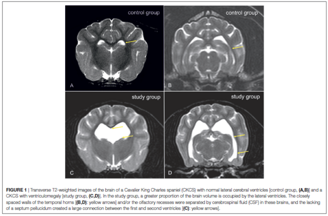
Dynamic Susceptibility Contrast Perfusion Magnetic Resonance Imaging Demonstrates Reduced Periventricular Cerebral Blood Flow in Dogs with Ventriculomegaly. Martin J. Schmidt, Malgorzata Kolecka, Robert Kirberger, Antje Hartmann. Vet. Neuro. & Neurosurgery. August 2017; doi: 10.3389/fvets.2017.00137. Quote: The nature of ventriculomegaly in dogs is still a matter of debate. Signs of increased intraventricular pressure and atrophy of the cerebral white matter have been found in dogs with ventriculomegaly, which would imply increased intraventricular pressure and, therefore, a pathological condition, i.e., to some extent. Reduced periventricular blood flow was found in people with high elevated intraventricular pressure. The aim of this study was to compare periventricular brain perfusion in dogs with and without ventriculomegaly using perfusion weighted-magnetic-resonance-imaging to clarify as to whether ventriculomegaly might be associated with an increase in intraventricular pressure. Perfusion was measured in 32 Cavalier King Charles spaniels (CKCS) with ventriculomegaly, 10 CKCSs were examined as a control group. ... The presence of ventriculomegaly was based on the following criteria. Dogs with a normal ventricular system have very narrow and slit-like horns of the lateral ventricles. In the finding of ventriculomegaly, the interpreter subjectively noted a greater proportion of the intracranial volume occupied by the lateral ventricles. The closely spaced walls of the temporal horns and/ or the olfactory recesses were separated by cerebrospinal fluid (Figures 1C,D: yellow arrows) in these brains and the lacking of a septum pellucidum created a large connection between the first and second ventricles (Figures 1C,D: blue arrows). CKCSs without these findings were used as a control (Figures 1A,B). ... Cerebral blood flow (CBF) was measured using free-hand regions of interest (ROI) in five brain regions: periventricular white matter, caudate nucleus, parietal cortex, hippocampus, and thalamus. CBF was significantly lower in the periventricular white matter of the dogs with ventriculomegaly but not in the other ROIs. Reduction of periventricular CBF might imply increase of intraventricular pressure in ventriculomegaly. ... Cerebral blood flow can be reduced in periventricular white matter in CKCSs with ventriculomegaly, which makes some increase of intraventricular pressure likely.
Hydrocephalus in Animals. Martin Schmidt, Nele Ondreka. Pediatric Hydrocephalus. January 2019; doi: 10.1007/978-3-319-27250-4_36. Quote: Naturally occurring internal hydrocephalus is diagnosed in all kinds of mammals including exotic species as well as in birds. The underlying pathomechanisms are extremely variable and species-specific. ... In dogs and cats, it can be congenital but also associated with impaired skull and vertebral growth. Reduced cranial capacity impairing cerebral compliance and malformations of the craniovertebral junction (atlantoaxial instability, occipito-atlantoaxial overlap syndrome, and "Chiari-like malformation") are the most common causes for an impaired CSF flow and communicating hydrocephalus in a high number of brachycephalic breeds. ... Some but not all dogs with CLM and SM do also have an internal hydrocephalus. Interestingly, some of these dogs only show increased volume of the lateral ventricles with normal dimensions of the third and fourth ventricles. It is unclear which factor influences the additional distension of the compartments and why not all parts of the ventricular system develop pre-stenotic dilation. ... A stenosis of the jugular foramen has been found in the brachycephalic Cavalier King Charles spaniels. This stenosis and a consecutive vascular compromise in the foramen may contribute to venous hypertension and increased intraventricular and intracranial pressure. ... With increasing knowledge and the increasing disposition of patient owners, veterinary specialists and researchers enduringly invest in the patient management; ventriculoperitoneal shunting techniques have become a reasonable treatment strategy in dogs.
Association between improvement of clinical signs and decrease of ventricular volume after ventriculoperitoneal shunting in dogs with internal hydrocephalus. Martin J. Schmidt, Antje Hartmann, Daniela Farke, Klaus Failling, Malgorzata Kolecka. J. Vet. Intern. Med. April 2019; doi: 10.1111/jvim.15468. Quote: Background: One of the remaining questions in treating dogs with internal hydrocephalus is the association between the decrease of ventricular volume and re-expansion of cerebral parenchyma with clinical improvement. Hypothesis: A decrease in ventricular volume and re-expansion of brain tissue occur after ventriculoperitoneal shunting (VPS). Clinical improvement defined by resolution of >1 clinical signs is associated with decreased size of cerebral ventricles and that the extent of change in ventricular size is associated with clinical improvement. Animals: Forty-five client-owned dogs [including 1 cavalier King Charles spaniel] with newly diagnosed communicating internal hydrocephalus. Methods: Ventricular volume, brain volume, and clinical status of dogs that underwent VPS were measured before and 3 months after surgery. Multiple logistic regression analysis was performed to assess the influence of decrease in ventricular size in addition to the covariates "age of the animal" and "duration of clinical signs before surgery" on improvement of clinical signs. Results: Decreased volume of cerebral ventricles was associated with resolution of >1 preoperative clinical sign. The covariates "age of the animal" and "duration of clinical signs" were not associated with improvement of clinical signs. The percentage decrease in ventricular size was associated with resolution of ataxia and obtundation. Conclusion and Clinical Importance: The decrease in ventricular volume and increase in brain parenchyma after VPS are associated with improvement in clinical signs.
Results of oral prednisolone administration or ventriculoperitoneal shunt placement in dogs with congenital hydrocephalus: 40 cases (2005-2016). Sabrina Gillespie, Zoe Gilbert, Steven De Decker. JAVMA. April 2019; doi: 10.2460/javma.254.7.835. Quote: Objective: To evaluate signalment, clinical findings, and outcomes of dogs with congenital hydrocephalus treated medically with orally administered prednisolone or surgically by ventriculoperitoneal shunt placement. Design: Retrospective case series. Animals: 40 client-owned dogs [including 1 cavalier King Charles spaniel]. Procedures: Medical records from 2005 to 2016 were searched to identify dogs with congenital hydrocephalus confirmed by MRI examination. Patients were categorized by treatment (medical vs surgical). Signalment, clinical signs, neurologic examination findings, results of diagnostic tests, duration of hospitalization, complications potentially related to treatment, and follow-up information were recorded. Outcome was categorized on the basis of clinical (neurologic) signs as improved, stabilized, or deteriorated. Variables of interest were compared between groups by Fisher exact or Mann-Whitney U tests. Results: 28 and 12 dogs had surgical and medical treatment, respectively; 3 medically treated dogs subsequently underwent ventriculoperitoneal shunt placement. No significant differences were noted in clinical or imaging findings between surgically and medically treated dogs. Median follow-up time was 9 months and 15.5 months for medically and surgically treated dogs, respectively. Of 12 medically treated dogs, 6 improved and 6 deteriorated. Of 26 surgically treated dogs with data available, 14 (54%) improved, 1 (4%) stabilized, and 11 (42%) deteriorated; 4 (15%) had known postoperative complications. Conclusions and Clinical Relevance: Approximately half of the dogs treated with prednisolone in this population had neurologic improvement at last follow-up; results of surgical treatment were comparable to those in previous studies. Further research is needed to assess factors associated with acceptable outcomes for dogs with congenital hydrocephalus.
Medical therapy using omeprazole in 12 hydrocephalic dogs: clinical, diagnostic, and therapeutic findings. Lidianne F. Pelegrini, Natalino F. Silva, Olga P.S. Campos, Clazielen C. Nery, Felipe M. Silva,, Raquel S. Lemos, Kelly C.I. Yamauchi, Alexandre M. Amude. Brazilian J. Vet. Res. October 2019; doi: 10.1590/1678-5150-PVB-6332. Quote: According to experimental studies with healthy dogs, omeprazole might decrease the CSF production by about 26%; therefore, book texts have been suggested the usage of omeprazole in medical protocols for hydrocephalus treatment. However, to the best knowledge of the authors, the usage and medical response of the omeprazole with substantial group of illness dogs, such as hydrocephalic animals, was lacking. This report describes clinical, diagnostic, and therapeutic findings in 12 dogs with hydrocephalus in which omeprazole were used for medical treatment. The diagnosis of hydrocephalus was accomplished by transcranial sonography (TCS) and/or computed tomography. The ventricular measurement was assessed periodically by TCS during medical treatment. Six dogs were diagnosed with non-obstrutive hydrocephalus and in the other 6 cases hydrocephalus occurred with other concomitant anomalous encephalic disease often related with obstructive hysdrocephalus, such as quadrigeminal cist, arachnoid cyst, chiary-like malformation, and syringomyelia. All of them had medical improvement after the use of omeprazole and the most of the cases had ventricular size reduction. In 10 dogs, the omeprazole was used as single drug, and in 2 dogs medical treatment with steroids and/or diuretics was previously being performed, and omeprazole was added because conventional treatment was resulting in mild to unsatisfactory medical control of the neurological status. The results of this paper shown that omeprazole may be used to ameliorate the neurological status in symptomatic hydrocephalic dogs. This work may represent the first description about the use of omeprazole in order to treat a substantial group of affected dogs with suspected increased intracranial pressure by hydrocephalus, probably due to limitation of CSF production.
The Need for Head Space: Brachycephaly and Cerebrospinal Fluid Disorders. Clare Rusbridge, Penny Knowler. Life. February 2021; doi: 10.3390/life11020139. Quote: Brachycephalic dogs remain popular, despite the knowledge that this head conformation is associated with health problems, including airway compromise, ocular disorders, neurological disease, and other co-morbidities. There is increasing evidence that brachycephaly disrupts cerebrospinal fluid movement and absorption, predisposing ventriculomegaly, hydrocephalus, quadrigeminal cistern expansion, Chiari-like malformation, and syringomyelia. In this review, we focus on cerebrospinal fluid physiology and how this is impacted by brachycephaly, airorhynchy, and associated craniosynostosis. ... Cavalier King Charles spaniels with syringomyelia associated with Chiari malformation have reduced volume caudal cranial fossa dorsal sinuses.
Ventriculomegaly in Cavalier King Charles Spaniels with Chiari-like Malformation: Relationship with Clinical Signs and Imaging Findings. F. Tirrito, F. Cozzi, S. Corazzo, B. Contiero, R. Lombardo. 33d ESVN-ECVN Symposium 2021, #P-30. September 2021. Vet. Intern. Med. December 2021; doi: 10.1111/jvim.16332. Quote: Chiari-like malformation (CM) frequently occurs in Cavalier King Charles Spaniels (CKCS). Ventriculomegaly, secondary to disturbance of cerebrospinal fluid flow at the craniocervical junction, has been reported in CKCS with CM. However, its clinical relevance is unclear. The aim of the study was to calculate lateral ventricles dimension in CKCS with CM and to investigate the association between ventriculomegaly and signalment, clinical signs, ventricular asymmetry, CM and syringomyelia (SM) grade, medullary kinking index, follow-up, and epileptic seizures. A retrospective study was performed enrolling forty-three client-owned CKCS with magnetic resonance imaging diagnosis of CM. Initial and follow-up (up to 36 months) clinical status was graded. Images were reviewed to quantify dimension of lateral ventricles, to evaluate ventricular symmetry, CM and SM grade, and medullary kinking index. Cases presenting epileptic seizures were also recorded. The most common initial clinical signs were scratching and neck pain. Ventriculomegaly was identified in 70% of dogs, CM grade 2 was observed in 77% of cases, ventricular asymmetry and syringomyelia were identified in 54% and 80% of dogs, respectively; median medullary kinking index was 37.77%. Moreover, 28% of dogs presented epileptic seizures. No significant association was identified between dimension of lateral ventricles and signalment, clinical signs, and imaging findings; no significant association was identified between ventriculomegaly and epilepsy. In conclusion, the prevalence of ventriculomegaly in CKCS with CM is high but this finding does not seem related to the severity of clinical signs, to CM/SM, to medullary kinking index, and to epileptic seizures.
Myoclonus in older Cavalier King Charles Spaniels. Carina Rotter, Danielle Whittaker, Clare Rusbridge. J. Vet. Intern. Med. March 2022; doi: 10.1111/jvim.16404. Quote: Background: Myoclonus is observed in older Cavalier King Charles Spaniels (CKCS) but a full description is lacking. Objectives: The presence, age of onset, characteristics and treatment of myoclonic episodes were retrospectively evaluated in a cohort of CKCS which presented to 1 board-certified neurologist. Clinical data, imaging studies, presence of seizures and their management, as well as other comorbidities were noted. Animals: Thirty-nine CKCS that were presented to 2 institutions between 2001 and 2018 with signs consistent with myoclonus. Clinical examination, blood sampling, advanced diagnostic imaging, cerebrospinal fluid analysis, and record keeping of other comorbidities was performed. ...Magnetic resonance imaging was performed in 30/39 cases, all of which had CM, 23/30 had lateral ventriculomegaly, which was mild in 9/23 dogs, moderate in 11 and marked in 3 dogs. One dog had hydrocephalus which was managed with placement of a ventriculo-peritoneal shunt. Twenty of 30 cases showed signs of SM [syringomyelia], 3 a presyrinx and 4 a central canal dilatation. OME [otitis media with effusion (primary secretory otitis media -- PSOM)] was visible in 13 dogs. ... Methods: This is a retrospective case series, describing the presence of myoclonus in CKCS. Results: Clinical signs reported were spontaneous in onset, lasted a few seconds and consisted of rapid blinking with head nodding and variable extension down the thoracic limbs. Myoclonus occasionally led to stumbling of the thoracic limbs or collapse. Mean age of onset was 8.38 years (SD ±1.96). Thirteen of 39 dogs with myoclonus had paroxysmal events, such as generalized seizures (9/13). ... Twenty cases showed signs of CM-associated pain (CM-P) [Chiari-like malformation associated pain] and 9 dogs had SM specific signs in including phantom scratching (SM-S) [syringomyelia associated phantom scratching]. Two dogs displayed signs of cognitive dysfunction, with 1 dog described as tending to stare blankly into space, showing less interaction with the owner, wandering aimlessly when left alone and signs suggesting anxiety. Both dogs had seizures which were managed with imepitoin. Four dogs had pelvic limb orthostatic tremor, 1/4 involved 1 of the thoracic limbs as well. The tremor was present while standing, disappeared when walking. ... Twenty-three of 39 dogs were prescribed gabapentin/pregabalin due to CM-P and/or SM-S. It is questionable if gabapentin or pregabalin worsened the myoclonus; however, no deterioration of myoclonus after start of gabapentinoids was documented. Two of the cases were prescribed gabapentin after the myoclonus occurred and the myoclonus did not worsen but the nature of this study does not help to draw a conclusion and it remains questionable if myoclonus was drug-induced in these cases. ... Conclusions and Clinical Importance: Myoclonus occurs in middle-aged to older CKCS and seems to be another epiphenomena of this breed. A link to epilepsy might be present. ... 100% of dogs that underwent MRI showed signs of CM and 66% SM, this resembles the overall reported breed prevalence in the general CKCS population. In other words, a link between CM and SM was neither proven nor disproven. The frequency of CM and SM was that expected for the age of the population and dogs could have myoclonus without CM or SM. ... There might be a link with generalized tonic-clonic seizures. It seems to be another epiphenomena for this breed Although the number of CKCS treated successfully with levetiracetam was low this could be a possible treatment for myoclonus in this breed. Further research to substantiate this is needed. (See video #1 and video #2.)
Brain magnetic resonance imaging and histopathology findings in a dog with global brain ischaemia following cardiopulmonary arrest. Jiah Goh, L. M. Eramanis, M. Milne, M. Boller. Australian Vet. J. June 2022; doi: 10.1111/avj.13178. Quote: Background: Global brain ischaemia following cardiopulmonary arrest is uncommonly reported in veterinary medicine yet neurologic injury after arrest is a known morbidity. Case report: An 18-week-old male entire Cavalier King Charles Spaniel-Poodle was referred following 3 days of neurologic abnormalities after cardiopulmonary arrest. After resuscitation, the animal had decerebrate rigidity, a stuporous mentation and intermittent episodes of vocalisation and apnoea. A brain magnetic resonance imaging (MRI) was undertaken 4 days after cardiopulmonary arrest, with standard sequences (T1-weighted, T2-weighted and fluid-attenuated inversion recovery) as well as diffusion-weighted imaging to better discern ischaemic injury and cytotoxic oedema for prognostic reasons. MRI findings were consistent with global brain ischaemia [GBI] affecting the hippocampus, cerebellum and substantia nigra, the latter two not previously identified in canine cases of global brain ischaemia. ... In conjunction with patient history, MRI findings were interpreted as GBI with cytotoxic oedema, cerebellar herniation and obstructive hydrocephalus. Cerebrospinal fluid analysis was not performed due to the presence of cerebellar herniation and associated high risk of morbidity. ... The patient was euthanased on day eight post-cardiopulmonary arrest due to a lack of neurological improvement and developing sepsis as a complication. Ante-mortem identification of affected areas of the brain was confirmed on histological examination, with evidence of ischaemic injury seen in the cerebrum, hippocampus, cerebellum, basal nuclei and thalamus. Conclusion: This report describes ante-mortem MRI and postmortem findings in a dog with global brain ischaemia following cardiopulmonary arrest. A multimodal approach to neuroprognostication in patients recovering from cardiopulmonary arrest is recommended.
Reliability and interobserver variability of a grading system of ventricular distension in dogs. Adriana Czerwik, Martin Jürgen Schmidt, Agnieszka Olszewska, Steven Hinz, Kathrin Büttner, Daniela Farke. Front. Vet. Sci. November 2023; doi:10.3389/fvets.2023.1271545. Quote: Introduction: Internal hydrocephalus is the most common malformation of the central nervous system in dogs. Although the grades of ventricular distension have importance for long-term prognosis, there is no standard classification scheme describing the grade of the ventricular distension in dogs. Materials and methods: Magnetic resonance imaging (MRI) scans from 147 dogs [including 13 cavalier King Charles spaniels (9%)] of various breed, sex, skull conformation, and weight were reviewed retrospectively and blinded between three observers. Based on objectively assessable morphologic characteristics, the lateral cerebral ventricles were graded as normal, minimally, mildly, moderately, severely enlarged or end stage (grade 0 to grade 5), respectively. Evans' index or the ventricle brain index was also measured in all animals. Interobserver agreement between a very experienced, experienced, and unexperienced person was evaluated by the Spearman coefficient and kappa tests. Additionally, correlation to the ventricle brain index was determined using the Spearman coefficient and F-tests. Results: The Spearman correlation coefficient reached a very strong correlation (r = 0.97) between the experienced and very experienced observer and a strong correlation (r = 0.91) between the very experienced and unexperienced observer. The kappa value revealed excellent interobserver agreement between the very experienced and experienced observers (weighted kappa 0.91) and moderate between the very experienced and unexperienced observers (weighted kappa 0.75). The ventricular-brain index correlated (r = 0.94, Spearman coefficient test) with the grading system, indicating that a more elevated ratio was related to a more advanced degree of ventricular enlargement. The interobserver agreement with regard to the grade between the neurologist in training and a board-certified neurologist was excellent and between the board-certified neurologist and general practitioner achieved lower values. Conclusion: The presented MRI-based grading of ventricular enlargement is a reliable and functional method for an objective grading of the ventricular system in dogs. Some experience in MRI and brain anatomy is needed for interpretation and grading.
Surgical management of primary and idiopathic internal hydrocephalus in dogs and cats. Martin Jürgen Schmidt, Daniela Farke. Frontiers in Vet. Sci. July 2024; doi: 10.3389/fvets.2024.1435982. Quote: Ventriculoperitoneal shunt placement is an effective method to treat internal hydrocephalus in dogs and cats. Although it has a long history in veterinary medicine, the technique continues to evolve. Despite continuing attempts to reduce the incidence of associated complications, shunt failure remains a major problem, and often leads to multiple hospital admissions. This review gives an overview about current knowledge of ventriculoperitoneal shunting techniques in animals, applicable shunt hardware as well as shunt-associated complications and their prevention and treatment.
CONNECT WITH US